-
Paper Information
- Next Paper
- Paper Submission
-
Journal Information
- About This Journal
- Editorial Board
- Current Issue
- Archive
- Author Guidelines
- Contact Us
American Journal of Medicine and Medical Sciences
p-ISSN: 2165-901X e-ISSN: 2165-9036
2021; 11(1): 7-11
doi:10.5923/j.ajmms.20211101.02
Received: Oct. 21, 2020; Accepted: Nov. 16, 2020; Published: Dec. 28, 2020

Influence of Polyprenols from Alcea Nudiflora and Vitis Vinifera to the Healing Process of Experimental Skin Wounds in Laboratory Animals
 Abstract
 Reference
 Full-Text PDF
 Full-text HTML
Ways Y. V.1, Yusupova S. M.1, Shakhmurova G. A.2, Khushbaktova Z. A.1, Syrov V. N.1
1Institute of Plant Chemistry Named after Academician S.Yu. Yunusov of the Academy of Sciences of the Republic of Uzbekistan, Tashkent, Uzbekistan
2Tashkent State Pedagogical University Named after Nizami, Tashkent, Uzbekistan
Correspondence to: Shakhmurova G. A., Tashkent State Pedagogical University Named after Nizami, Tashkent, Uzbekistan.
| Email: |  |
Copyright © 2021 The Author(s). Published by Scientific & Academic Publishing.
This work is licensed under the Creative Commons Attribution International License (CC BY).
http://creativecommons.org/licenses/by/4.0/

Polyprenols, isolated from Alcea nudiflora and Vitis vinifera, when ones applied regularly to flat, full-thickness wounds on the skin of mice and rats, have a pronounced stimulating effect to the healing process. In this position, they are not inferior in activity, and in some cases, they are superior to well-known medicinal preparations with regenerative activity: sea buckthorn oil and 10% methyl uracil ointment.
Keywords: Polyprenols from Alcea nudiflora, Polyprenols from Vitis vinifera, Wound healing action
Cite this paper: Ways Y. V., Yusupova S. M., Shakhmurova G. A., Khushbaktova Z. A., Syrov V. N., Influence of Polyprenols from Alcea Nudiflora and Vitis Vinifera to the Healing Process of Experimental Skin Wounds in Laboratory Animals, American Journal of Medicine and Medical Sciences, Vol. 11 No. 1, 2021, pp. 7-11. doi: 10.5923/j.ajmms.20211101.02.
1. Introduction
- Polyprenols are one of the most frequently found biologically active substances in plants. They are unsaturated acyclic-branched spirits with a primary hydroxyl group in the terminal isoprene residue. The number and geometric configuration of these residues varies depending on the plant family [1]. Among the various biological activities of polyprenols [2,3,4,5], especially their ability stands out to accelerate regenerative processes in the body [6]. If we think that regeneration is one of the key of mechanisms for the restoration of the organism, both in the course of normal life, and when exposed to traumatic, chemical, thermal and other factors, threatening the viability of individual organs, systems and the organism as a whole, then a full-scale study of polyprenols in this regard will open up great possibilities for the creation of new effective preparations for use in the corresponding pathological conditions. This work presents the results of studying the wound-healing effect of polyprenols isolated from Alcea nudiflora and Vitis vinifera on various experimental animals.Purpose: To evaluate the study of the early healing effect of polyprenols isolated from plants Alcea nudiflora and Vitis vinifera in laboratory animals.
2. Materials and Methods
- We used a outbred white mice - males (12-20 grams) and rats - males (190-200 grams) in the experiments. All animals were kept in stationary vivarium conditions on a normal food ration with free access to water. Experiments with them were carried out in accordance with the rules adopted by the International Convention for the protection of vertebrate animals, used for experimental purposes (Strsburg, 1986). Polyprenols isolated from the leaves of Alcea nudiflora L. [7] and from the leaves of Vitis vinifera subsp. Silvestris [8], conventionally named by us prenalon and vitaprenol, respectively. A pharmacological assessment of the effectiveness of polyprenols as agents, stimulating regenerative processes in the organism was carried out on models of destructive formations on the skin. In animals, at the site of the alleged application of the skin defect, the wool was previously cut, and the remnants of the wool cover were removed with 10% sodium sulfide (moistened for 2 - 3 minutes, followed by rinsing with warm running water).All traumatic procedures in animals were carried out under aseptic conditions under light ether anesthesia. Flat, full-thickness skin wounds in mice were reproduced in the dorsal region, using a special round stamp 9 mm in diameter, in rats – by excision of a skin flap with an area of 230 ± 1.2 mm2 to the fascia [9]. The observation of the healing process of experimental wounds was carried out until the complete recovery of skin defects. The state of the wound surface, the quality and maturity of granulations and the time of epithelialization were recorded. The area of wounds was measured according to Yupatov [10], the morphological assessment of their healing was carried out according to the description of Merkulov [11]. An integral quantitative assessment of the intensity of reparative processes was conducted by determining the value of the so-called reparative index, which takes into account in points the total severity of nine main morphological nonspecific signs of wound healing [12]. In some rats, Teflon rings were implanted into full-thickness circular skin defects on the back to prevent contraction and epithelialization. On the 7th day, the granulation tissue, developed in the defect (the moment of maximum development) was isolated, weighed and examined biochemically (deoxyribonucleic and ribonucleic acids, oxyproline, hexosamines, hexuronic acids, hexoses, sialic acids were determined [13,14]).The studied polyprenols (prenalon and vitaprenol), which are an oily liquid and reference preparations: sea buckthorn oil and 10% methyl uracil ointment (with high regenerative activity [15]), were applied to wound skin defects with a uniform layer once a day throughout the experiment. In parallel, there was a group of rats, the wound surface of which was not subjected to any treatment or was lubricated with sunflower oil (control). The obtained data during the experiments were processed by the method of variation statistics using t-criteria of the Student.
3. The Results of Research
- The carried out experiments gave possibility to establish clearly that the regular application of the studied polyprenols to wound skin defects in mice and rats significantly accelerates their healing. Thus, in mice with clean planar skin wounds, from the first days of observation, the inflammatory reaction around wounds in animals, treated with prenalon and vitaprenol was less pronounced, and regenerative processes from the very beginning were more active than in control. They had less swelling, more meager wound exudate, active development of granulations. At the same time, in parallel with the development of connective tissue and vascularization of wounds, pronounced epithelialization of the wound defect was observed (the growing epithelium was clearly visible as a white rim against a dark red background of forming granulations). Most clearly, all of these processes took place on the wound surface of mice treated with vitaprenol. As a result, the healing of wound defects under its influence occurred on average 10 days earlier than in the control. Prenalon acted somewhat weaker, under its influence the wounds healed only for 7,8 days faster than controls.Prenalon and vitaprenol were superior in activity to sea buckthorn oil. In relation to methyl uracil ointment, their effect was different. Vitaprenol was significantly more active; prenalon had a similar effect (Table 1).
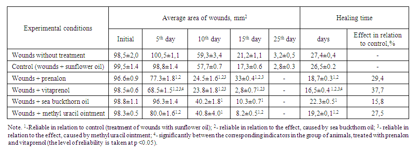 | Table 1. An influence of prenalon and vitaprenol in comparison with sea buckthorn oil and methyl uracil ointment on the healing of experimental skin wounds in mice (M ± m, n = 6) |
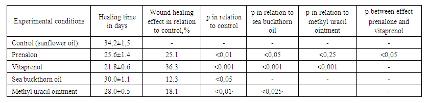 | Table 2. The influence of prenalon and vitaprenol in comparison with sea buckthorn oil and methyl uracil ointment on the timing of healing of full-thickness skin wounds in rats (M ± m, n = 6) |
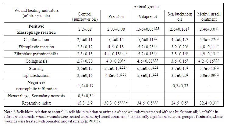 | Table 3. Indicators of the severity of reparative processes in planar full-thickness skin wounds of rats after their regular treatment with prenalon and vitaprenol in comparison with sea buckthorn oil and methyl uracil ointment on the 15th day of observation (M ± m, n = 6) |
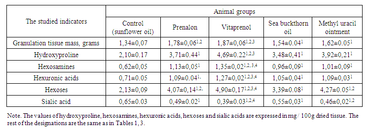 | Table 4. An influence of prenalon and vitaprenol in comparison with sea buckthorn oil and methyl uracil ointment on the development of granulation tissue in planar skin wounds of rats on the 7th day of observation and some indicators of its biochemical composition (M ± m, n = 6) |
4. Conclusions
- The investigated polyprenols isolated from Alcea nudiflora and Vitis vinifera have a pronounced stimulation of regenerative processes and may have an early healing effect.