American Journal of Medicine and Medical Sciences

p-ISSN: 2165-901X    e-ISSN: 2165-9036

2021;  11(1): 1-6

doi:10.5923/j.ajmms.20211101.01

Received: Nov. 22, 2020; Accepted: Dec. 15, 2020; Published: Dec. 26, 2020


New Pathogenetic Mechanisms of Alopecia Areata

Azimova F. V., Sharakhmedov Sh. Sh.

Republic Specialized Scientific-Practical Medical Center of Dermatology and Venerology Ministry of Health of the Republic of Uzbekistan, Tashkent

Correspondence to: Azimova F. V., Republic Specialized Scientific-Practical Medical Center of Dermatology and Venerology Ministry of Health of the Republic of Uzbekistan, Tashkent.

Email:

Copyright © 2021 The Author(s). Published by Scientific & Academic Publishing.

This work is licensed under the Creative Commons Attribution International License (CC BY).
http://creativecommons.org/licenses/by/4.0/

Abstract

The following studies are aimed at clarify the role of stem cells of the hair follicle that determine the rhythm of the latter during circular alopecia. Two important signal molecules, Bone Morphogenetic Protein (BMP) and Wingless-type (WNT), play a key role in the hair cycle during the telogen and anagen stages. We observed 54 patients with circular alopecia aged 18 to 46 years, of whom 12 were males and 42 females. The selection of patients and clinical part of the work was carried out on the basis of RSSPMCDV of MHRUz. The presented data indicate the leading role of BMP and Wnt signaling pathways in the pathogenesis of circular alopecia, which determines mitosis and differentiation of hair follicle cells, and open the perspectives of developing new effective drugs.

Keywords: Alopecia, Erythema, Dermal papilla, Hair follicle

Cite this paper: Azimova F. V., Sharakhmedov Sh. Sh., New Pathogenetic Mechanisms of Alopecia Areata, American Journal of Medicine and Medical Sciences, Vol. 11 No. 1, 2021, pp. 1-6. doi: 10.5923/j.ajmms.20211101.01.

At present, thanks to fundamental research in cytogenetics and biochemistry, the morphology and physiology of the hair follicle has been significantly enhanced. The attention of researchers is drawn to the so-called bulge zone, a thickening located under the sebaceous gland, which some authors called the secondary hair germ. The biochemical and proliferative activity in the bulge zone even during the telogen phase (rest period) has been established and it has been proved that the hair growth cycle in the follicles occurs not only asynchronously, but also independently of the neighboring follicles, etc. [1,2,10,16,24]. During the whole hair cycle the repeated process of regress and regeneration can be observed only in the lower part of the follicle including the suprabulbar and bulbar areas. The internal regulation of the hair cycle is based on the interaction of two key populations of cells in the hair follicle - epithelial cells of the outer root vagina and mesenchymal cells of the dermal papilla and the dermal (connective tissue perifollicular) vagina. However, the location and type of inductors and anagen phase inhibitors have not yet been fully studied. At present, two important signal molecules - (Bone Morphogenetic Protein) (BMP) and (Wingless-type) WNT play a key role in the hair cycle during the telogen and anagen stage [3,5,15,29].
Animal model studies have shown that activation of the Wnt-track is crucial to trigger follicle morphogenesis through activation of hair follicle stem cells. For stimulation of the anagen phase, there are always factors inhibiting the BMP signaling pathways [4,7,25] in addition to the activating Wnt-track signals. The increased activity of BMP-signals has been proved to preserve the hair follicle stem cells at rest during the period of telogen [8,11,13]. Scientists from different countries have carried out experimental studies that revealed that WNT signaling pathway is a key component in activation of Hair Follicle Stem Cells (HFSCs). HFSCs stimulate the regeneration of hair follicle cells, resulting in the growth of new hair. At the same time, the Hair Follicle Stem Cells (HFSCs) stay at rest for a few weeks before the signal for proliferation is received. Studies show that hair loss occurs when hair follicle stem cells (HFSCs) reach their end of life. HFSCs were found to be able to initiate WNT signals on their own, unlike most stem cells in other tissues [9,12,18,23]. The breakdown of the WNT Wnt signaling causes the hair follicle to delay in the telogen phase, which is manifested by the absence of hair growth. The researchers also found that the transmission of signals BMP (Bone Morphogenetic Protein - signal molecule of hair loss) can control the transmission of WNT signals. Uncontrolled transmission of WNT Wnt signals can stimulate premature activity of Hair Follicle Stem Cells (HFSCs), which in turn causes a premature transition to the anagen, which leads to disruption of hair chronobiology. The balanced interaction between the two signaling pathways makes it possible for HFSCs to stay at rest until the pre-programmed anagen stage occurs. Therefore, understanding the basic mechanisms of the hair growth cycle will open the way to discover new methods of hair growth recovery [17,20,26,27].
One of the most difficult-to-treat and recurring diseases is circular alopecia, which is currently considered a tissue specific autoimmune disease mediated by autoactivated T-lymphocytes in conditions of immune tolerance disorders by hair follicles in the anagenic stage. The conditions of manifestation, clinical diversity of circular alopecia are a complex combination of external and genetic factors [6,14,19,21,22,28].

1. The Aim of the Study

Is to identify key molecules involved in initiation and suppression of the hair growth cycle in circular alopecia patients.

2. Study Materials and Methods

We observed 54 circular alopecia patients aged 18 to 46 years, of whom 12 were males and 42 females. Patients were selected and the clinical part of the work was performed on the basis of RSSPMCDV of MHRUz. Inpatient records and outpatient records were used to study the clinical picture of alopecia, which took into account complaints, anamnestic data, general clinical trials and the results of special testing methods.
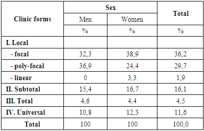
As can be shown from Table 1, the main number of patients was with the local form of circular alopecia, among which there were 36.2% of patients with the focal form, 29.7% with the poly-focal form, and 1.9% with the linear form. Patients with subtotal form of circular alopecia were registered -16.1%. Patients with total form of diseases were 4.5%, universal form - 11.6%.
Table 1. Showed distribution of patients with circular alopecia according to clinical form type
     
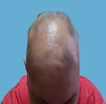
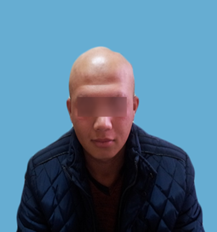
Clinically local (focal) form was characterized by the presence of isolated foci of hair loss ranging in size from 1X1.5 cm to 6X8 cm on the scalp of the rounded-oval shape, with clear boundaries. Skin in the centers was of usual color, without signs of hyperemia and peeling. Around the foci was marked "zone of loose hair". The poly-focal shape was diagnosed in the presence of more than 3 foci of hair loss on the whole surface of the scalp, but the hair in the area of eyebrows and eyelashes were still intact. In the linear-like form (Officis Celsius), the lesions were localized in the occipital region and spread upwards to the temporal regions as a ribbon. There are typical transition of the lesion area to smooth skin and prognostically unfavorable course of the disease. The subtotal form was characterized by the presence of extensive lesions on the scalp, formed by the fusion of smaller lesions. And also in this form a single hair loss was observed in the eyebrows and eyelashes area. The most severe forms of circular alopecia are total and universal forms. Thus, in the total form there was no hair on the whole surface of the head, including eyelashes, eyebrows, and - and beards in men. In universal alopecia, there were no hairs on the whole surface of the patient's skin - scalp and torso (Picture 1, 2).
Picture 1. Total form
Picture 2. Universal form
Affection of nail plates of the hands was observed in 38.6% of patients: dystrophic changes, small point changes in the type of "thimble", expressed longitudinal depletion in combination with dull color of the plates themselves. They depended on the severity of alopecia and were more often observed in patients with total and universal forms of disease.
In 71.6% of patients with circular alopecia there was a progressive stage of the disease, which was characterized by the "zone of loose hair" around the foci (with a slight hair pulling along the edge of the foci is painless epilation). In 27.1% of the patients there was observed the growth of vellus hair (Vellus) in the focus of alopecia with the progress of the pathological focus. Inpatient stage of circular alopecia was observed in 23.9% of patients, regression - in 4.5%. In 7.5% of patients there were subjective sensations in the form of hypersensitivity of the skin, Paraesthesias and erythema preceding the appearance of a new focus of hair loss. Recurrence of the disease occurred in 68.2% of patients, the duration of intervals between the appearances of new foci is not constant.
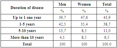
Analyzing the data on the duration of the disease (Table 2), we received the following results: the majority of patients suffered from circular alopecia up to 1 one year (43.9%) and from 1 one to 5 years - 38.7%. Longer duration of the disease was observed in 1/5 of patients (17, 5%).
Table 2. Showed distribution of patients with circular alopecia according to the duration of the disease
     
It is known that the onset and course of the primary disease is influenced by accompanying diseases. Thus, at patients with circular alopecia thyroid diseases in the form of diffuse goiter of various degrees (51%), iron deficiency anemia (32.9%), gastrointestinal tract diseases (27.7%) were registered (Table 3).
Table 3. Showed accompanying pathology in patients with circular alopecia
     
Skin fragments from lesions in the form of biopsies were taken from all patients with circular alopecia with informed consent. Immunohistochemical studies of signaling pathways BMP (Bone Morphogenetic Protein) and Wnt (Wingless-type) were carried out in patients of this group.
Immunofluorescence: The cultures were fixed in 4% paraformaldehyde solution (PFA), then after washing with Phosphate Buffer Solution (PBS) they were permeabilized in 0, 5% Triton X-100 solution. Then PBS was washed and a blocking solution or normal serum solution was applied and incubated with the first antibodies for 18 hours at 40 C. After washing in PBS the second antibodies conjugated with fluorochromes (Alexa) were applied and the preparations were studied by fluorescence and confocal microscopy (Leica TCS SP with Leica LCS software, Leica DM RXA2 fluorescence microscope). Immunofluorescent investigation of cross sections of keratinocyte colonies was performed on collagen gel grown colonies. The cultures were fixed as described above and impregnated with 20% Saccharose solution for 12 hours. Then they were frozen in nitrogen vapors. Cuts with thickness of 10 microns were made on cryostat. Then the cuts were treated as described above, then coloring with DiAminoBenzidine (DAB). Dewaxed cuts were washed in PBS and incubated in 3% H2O2 solution for 10 min at room temperature. Then they were washed in PBS and placed in 0, 5% Triton-X100 solution (in PBS) for 20 min at room temperature. After repeated washing in PBS 20 min were incubated in normal serum solution. Then the first antibodies were applied for 12 hours at +40 C. After washing in PBS the second antibodies were applied for 30 min at room temperature. Washed in PBS 3 times for 5 minutes and stained with DAB (DAB, BD Pharmingen) until coloring occurs, the reaction was stopped by transferring to distilled water. If necessary, was dyed by Haematoxylin.
Histochemical coloring: For staining the culture the cells at reaching the confluent monolayer were fixed with 10% formalin solution during 10 minutes at room temperature; washed with tap water. For dyeing skin slices, the slices were Dewaxed in xylene and rehydrated, incubating in alcohol of decreasing concentration one after another. The material was dyed with 0, 1% solution of Toluidine blue (pH-2.0) in 30% ethanol for 20 minutes; 1% solution of Acetic acid in 3% (pH-2.84) in Acetic acid for 30 minutes; then it was rinsed with distilled water. Dehydrated preparations through alcohol of increasing concentration and Xylene were placed in Canadian balsam. For induction of osteogenic differentiation, cells were cultivated until they reached the confluent layer. Then the medium was changed to the following composition: medium DMEM/F-12, 10% ETS, 0.01 micron 1.25-dihydroxy vitamin D3 (Sigma), 50 micron ascorbate-2-phosphate (Sigma), 10 mM -Glycerophosphate (Sigma). The cells were cultivated for 21 days; the medium was changed every 3 days. Calcification of extracellular matrix was determined by coloring with Alizarin red. For this purpose the cells were washed in PBS, then fixed 4% PFA for 30 minutes at room temperature, after that the cells were washed with PBS and stained with 1% solution of Alizarin red (Sigma) for 2 minutes, washed with distilled water, then in acidic ethanol. For induction of adipogenic differentiation the cells were cultivated in the following medium: DMEM/F-12 medium, 10% ETS, 0.5 mM Isobutyl Methylxanthin, 1 µM Dexamethasone, 10 µM Insulin, 200 µM Indomethacin. The medium changes were carried out every 3 days. For cell coloring with Oil Red-O (Sigma) lipophilic dye they were washed in PBS, fixed in 4% PFA 30 min at room temperature, incubated for 15 min in 60% Isopropanol and then colored for 15 min with filtered Oil Red-O solution. After that the cells were rinsed in 60% Isopropanol, thoroughly washed in distilled water and dyed with Haematoxylin.
Detection of Alkaline Phosphatase activity: The cells were fixed in 70% ethanol for 10 min, washed in PBS and incubated in NBT/BCIP (Roche) solution for 15 min at room temperature in the darkness and washed with distilled water.
Statistical processing of the results: Average value and standard deviation were evaluated during results processing. Scheduling was performed in Excel. The difference was considered statistically reliable if the significance level was p<0.05, which is a measure of sufficient reliability of biological results.

3. Results and Discussion

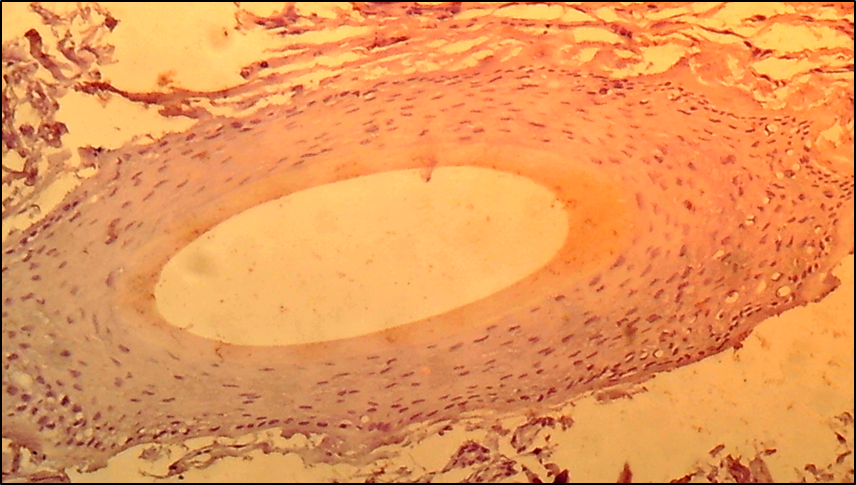
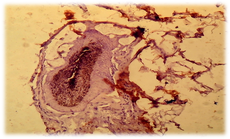
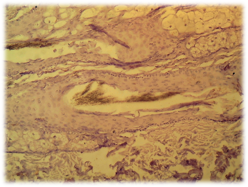
The cells of the dermal papilla isolated by us in the culture were proliferated, organized groups and even when the monolayer was reached, formed multilayer aggregates (so-called pseudopapillae). A number of functional features of the dermal papilla cells have also been revealed. There are indications in the literature that the hair follicle is the source of mesenchymal stem Cells (MSCs) that are involved in repairing the dermal papilla when it is damaged (Jahoda & Reynolds, 2001). We investigated the expression of dermal papilla surface markers, the BMP and Wnt proteins that carry the signal from the cell membrane to the nucleus. The deposition of lipid droplets in the cytoplasm and the expression of Leptin (a marker of mature adipocytes) were observed only in 20-30% of cells of the dermal papilla. Adipogenic differentiation of the dermal papilla cells was less expressed in patients with alopecia than in the control group. Spontaneous differentiation of the dermal papilla cells was not observed in patients with alopecia. The study results showed that patients with more severe forms of circular alopecia had positive BMP expression and negative Wnt1 expression. In these cases, morphogenetic protein signals transfer the hair follicle. Patients with mild forms of circular alopecia (focal, poly-focal) showed positive expression of Wnt1 and negative expression of BMP, proving the presence of mitosis and differentiation of the hair follicle cells, and lengthening of the hair follicle anagen stage. Undoubtedly, further studies based on the model will provide valuable data on the regulation of folliculogenesis induction in humans and prospects for finding agents that effectively regulate this process. (Pic. 3,4,5,6).
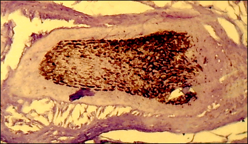
Picture 3. Subtotal form of circular alopecia - hair follicle BMP-4: positive reaction of cells, cells are colored brown. V.40xСir. 20
Picture 4. Focal form of circle alopecia - hair follicle BMP-4: negative reaction of cells, cells are not colored. V. 40 x Cir. 20
Picture 5. Focal form of circle alopecia - hair follicle WNT1: positive reaction of hair follicles, cells are colored brown. V. 40 x Cir. 20
Picture 6. Total form of circle alopecia - hair follicle WNT1: negative reaction of hair follicles, cells are not colored. V.40 x Cir. 20
The presented data indicate the leading role of BMP and Wnt signaling pathways in the pathogenesis of circle alopecia, which determine mitosis and differentiation of hair follicle cells, and open new prospects for the development of new effective drugs.
Conclusions: The key role of inductors and inhibitors of anagen phase - BMP (Bone Morphogenetic Protein) and Wnt (Wingless-type) signaling protein pathways in the rhythm of the hair follicle during circle alopecia has been proved and determine the severity of the pathological process. Undoubtedly, further studies of the pathogenesis of circle alopecia in the direction of signaling pathways will provide valuable data on the regulation of the rhythm of the hair follicle in this disease and open up prospects for finding agents that effectively regulate the pathological process.

References

[1]  Adaskevich V.P., Myadelets O.D., Tikhonovskaya I.V. Alopecia. M -2000. -187 p.
[2]  Birch M. P., Messenger J. F., Messenger A. G. Hair density, hair diameter and the prevalence of female pattern hair loss // Br. J. Dermatol. – 2001. – Vol. 144. – P. 297–304.
[3]  Birch M. P., Messenger A. G. Genetic factors predispose to balding and non–balding in men // Eur. J. Dermatol. – 2001.– Vol. 11. – P. 309–314.
[4]  Blaumeiser B, van der Goot I, Fimmers R, et al. Familial aggregation of alopecia areata. // J Am Acad Dermatol. 2006; 54(4): 627–632.
[5]  Cetin ED, Savk E, Uslu M, Eskin M, Karul A. Investigation of the inflammatory mechanisms in alopecia areata. // Am J Dermatopathol. 2009; 31(1): 53–60.
[6]  Christoph T, Muller-Rover S, Audring H, et al. The human hair follicle immune system: cellular composition and immune privilege // Br J Dermatol. 2000; 142(5): 862–873.
[7]  Chu SY, Chen YJ, Tseng WC, et al. Comorbidity profiles among patients with alopecia areata: the importance of onset age, a nationwide population-based study. // J Am Acad Dermatol. 2011; 65(5): 949–956.
[8]  Colombe L., Michelet J.F., Bernard B.A., Prostanoid receptors in anagen human hair follicles. Experimental Dermatology, 2008. 17(1): p. 63-72.
[9]  Cross M.J., et al., VEGF-receptor signal transduction. Trends in Biochemical Sciences, 2003. 28: 488-94.
[10]  Chu SY, Chen YJ, Tseng WC, et al. Psychiatric comorbidities in patients with alopecia areata in Taiwan: a case-control study // Br J Dermatol. 2012; 166(3): 525–531.
[11]  Conteduca G, Rossi A, Megiorni F, et al. Single nucleotide polymorphisms in the promoter regions of Foxp3 and ICOSLG genes are associated with alopecia areata. // Clin Exp Med. 2014; 14(1): 91–97.
[12]  Ding, L., Saunders, T. L., Enikolopov, G., Morrison, S. J. Endothelial and perivascular cells maintain haematopoietic stem cells. Nature 481: 457-462, 2012.
[13]  Gilhar A, Paus R, Kalish RS. Lymphocytes, neuropeptides, and genes involved in alopecia areata. // J Clin Invest. 2007; 117(8): 2019–2027.
[14]  Goh C, Finkel M, Christos PJ, Sinha AA. Profile of 513 patients with alopecia areata: associations of disease subtypes with atopy, autoimmune disease and positive family history.// J Eur Acad Dermatol Venereol. 2006; 20(9): 1055–1060.
[15]  Guttman-Yassky E(1), Ungar B(2), Noda S(3), Suprun M(4), Shroff A(5), Dutt R(5), Khattri S(5), Min M(5), Mansouri Y(5), Zheng X(3), Estrada YD(5), Singer GK(5), Suarez-Farinas M(6), Krueger JG(3), Lebwohl MG(5). Extensive alopecia areata is reversed by IL-12/IL-23p40 cytokine antagonism.// J Allergy Clin Immunol. 2016 Jan; 137(1): 301-4. doi: 10.1016/j.jaci.2015.11.001. Epub 2015 Nov 20.
[16]  Guzmán-Sánchez DA, Villanueva-Quintero GD, Alfaro Alfaro N, McMichael A. A clinical study of alopecia areata in Mexico. Int J Dermatol. // 2007; 46(12): 1308–1310.
[17]  Happle R., Diphencyprone for the Treatment of Alopecia Areata-More Data and New Aspects. Archives of Dermatolology, 2002. 138: p. 112-113.
[18]  Heissig, B., Hattori, K., Dias, S., Friedrich, M., Ferris, B., Hackett, N. R., Crystal, R. G., Besmer, P., Lyden, D., Moore, M. A. S., Werb, Z., Rafii, S. Recruitment of stem and progenitor cells from the bone marrow niche requires MMP-9 mediated release of Kit-ligand. Cell 109: 625-637, 2002.
[19]  Huang KP, Mullangi S, Guo Y, Qureshi AA. Autoimmune, atopic, and mental health comorbid conditions associated with alopecia areata in the United States. // JAMA Dermatol. 2013; 149(7): 789–794.
[20]  Fricke Villasante AC, Miteva M. Epidemiology and burden of alopecia areata: a systematic review. // Clin Cosmet Investig Dermatol. 2015 Jul 24; 8: 397-403. doi: 10.2147/CCID.S53985.
[21]  Ортикбоев Ж.О., & Саипова Д.С. (2019). Влияние гипотериоза на ремоделирование миокарда левого желудочка у женщин. Евразийский кардиологический журнал, (S1), 271-272.
[22]  Kakourou T, Karachristou K, Chrousos G. A case series of alopecia areata in children: impact of personal and family history of stress and autoimmunity.// J Eur Acad Dermatol Venereol. 2007; 21(3): 356–359.
[23]  Karadag AS, Ertugrul DT, Bilgili SG, Takci Z, Tutal E, Yilmaz H. Insulin resistance is increased in alopecia areata patients. // Cutan Ocul Toxicol. 2013; 32(2): 102–106.
[24]  Kavak A, Yeşildal N, Parlak AH, et al. Alopecia areata in Turkey: demographic and clinical features.// J Eur Acad Dermatol Venereol. 2008; 22(8): 977–981.
[25]  Kawano M., Komi-Kuramochi A, Asada M, Suzuki M. Comprehensive analysis of FGF and FGFR expression in skin: FGF18 is highly expressed in hair follicles and capable of inducing anagen from telogen stage hair follicles. //J Invest Dermatol. 2005 May; 124(5): 877-85.
[26]  Kyriakis KP, Paltatzidou K, Kosma E, Sofouri E, Tadros A, Rachioti E. Alopecia areata prevalence by gender and age. // J Eur Acad Dermatol Venereol. 2009; 23(5): 572–573.
[27]  Lundin M, Chawa S, Sachdev A, Bhanusali D, Seiffert-Sinha K, Sinha AA. Gender differences in alopecia areata. J Drugs Dermatol //2014; 13(4): 409–413.
[28]  MacLean KJ, Tidman MJ. Alopecia areata: more than skin deep. // Practitioner. 2013; 257(1764): 29–32.
[29]  Ramos PM(1), Brianezi G(2), Martins AC(2), Silva MG(2), Marques ME(2), Miot HA(1). Apoptosis in follicles of individuals with female pattern hair loss is associated with perifollicular microinflammation. // Int J Cosmet Sci. 2016 May 10. doi: 10.1111/ics.12341.
[30]  Islam N, Leung PS, Huntley AC, Gershwin ME. The autoimmune basis of alopecia areata: a comprehensive review. // Autoimmun Rev. 2015; 14(2): 81–89.