American Journal of Medicine and Medical Sciences

p-ISSN: 2165-901X    e-ISSN: 2165-9036

2020;  10(12): 965-970

doi:10.5923/j.ajmms.20201012.08

Received: Nov. 11, 2020; Accepted: Nov. 28, 2020; Published: Nov. 30, 2020


Correction and Ways of Its Treatment with a High Risk of Thrombosis of the Vascular Access in Patients in Program Hemodialysis

B. T. Daminov, A. K. Sidikkhodzhaev

Tashkent Pediatric Medical Institute, Tashkent, Uzbekistan

Copyright © 2020 The Author(s). Published by Scientific & Academic Publishing.

This work is licensed under the Creative Commons Attribution International License (CC BY).
http://creativecommons.org/licenses/by/4.0/

Abstract

The aim is to study the effect of pentoxifylline on the pathogenetic aspects of vascular access thrombosis in patients requiring programmed hemodilysis. 187 patients with newly started hemodialysis were examined. All patients underwent an assessment of the risk of vascular access thrombosis according to the developed algorithm, as a result, 2 prognostic groups were formed: with a high risk of vascular access thrombosis (38 patients - 20.32%) and a low risk of access thrombosis (149 patients - 79.28%). The use of pentoxifylline during the year contributed to a significant (p <0.05 with baseline) decrease in the IR of the radial artery of the contralateral limb by 3.70%, while in patients in the low-risk group of diabetes mellitus thrombosis, an increase in IR by 1.55% was observed (p <0.05 with baseline data).

Keywords: Hemodialysis, Vascular access thrombosis, Duplex scanning of the vessels of the upper extremity, Endothelium-dependent vasodilation, Autoroset formation, Pentoxifylline

Cite this paper: B. T. Daminov, A. K. Sidikkhodzhaev, Correction and Ways of Its Treatment with a High Risk of Thrombosis of the Vascular Access in Patients in Program Hemodialysis, American Journal of Medicine and Medical Sciences, Vol. 10 No. 12, 2020, pp. 965-970. doi: 10.5923/j.ajmms.20201012.08.

1. Introduction

Today, about 0.1-0.15% of the world's population requires programmed hemodialysis (HD) [1,2,3]. Hemodialysis can improve the survival and quality of life of patients with CKD [4]. Hemodialysis is possible using a catheter, arteriovenous shunt and arteriovenous fistula. AV shunt thrombosis occurs in 0.5-2% of patients per year, AV fistula - 0.1-0.5% per year. Thrombosis of the AV vascular access (DM) is associated with the interruption of the hemodialysis program, hospitalization of patients, and the installation of a temporary catheter. Diabetes thrombosis is the cause of 65-85% of diabetic dysfunction [5].
In the aspect of diabetes mellitus and thrombus formation, Virchow's triad, including endothelial damage, blood stasis, and hypercoagulation, remains relevant [6]. DM endothelial damage is a complex problem. One component is a chronic abnormal increase in shear stress that causes endothelial dysfunction. Neointimal hyperplasia leads to disruption of the relationship between the endothelium and the excess connective tissue matrix on the basement membrane. Uremic toxins are also involved in the formation of endothelial dysfunction. Direct damage to the endothelium is caused by regular puncture of diabetes mellitus [7,8,]. Blood stasis in diabetes mellitus may be associated with stenosis of the efferent, efferent, or midsection (conduit). A small supply flow may be associated with low cardiac output, systemic hypotension, hypovolemia, and stenosis of the supply artery. In the area of the conduit, the localization of stenoses is predictable. In the case of an AV shunt, stenoses are observed proximal to the shunt-central vein anastomosis zone in more than 85% of cases [9], in the case of an AV fistula, it depends on the type of fistula. For example, in the case of a brachiocephalic fistula, the cephalic part is stenotic in 77% of cases; in a radiocephalic fistula, stenosis in 60% of cases is localized in the region of the superior anatomosing segment [10,11]. These stenoses result from a complex interaction of molecular and hemodynamic factors. Cytokines such as Hypoxia-Induced Factor-1alpha, Vascular Endothelial Growth Factor-A, Matrix Proteases and Platelet Growth Factor, produced by the endothelium under conditions of turbulent flow, abnormal shear stress, endothelial hypoxia, and trauma. These cytokines induce the migration of fibroblasts and vascular smooth muscle cells from the adventitia and media to the intima. Here they proliferate, leading to vascular stenosis [12]. Long compression times and overcompression after decanulation of diabetes can also cause stasis and thrombus formation.
The role of hypercoagulability in diabetes thrombosis has long been underestimated. Uremia is associated with a violation of the functional state of platelets, which places HD patients in a cohort of persons with a hypercoagulable status [13]. Terminal CKD is a syndrome characterized by chronic activation of systemic inflammation, as evidenced by high levels of C-reactive protein. A high concentration of C-reactive protein is also one of the predictors of the risk of diabetes thrombosis [14]. Other risk factors for thrombosis associated with end-stage CKD include hyperhomocysteinemia, hypoalbuminemia, and hyperlipoproteinemia [15]. Blood circulation in the dialyzer and contact with the dialysis membrane also activates the coagulation cascade and platelet aggregation activity [16,17,18]. To correct these effects, heparinization of diabetes is used, but the systemic prothrombotic status remains. Hereditary thrombophilia also plays a role in the dysfunction of diabetes mellitus. In one study, at least 55% of patients with DM thrombosis were diagnosed with one or more prothrombotic syndromes [19]. The most common variants are Leiden factor V and prothrombin G20210A [20]. In patients with recurrent thrombosis, especially in the absence of significant stenosis, examination for hereditary thrombophilia is recommended [21].
For the prevention of diabetes thrombosis, systemic antiplatelet agents and anticoagulants are used. In one meta-analysis, antiplatelet therapy was associated with a decrease in the risk of early thrombosis of AV fistulas (within the first month), but was ineffective in preventing long-term events, nor did it reduce the risk of thrombus formation and did not increase the duration of the so-called. "Mature" fistulas [22]. In a randomized trial, omega-3,6,9 polyunsaturated fatty acid preparations and dietary modification with the inclusion of seafood reduced the risk of AV shunt thrombosis by 50% [23]. In modern guidelines for the management of patients on HD, there are no recommendations regarding the use of anticoagulants.
Objective: to study the effect of pentoxifylline on the pathogenetic aspects of vascular access thrombosis in patients requiring programmed hemodialysis.

2. Materials and Methods

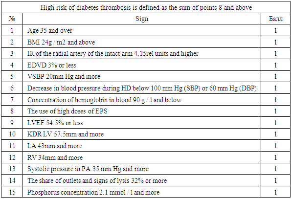
The study included patients who received programmed hemodialysis at the Tashkent City Center for Nephrology and Kidney Transplantation. All patients underwent hemodialysis using diabetes mellitus in the form of an arteriovenous fistula (AVF) formed in the surgical department of this center. The time from the moment of surgery to the start of hemodialysis (the period of "maturation" of the fistula) ranged from 3 to 6 months.
In our center, a risk scale for developing vascular access thrombosis was developed (Table 1).
Table 1. Scale for the identification of HCKD patients with a high risk of diabetes thrombosis
     
187 patients with newly started hemodialysis were examined. All patients underwent an assessment of the risk of vascular access thrombosis according to the developed algorithm, as a result, 2 prognostic groups were formed: with a high risk of vascular access thrombosis (38 patients - 20.32%) and a low risk of access thrombosis (149 patients - 79.28%). In high-risk patients, an antiplatelet agent (pentoxifylline 600 mg / day) was included in the therapy. The follow-up period was 1 year, by the end of which the incidence of the endpoint, diabetes thrombosis, was assessed in both prognostic groups. The clinical characteristics of the patients are presented in Table 2.
Table 2. Clinical characteristics of patients included in the second stage of the study
     
Ultrasound examination of the vessels of the upper limb included duplex scanning (ultrasound scanning modes and spectral pulse-wave Doppler mode) of the arteries and veins of the contralateral (without DM) arm. The study was carried out in the morning hours (8-11 am) on the second day after hemodialysis, in the supine position after 10 minutes of rest, using an ultrasound scanner equipped with a linear transducer with a frequency of 7.5 MHz. The following indicators were recorded:
- maximum systolic blood flow velocity in the radial artery;
- the final diastolic blood flow velocity in the radial artery;
- the resistivity index (Pursello index, IR) was calculated: IR = (systolic velocity-diastolic velocity) / systolic velocity;
- the diameter of the brachial artery is 2-5 cm above the elbow bend;
- a test with endothelium-dependent vasodilation (EDVD) of the brachial artery was performed, for which a tonometer cuff was applied to the shoulder, the pressure in which was pumped 50 mm Hg higher than the previously measured systolic blood pressure (SBP). The compression lasted 5 minutes, after which, after the release of compression, at 60 seconds, the diameter of the brachial artery was re-measured and the EZVD was calculated: EZVD = (diameter of the brachial artery after release of compression-diameter of the brachial artery initially) / diameter of the brachial artery initially * 100.
Echocardiography. To assess the characteristics of central hemodynamics, all patients included in the study underwent echocardiography (EchoCG) using an ultrasound scanner equipped with a sector transducer with a frequency of 4.2 MHz. The study was carried out on the 2nd day after hemodialysis, in the morning hours, in the position of lying on the left side and lying on the back, with the left hand located under the patient's head. During echocardiography, standard echocardiographic positions and projections were used. To determine the measurement time, the synchronization of the image with the electrocardiogram was used. The following indicators were recorded:
In the parasternal position, projection along the long axis of the left ventricle
- end diastolic size of the left ventricle (LV EDD);
- the final systolic size of the LV;
- anteroposterior diameter of the left atrium (LA);
- the diameter of the right ventricle (RV).
LV end-systolic and diastolic volumes (Teichholtz method) and LV ejection fraction (EF) were calculated. LVEF = (LV end-diastolic volume-LV end-systolic volume) / LV end-diastolic volume * 100.
In the case of a change in the geometric shape of the LV (spherical deformation of the cavity, the presence of an aneurysm or regional dyskinesis), vertical position of the heart or LV angulation, or LV EDD exceeding 54 mm, LV volumes for calculating LVEF were measured by the Simpson method using apical 2 and 4- x chamber echocardiographic position.
- systolic pressure in the pulmonary artery (PA systolic) was calculated using tricuspid regurgitation, which was recorded in the parasternal position along the short axis at the level of the aortic valve with focus on the tricuspid valve or in the apical 4-chamber position (the maximum recorded velocity was used). After mapping tricuspid regurgitation by color pulse-wave Doppler sonography, registration of the peak velocity of tricuspid regurgitation was performed by non-first-wave Doppler sonography. The Bernoulli equation (pressure gradient = 4 * peak velocity 2) was used to calculate the peak systolic pressure gradient across the tricuspid valve. SystR PA was defined as the sum of the peak systolic pressure gradient across the tricuspid valve and the pressure in the right atrium. The pressure in the right atrium was determined by a semi-quantitative method using ultrasound scanning of the inferior vena cava with determination of its diameter during calm breathing and its collapse during deep inspiration (subcostal position, projection of the inferior vena cava), as well as measuring the diameter of the right atrium (apical 4-chamber position) (45).
24-hour blood pressure monitoring (ABPM) was carried out on the second day after hemodialysis using the BTL ABPM complex (UK). ABPM lasted for 24 hours, BP was measured every 30 minutes during the day and every 60 minutes at night. The measurement method is oscillometric. During the study, the following calculated data were used:
- the average daily level of systolic blood pressure (SBP) - the arithmetic mean of all registered indicators of systolic blood pressure;
- the average daily level of diastolic blood pressure (DBP) - the arithmetic mean of all registered indicators of diastolic blood pressure;
- daily index (SI) = (mean daytime hemodynamic blood pressure-mean nighttime hemodynamic blood pressure) / mean hemodynamic daytime blood pressure * 100, where mean hemodynamic blood pressure was the arithmetic mean of all hemodynamic blood pressure indicators for the corresponding period of time. Hemodynamic blood pressure = (SBP-DBP) / 3;
- variability of SBP (VSAP) - standard deviation of all SBP indicators recorded during the day;
- DBP variability (SDPP) - standard deviation of all DBP indicators recorded during the day.
In addition, in the course of the study, blood pressure was monitored during the hemodialysis procedure and episodes of arterial hypotension were recorded - a decrease in SBP below 100 mm Hg. or DBP below 60 mm Hg. Blood pressure was measured by the Korotkov method using a Riva-Rocchi tonometer every 20 minutes.
Laboratory research.
During the study, all patients underwent a general blood test using an automatic analyzer. The concentration of hemoglobin in the peripheral venous blood was analyzed. Blood sampling was carried out on the 2nd day after the hemodialysis procedure, on an empty stomach at 7-8 in the morning, after 10 minutes of rest, in a sitting or lying position. The cubitalipsilateral vein was used. Blood sampling was carried out in vacutainers containing an anticoagulant.
To study the state of mineral metabolism, the concentration of parathyroid hormone and phosphorus in serum was determined during the study. Venous blood was taken from the cubital vein of the ipsilateral arm, on an empty stomach, at 7-8 am after 10 minutes of rest, in a sitting or lying position, on the 2nd day after hemodialysis. Determination of the phosphorus concentration was carried out by the biochemical method, parathyroid hormone - by the enzyme immunoassay.
To determine the aggregation and rheological properties of blood in the course of the study, the phenomenon of auto-socket formation was studied. For this purpose, on the 2nd day after hemodialysis, on an empty stomach, in a lying or sitting position, in the morning hours (7-8 hours), after a 20-minute rest, capillary blood was taken. In a smear of native blood, the number of outlets was counted - an accumulation of cellular elements of blood, in the center of which there is a rosette-forming cell - a leukocyte, to which erythrocytes and platelets are attached. The number of outlets per 200 leukocytes was counted. In addition, among the outlets, the proportion of lysed outlets was recorded with signs of damage to the cell membrane and disruption of the integrity of the cells forming the outlet.
All received data were entered into Excell summary tables. After the formation of groups, all parameters were described in the form of the arithmetic mean and its standard deviation. The significance of intergroup differences was determined using the Student's t test. Comparison of the frequency of occurrence of signs between groups was carried out using the tabular chi-square test and checking its reliability against the tables depending on the number of degrees of freedom.

3. Results and It’s Discussion

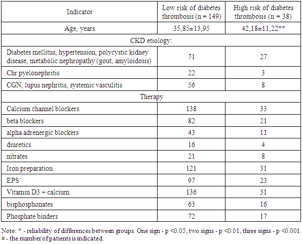
The average age of the patients was 37.13 ± 13.66 years. Patients with a high score for the risk of thrombosis were, on average, older than those with a low score (p <0.01, Table 3).
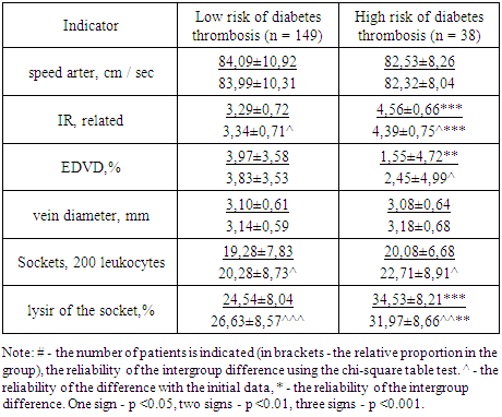
Table 3. Comparative dynamics of pathogenetic triggers of diabetes thrombosis in patients with CKD V, depending on the risk score
     
Duplex scanning of the vessels of the intact (contralateral diabetes) upper limb revealed that the mean peak systolic velocity in the radial artery was 83.77 ± 10.44 cm / sec and did not depend on the score for the risk of thrombosis. IR of the radial artery averaged 3.55 ± 0.87 and was significantly increased in patients with a high risk of diabetes thrombosis (p <0.001). Thus, in the group of patients with a high risk of thrombosis, a score for this criterion was scored by 30 patients (78.95%), and in the group with a low risk - 20 (13.42%, chiradrat = 65.48, p <0.001). The diameter of the cephalic vein averaged 3.10 ± 0.62 mm and did not depend on the score for the risk of diabetes thrombosis.
The indicator of structural and functional disorders of the membranes of blood cells, autoroset formation was 19.44 ± 7.60 sockets per 200 leukocytes and did not depend on the score for the risk of diabetes thrombosis. However, the proportion of outlets with signs of lysis (26.57 ± 9.01%) was significantly higher in the high-risk group (p <0.001). A score for this criterion of the risk scale was assigned to 26 patients (68.42%) in the high-risk group and only 39 patients (23.49%) in the low-risk group (chi square = 27.43, p <0.001).
A decrease in the degree of EDVD was found in 176 patients (94.12%) included in the study, and in 40 patients (21.39%) paradoxical vasoconstriction was observed. The functional state of the endothelium was more impaired in the group of patients with a high risk of thrombosis (the significance of the difference in the degree of EDVD between the groups depending on the risk score was p <0.01). Patients who scored on this criterion of the diabetes thrombosis risk scale were 26 (68.42%) in the high-risk group, and 60 (40.27%, chi square = 9.64, p <0.01) in the low-risk group.
All patients allocated to the high-risk group were prescribed pentoxifylline in a daily dose of 600 mg. The drug combines the properties of an antiplatelet agent and a venotonic agent. The use of pentoxifylline during the year contributed to a significant (p <0.05 with baseline) decrease in the IR of the radial artery of the contralateral limb by 3.70%, while in patients in the low-risk group of diabetes mellitus thrombosis, an increase in IR by 1.55% was observed (p <0.05 with baseline data). However, despite the multidirectional dynamics, the intergroup comparison revealed that, as at baseline, at the end of the year of follow-up, the IR of the radial artery in patients with a high risk of thrombosis of diabetes remained significantly higher than in patients with a low risk of thrombosis (p <0.001 significance of the difference in IR between groups at the end of the observation period). The maximum systolic blood flow velocity in the radial artery did not change in any of the groups and was comparable regardless of the score for the risk of thrombosis. The degree of endothelium-dependent vasodilation in the group of patients with a high risk of vascular access thrombosis increased by 57.63% (p <0.05 with the baseline data), while in the group of low-risk patients this indicator did not significantly worsen (decreased by 3.72%) As a result, the initially less favorable level of EZVD in patients with a high risk of thrombosis by the end of the observation period became equal to the indicator typical for patients with a low risk of thrombosis. The diameter of the cephalic vein did not change in both groups of patients and remained comparable, regardless of the score for the risk of thrombosis.
The study studied the dynamics of rosette formation in the peripheral blood in patients with HCKD. In both groups of patients, an increase in the activity of the phenomenon of rosette formation was found by 6.85%: by 5.19% in the group of patients with a low risk of vascular access thrombosis (p <0.05 with baseline data) and by 13.11% in the group of patients with high risk of vascular access thrombosis (p <0.05 with baseline data). At the same time, it was found that in the group of patients with a low risk of thrombosis, the proportion of lysed outlets also increased (by 8.50%, p <0.001 with the initial data), while in the group of patients with a high risk of thrombosis of the vascular access, the use of pentoxifylline contributed to a decrease in the share of lysed outlets by 7.39% (p <0.01 with initial data).

4. Consclusions

Despite the multidirectional dynamics of the indicator, in the group of patients with a high risk of thrombosis of the vascular access, the proportion of outlets with signs of lysis at the end of the observation period remained significantly higher than in the group of patients with a low risk of thrombosis (p <0.01 reliability of intergroup differences). Long-term use of pentoxifylline at a dose of 600 mg / day in patients with a high risk of diabetic thrombosis contributes to a decrease in the IR of the radial artery, an increase in the degree of EDVD, and a decrease in the formation of lysed outlets. As a result, in the course of 12-month therapy with pentoxifylline, the score for the risk of diabetes thrombosis significantly decreased.

References

[1]  Saran R, Robinson B, Abbott KC, et al. US Renal Data System 2016 Annual Data Report: Epidemiology of Kidney Disease in the United States. Am J Kidney Dis 2017; 69: A7-8. 10.1053/j.ajkd.2016.12.004.
[2]  Zachara BA. Selenium and selenium-dependent antioxidants in chronic kidney disease. AdvClin Chem. 2015; 68: 131–151.
[3]  Farrington K, Rao R, Steenkamp R, Ansell D, Feest T. Chapter 4: All Patients Receiving Renal Replacement Therapy in the United Kingdom in 2005. In: Ansell D, Feest T, Tomson C, Williams A, Warwick G, editors. UK Renal Registry: The Ninth Annual Report 2006. Bristol: UK Renal Registry; 2006. p. 43-76.
[4]  NKF-K/DOQI. Hemodialysis Adequacy Guideline 2: Methods for Measuring and Expressing the Hemodialysis Dose. 2006 [updated 2006; cited]; Available from: http://www.kidney.org/professionals/KDOQI/guideline_upHD_PD_VA/hd_guide2.htm.
[5]  Quencer KB, Friedman T. Declotting the Thrombosed Access. Tech VascIntervRadiol 2017; 20: 38-47.
[6]  Mammen EF. Pathogenesis of venous thrombosis. Chest 1992; 102: 640S-4S.
[7]  Gibson KD, Gillen DL, Caps MT, et al. Vascular access survival and incidence of revisions: a comparison of prosthetic grafts, simple autogenous fistulas, and venous transposition fistulas from the United States Renal Data System Dialysis Morbidity and Mortality Study. J VascSurg 2001; 34: 694-700.
[8]  Young EW, Dykstra DM, Goodkin DA, et al. Hemodialysis vascular access preferences and outcomes in the Dialysis Outcomes and Practice Patterns Study (DOPPS). Kidney Int 2002; 61: 2266-71.
[9]  Kanterman RY, Vesely TM, Pilgram TK, et al. Dialysis access grafts: anatomic location of venous stenosis and results of angioplasty. Radiology 1995; 195: 135-9.
[10]  Sivanesan S, How TV, Bakran A. Sites of stenosis in AV fistulae for haemodialysis access. Nephrol Dial Transplant 1999; 14: 118-20.
[11]  Hammes M, Funaki B, Coe FL. Cephalic arch stenosis in patients with fistula access for hemodialysis: relationship to diabetes and thrombosis. HemodialInt 2008; 12: 85-9.
[12]  Misra S, Fu AA, Rajan DK, et al. Expression of hypoxia inducible factor-1 alpha, macrophage migration inhibition factor, matrix metalloproteinase-2 and -9, and their inhibitors in hemodialysis grafts and arteriovenous fistulas. J VascInterv Radiol 2008; 19: 252-9.
[13]  Weigert AL, Schafer AI. Uremic bleeding: pathogenesis and therapy. Am J Med Sci 1998; 316: 94-104.
[14]  Chou CY, Kuo HL, Yung YF, et al. C-reactive protein predicts vascular access thrombosis in hemodialysis patients. Blood Purif 2006; 24: 342-6.
[15]  Montagnana M, Meschi T, Borghi L, et al. Thrombosis and occlusion of vascular access in hemodialyzed patients. SeminThrombHemost 2011; 37: 946-54.
[16]  Ando M, Iwata A, Ozeki Y, et al. Circulating platelet-derived microparticles with procoagulant activity may be a potential cause of thrombosis in uremic patients. Kidney Int 2002; 62: 1757-63.
[17]  Cianciolo G, Stefoni S, Donati G, et al. Intra- and post-dialytic platelet activation and PDGF-AB release: cellulose diacetate vs polysulfone membranes. Nephrol Dial Transplant 2001; 16: 1222-9.
[18]  Lucchi L, Ligabue G, Marietta M, et al. Activation of coagulation during hemodialysis: effect of blood lines alone and whole extracorporeal circuit. Artif Organs 2006; 30: 106-10.
[19]  Knoll GA, Wells PS, Young D, et al. Thrombophilia and the risk for hemodialysis vascular access thrombosis. J Am SocNephrol 2005; 16: 1108-14.
[20]  Atac B, Yakupoglu U, Ozbek N, et al. Role of genetic mutations in vascular access thrombosis among hemodialysis patients waiting for renal transplantation. Transplant Proc 2002; 34: 2030-2.
[21]  Eleftheriadis T, Antoniadi G, Akritidou A, et al. A case report of recurrent vascular access thrombosis in a hemodialysis patient reveals combined acquired and inherited thrombophilia. Ther Apher Dial 2008; 12: 190-2.
[22]  Palmer SC, Di Micco L, Razavian M, et al. Antiplatelet therapy to prevent hemodialysis vascular access failure: systematic review and meta-analysis. Am J Kidney Dis 2013; 61: 112-22.
[23]  Lok CE, Moist L, Hemmelgarn BR, et al. Effect of fish oil supplementation on graft patency and cardiovascular events among patients with new synthetic arteriovenous hemodialysis grafts: a randomized controlled trial. JAMA 2012; 307: 1809-16.