American Journal of Medicine and Medical Sciences

p-ISSN: 2165-901X    e-ISSN: 2165-9036

2020;  10(11): 858-865

doi:10.5923/j.ajmms.20201011.06

Received: Oct. 20, 2020; Accepted: Oct. 28, 2020; Published: Oct. 30, 2020


Simultaneous Laparoscopic Operations in Patients with a High Degree of Operational Risk with Chronic Calculous Cholecystitis

Batirov D. Yu., Yangibaev Z. R., Yangibaev O. Z., Rakhimov A. P., Yusupov D. D.

Urgench Branch of the Tashkent Medical Academy, Urgench, Uzbekistan

Copyright © 2020 The Author(s). Published by Scientific & Academic Publishing.

This work is licensed under the Creative Commons Attribution International License (CC BY).
http://creativecommons.org/licenses/by/4.0/

Abstract

There have been analyzed results of clinical examination of 168 patients with chronic calculous cholecystitis with associated pathology of abdominal cavity organs, of them 88 patients with high operative risk who were performed various simultaneous laparoscopic operations. Aim of the inquiry: to improve results of the treatment of patients with chronic calculous cholecystitis with associated diseases of the abdominal cavity and high operative risk with improvement of laparoscopic methods of operations. Methods of investigation: general clinical analyses of blood, urine and feces; biochemical blood analyses; ECG; ultrasonography of the liver, gall bladder, biliary ducts, pancreatic gland, spleen and small pelvis; esophagofibrogastroduodenoscopy. The results achieved and their novelty: improved methods of simultaneous laparoscopic operations which increase diagnostic and therapeutic possibilities in associated pathology of abdominal cavity organs, particularly in the patients with higher operative risk factors that increased range of indications for simultaneous laparoscopic operations. Use of our improved methods of simultaneous laparoscopic operations in the patients with higher operative risk factors allow maximum reduction of the frequency of intra- and postoperative complications (bleeding, peritonitis, thromboembolic complications, intestinal paresis, commissural ileus, wound infection, ligature fistula, postoperative hernia, deforming scars and others) and provide high economical effect due to decrease in the costs for the following in-patients and ambulatory treatment and payment of the day of disability. Practical value: improved methods of simultaneous laparoscopic operations are safe, mini-invasive and reliable that allows their performance even in the patients with high operative risk factors (with associated therapeutic diseases).

Keywords: Laparoscopic cholecystectomy, Eppendectomy, Hernioplasty, Chronic calculous cholecystitis, Ovarian cycts, Adhesive disease

Cite this paper: Batirov D. Yu., Yangibaev Z. R., Yangibaev O. Z., Rakhimov A. P., Yusupov D. D., Simultaneous Laparoscopic Operations in Patients with a High Degree of Operational Risk with Chronic Calculous Cholecystitis, American Journal of Medicine and Medical Sciences, Vol. 10 No. 11, 2020, pp. 858-865. doi: 10.5923/j.ajmms.20201011.06.

1. Introduction

In recent years, an increase in the number of combined pathologies of the abdominal organs requiring surgical intervention has been observed everywhere. Mass surveys of the population of the USA, Germany, Great Britain, Japan, Sweden, etc. (WHO, 1985) showed that 30% of patient’s subject to surgical treatment suffer from one or more diseases. However, simultaneous operations are performed only in less than 6% of cases (in the world's leading clinics by experienced specialists). This means that more than 94% of them in the future expect repeated surgical interventions or continue to suffer from these diseases (Malinovsky N.N. et al., 1983; Potashov L.V. et al., 1989; Stebunov S.S. and et al., 1998 and others, Brekhov E.I., 2010; Surico D., 2010).
The reason for refusing to perform simultaneous operations on the abdominal organs using traditional open methods is the high risk of complications such as bleeding, peritonitis, thromboembolic complications, intestinal paresis, adhesive intestinal obstruction, wound suppuration, ligature fistulas, postoperative hernias, gross deforming scars, etc. (up to 25.5%). Mortality after such operations ranges from 5 to 26% (Krotov N.F. et al., 1997; Prudkov I.D. et al., 1997; Gallinger Yu.I. et al., 2002; Kubyshkin V.A. and et al., 2002; Karimov Sh.I. et al., 2004; Akilov Kh.A. et al., 2004; Geshelin S.A. et al., 2008; Zieniewicz K. et al., 1993; Adrales GL et al., 2003; Griffin S., 2006).
The presence of concomitant therapeutic diseases, such as severe anemia, obesity III-IV degree, hypertension (HD) and coronary heart disease (СHD), as well as previous open abdominal surgery (due to adhesions) complicate even routine operations.
Since the 90s of the last century, with the introduction of laparoscopic technologies in surgery, it became possible to simultaneously correct several surgical pathologies. However, despite the undeniable advantages of laparoscopic surgical methods over traditional surgical interventions (Gallinger Yu.I. et al., 1997; Balalykin A.S., 1997; Yangiev A.Kh. et al., 1997; Karimov Sh.I. and et al., 2000; Kim V.L., 2002; Hart S., 2010), many aspects of this problem have not received proper coverage.
The experience accumulated in recent years, the improvement of surgical techniques, anesthetic and resuscitation care have changed not only the psychology of surgeons, but also the attitude of patients towards operations. Many of them want to simultaneously get rid of several diseases (Strekalovsky V.P. et al., 1999; Zuker KA et al., 1991; Khatkov I.E. et al., 1997; Dronova V.L. et al., 2013; Zaniotto G. et al., 1993; Jocko JA, 2013).
It is known that women over 40 years of age often suffer from Chronic calculous cholecystitis (CCC), which in many of them is combined with gynecological diseases. It is in these patients that chronic anemia of moderate and severe severity is detected with a high frequency. Severe anemia is still a risk factor for even simple operations (Baulina N.V., 2005; Lebedeva E.A., 2010; Olivari N. et al., 2007).
In this regard, it is of particular importance to develop, improve and introduce into surgical practice minimally invasive, reliable and easily tolerated patients with combined pathologies of the abdominal organs with a high degree of surgical risk due to concomitant therapeutic diseases (coronary artery disease, hypertension, severe chronic anemia, obesity III -IV degree), simultaneous operations using laparoscopic technologies. All this is currently acquiring not only theoretical, but also enormous practical significance.

2. Purpose of the Research

Improvement of the results of surgical treatment of patients with chronic calculous cholecystitis and combined pathologies of the abdominal organs with a high degree of operational risk (with concomitant therapeutic diseases) by improving simultaneous laparoscopic operations.

3. Material and Research Methods

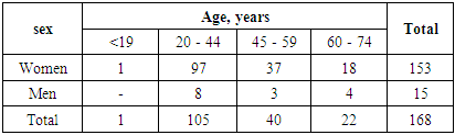
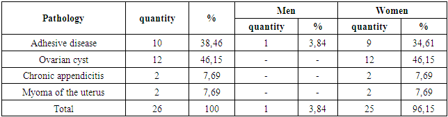
The work is based on the results of a clinical examination of 168 patients who underwent various versions of simultaneous laparoscopic operations (SLO).
The data on the distribution of patients by sex and age, made according to the WHO classification (1962), are presented in the table.
As can be seen from Table 1, the majority of patients (62.50%) were at the most working age - from 20 to 44 years old.
Table 1. Distribution of patients by sex and age
     
Depending on the nature of the identified surgical diseases and concomitant therapeutic pathologies, the patients were divided into two groups. The first group consisted of 80 (47.62%) patients in whom, apart from the combined surgical pathology of the abdominal cavity organs, no significant concomitant therapeutic diseases were identified.
The second group included 88 (52.38%) patients who, in addition to concomitant diseases of the abdominal cavity with CCC, had concomitant therapeutic diseases such as coronary artery disease, hypertension, chronic anemia, obesity of III-IV degree, as well as patients, previously undergone open operations on the abdominal organs, that is, patients with a high degree of operational risk (diagram).
Diagram 1
Performed simultaneous laparoscopic operations were divided into 3 groups according to the classification of Lohlein D., Pichlmayr (1978). In the control group, 42 operations were classified as small and 38 - as medium simultaneous operations. In the main group, due to concomitant therapeutic pathologies, all 88 operations were classified as high-risk operations.
General characteristics of the identified simultaneous pathologies of the abdominal organs are presented in Table 2.
Table 2. General characteristics of simultaneous pathologies of the abdominal organs
     
During the study, all patients underwent a general analysis of blood, urine, feces and biochemical blood tests (the fractions of bilirubin, sugar, diastase, creatinine, urea, total protein, ALT, PTI, coagulogram were determined). Ultrasound scanning of the abdominal organs, esophagogastroduodenofibroscopy, X-ray examination were performed.
Research results
In all cases, the main stage of the SLO was laparoscopic cholecystectomy (LChE). Table 3 shows the average duration of the AO (table 3).
Таble 3. Average duration of simultaneous laparoscopic operations
     
It should be noted that in 30% of patients, combined surgical pathology was an intraoperative finding, but this did not become a reason for refusing to perform the simultaneous stage of the operation (Table 4).
Table 4. Simultaneous pathologies identified in the intraoperative period
     
Each of the groups consisted of 21 patients with associated pathology of adhesions of the abdominal cavity. In 15 (17.04%) patients of the second group, adhesions of the abdominal cavity were observed after previous open operations. Considering that in patients with a high degree of operational risk, in particular with moderate and severe chronic anemia, the dissection of adhesions is of particular importance, we have improved the "movable" L-shaped hook-electrode of a special modification. With a pronounced adhesive process, its use several times reduces the time for performing this stage of the operation, providing reliable hemostasis, preventing rupture of adhesions, deserosing the intestinal wall, bleeding and other complications (this hook, collecting adhesions together, compresses them, dissects them in place using coagulation) ... With the help of a "movable" L-shaped hook-electrode, adhesions were dissected in 21 patients of the second group.
LCE with laparoscopic appendectomy (LAE) was performed in 11 (13.75%) patients in the first group and in 14 (15.90%) patients in the second group.
In the first version of LAE, 2 clips or endoploops were placed on the base of the appendix, dissecting between them. In this case, the stump of the appendix is not immersed in the dome of the cecum, and peritonization is not performed. We used this method at the beginning of our study in 4 patients of the first group with a fixed dome of the cecum or the presence of an adhesive process. Subsequently, despite the absence of postoperative complications associated with this particular method of LAE, we refused to use it, since when a clip and an end loop are applied to the appendix base, there is a risk of crush injury and tissue rupture (due to inflammation, edema, and infiltration of the appendix). In addition, it is not possible to immerse the stump and peritonize. Having improved the technique, we began to use safer methods of LAE.
In the second variant of LAE, in 4 patients of the first group, also with an immobile dome of the cecum or with an adhesive process, the base of the appendix and the mesentery were sutured using the endo GIA apparatus. With this method, the stump of the appendix also does not sink into the dome of the cecum, and peritonization is not performed. We also had to abandon this technique due to its inherent disadvantages and the high cost of the GIA endo apparatus.
In the third variant of LAE, using a bipolar electrode, the appendix was separated from the mesentery through the serous membrane, which ensured reliable hemostasis. Then, endoploops were placed on the base of the appendix in two places and dissected between them using bipolar coagulation. This technique operated on 2 patients of the second group, and in isolation more than 70 patients. However, this method also fell short of our expectations.
In the fourth version of LAE through a trocar (10 mm) in the right iliac region, expanded with a retractor, holding a clamp through the apical part, the appendix together with the dome of the cecum is brought outwards. After that, appendectomy is performed using the classical method: the stump of the appendix is immersed in the dome of the cecum with purse string and Z - shaped sutures, if necessary, peritonization is performed. Then the dome of the cecum is immersed in the abdominal cavity. Our experience has shown that this LAE method, regardless of whether it is performed in isolation or simultaneously, is the most reliable. This technique was used to operate 3 patients of the second group. At the same time, it can be used only in cases when the thickness of the abdominal wall does not exceed 5-6 cm, the dome of the cecum is mobile, and there is no adhesion around the process.
In the fifth version of LAE, all stages of the operation - dissection of the mesentery, stitching it, ligation from the base, immersion into the dome of the cecum using purse string and Z - shaped sutures, etc. - performed only with laparoscopic instruments. This method operated on 3 patients of the first group. Due to technical difficulties, we later abandoned the use of this technique.
Thus, as our experience shows, all known LAE techniques (typical, with the help of clips, loops, rings, GIA, coagulation, ultrasound, etc.) are not without drawbacks. Taking this into account, we have developed an improved version of LAE, devoid of these drawbacks. The advantages of this 6th variant of LAE are especially evident in cases where the thickness of the abdominal wall exceeds 6 cm. In patients suffering from grade III-IV obesity, the dome of the cecum is deeply located, its mobility is limited. In this situation, in the right iliac region around the trocar incision, we applied 2 U-shaped sutures through all layers of the abdominal wall and tied them tightly, due to which the abdominal wall was compressed up to 5-10 times. With the help of these threads, the abdominal wall can be moved in the desired direction (up, down, right, left, etc.). After that, holding by the apical part, the appendix is brought outwards together with the dome of the cecum. The rest of the stages of appendectomy are performed as in the classic 4 variants. This method has proven itself especially well in 9 obese patients.
All 24 patients (9 patients of the first, 15-second group) CCC with hernias of the anterior abdominal wall (white line of the abdomen and umbilical hernias) LCE was performed with hernioplasty (HP).
Currently, in most cases, hernia repairs are performed using traditional methods. That is, the share of laparoscopic technologies in this section of surgery remains small, since laparoscopic hernioplasty requires the surgeon to have appropriate skill and experience in this technique. Therefore, the development of new improved laparoscopic GP methods is an urgent task.
Often in patients with CCC, hernias of the white line of the abdomen or around the navel are observed. In such situations, it is possible to perform both stages of the operation using laparoscopic technology. With LCE, we used 2 variants of the GP.
In the first variant, in 9 patients of the first group with small hernias (hernia orifices less than 2-4 cm), a 10-15 mm long skin incision was made above the hernial protrusion. Having found the hernial sac and opened it, after determining the viability of the hernial contents, it was set into the abdominal cavity. Then a laparoscope was inserted through the hernia orifice into the abdominal cavity, and sealing was performed using two clamps. Further, LCE was produced according to the usual procedure. Subsequently, the port of the laparoscope was moved into the epigastrium and, under the control of the laparoscope, the muscular-aponeurotic layers were sutured with a special needle, eliminating the defect.
The second variant of the HP was used in 15 patients of the second group with an abdominal wall defect of more than 4 cm. For hernioplasty of such hernias, we proposed an improved technique (patent application IDP 2002 0079), in which the hernial orifice was eliminated with a special polypropylene net. Above the hernial protrusion, a transverse or longitudinal skin incision with a length of 1.5-2 cm was made. After identification, the hernial sac was opened, the viability of its contents was determined, and then immersed in the abdominal cavity. Through this place, a 10 mm trocar was inserted into the abdominal cavity, and a seal was made with 2-4 clamps. After performing the LCE stage, a previously prepared polypropylene mesh was introduced into the abdominal cavity, the area of which was 2-4 cm larger than the size of the hernial collar. At the edges of the net there were 8-12 lavsan threads 15-20 cm long, the distance between which did not exceed 15-20 mm. To prevent the threads from getting tangled, they were carefully fixed with nylon threads to the edge of the mesh. In the abdominal cavity, the mesh is expanded and brought to the defect of the abdominal wall. Then, with a special needle, all the threads are taken out in turn and tightly tied over the aponeurosis. All manipulations are performed strictly under the control of a laparoscope. This allows you to fix the mesh and reliably eliminate the defect of the abdominal wall without tension, and also greatly facilitates the performance of all stages of the operation.
It is known that women who suffer from CCC are more likely to suffer from various gynecological diseases. Operations laparoscopic cystectomy (LCE) from the ovary, conservative laparoscopic myomectomy (LME) and voluntary surgical sterilization (DCS) are general surgical procedures. We believe that these operations can be performed by a surgical team well-versed in laparoscopic techniques, especially since they are simultaneous stages. Having gained experience, we also began to independently perform such operations.
LCE with LCE were performed in 35 patients (15 in the first group and 20 in the second group). In this case, 5 variants of LCE were used.
Option 1 was used in women (in 3 patients of the first group), in whom the size of ovarian cysts did not exceed 3-5 cm, their mobility was limited, in the presence of polycystic ovaries, in girls and young women. After the LCE stage, ovarian cysts were drilled using bipolar coagulation, the fluid was aspirated by suction. The free part of the cyst wall was removed by coagulation. Despite a number of advantages, this technique is not without drawbacks, since the likelihood of relapses is high.
Option 2 was used in 8 patients (4 patients of the first group and 4 - of the second group) with cysts on a short stem, limitation of ovarian mobility. After LCE, in 4 patients, 2 endo-loops were placed on the base of the cyst pedicle, bandaged and dissected between them. In similar situations in 4 patients, the base of the cyst pedicles was sutured with the ENDOGIA 30, 60 apparatus, and then removed.
Option 3 was used in 5 patients (3 patients of the first group and 2 - of the second group) in the presence of adhesions with surrounding tissues and severely limited mobility of the cyst. Using an L-shaped hook-electrode, the adhesions were dissected by bipolar coagulation as close as possible to the cyst wall. The base of the cyst was sutured with an ENDOGIA 30, 60 apparatus and then removed.
Option 4 was performed in 5 patients of the first group and in 2 - of the second group with large cysts and good mobility. First, the cyst was punctured, the contents were aspirated by suction, then a small incision (1 cm) was made above the bosom, and a 10 mm trocar was inserted into the abdominal cavity. The edges of the cyst wall were fixed with a clamp and through this port they were brought outwards together with the pedicle. Cystectomy was performed outside the abdominal cavity according to the traditional technique, after which the ovarian stump was immersed in the abdominal cavity (extracorporeal cystectomy). This technique is reliable and easy.
In the course of the study, taking into account the disadvantages of all 4 options, we had to develop an improved 5th option LCE, which was used in 12 patients of the second group (with obesity of III-IV degree). To facilitate cystectomy in obese patients, 2 U-shaped sutures were applied from both edges of the trocar, passing through all layers of the abdominal wall, and they were tightly tied. This made it possible to compress the abdominal wall as much as possible. Then the edges of the cyst wall were fixed with a clamp and through this port they were brought outwards together with the leg. Cystectomy was performed outside the abdominal cavity according to the traditional technique, after which the ovarian stump was immersed in the abdominal cavity (extracorporeal cystectomy). This technique greatly facilitates the implementation of all stages of cystectomy.
Thanks to the trocars introduced for LCE, DHS is possible in 90% of cases. Only in patients with grade III-IV obesity (10%) it is difficult to perform DCS without introducing an additional trocar. We performed LCE in combination with DCS in 37 patients (22 in the first group and 15 in the second group). All pre-planned DCSs were performed using laparoscopic methods. We performed the DHS stage in 5 versions.
Option 1. At the beginning of our study, in patients without signs of salpingitis (3 patients of the first group), 2 clips were applied to the uterus near the fallopian tubes and dissected between them with scissors. In the future, given the possibility of rupture and crushing of tissues in the presence of an inflammatory process, we refused to use this technique.
Option 2. In 4 patients of the first group and in 2 - of the second group, clips were placed in the parts of the fallopian tube close to the uterus in two places, but the dissection was performed not with scissors, but with the help of bipolar coagulation. Subsequently, we also abandoned this technique.
Option 3. In this variant, DCS in 5 patients (in 3 patients of the first and 2 - the second group) was performed using an end loop. However, this technique cannot be used in the presence of adhesions with surrounding tissues and organs, inflammation (salpingitis).
Option 4. The fallopian tubes in 5 patients of the first group and in 4 of the second group were taken out through the trocar above the bosom (additional) and tied with lavsan or nylon threads or with the help of staplers, coagulation. The dissection between them was performed extracorporeally. This method has worked well in the absence of adhesions and inflammation.
The most reliable and simpler version was improved by us. At the same time, in 7 patients of the first group and in 7 patients of the second group, at points close to the uterus, the fallopian tubes were coagulated with a bipolar coagulator, and a small segment of the tube was removed.
Thus, DHS with LCE can be performed as a simultaneous stage of the operation. Of all the options, we consider 4 and 5 to be the most reliable and lightweight.
LCE with myomectomy was performed in 5 patients (in 2 - in the first group and in 3 - in the second group), in whom myomatous nodes did not exceed 9 weeks. LME was performed using bipolar coagulation, but in 2 patients of the second group, due to bleeding, the node bed had to be sutured with laparoscopic instrumentation.
As you know, the presence of therapeutic diseases in patients often serves as a contraindication to surgery. One of these diseases is chronic anemia. The risk of simultaneous surgery in patients with chronic anemia increases the risk of bleeding. In these patients, significant blood loss is possible during surgery due to a violation of the coagulation system. Even with banal cholecystectomy, hernioplasty and appendectomy, blood loss averages from 50 to 300 ml. And with simultaneous operations, especially in patients with moderate and severe chronic anemia, blood loss increases significantly. In our observations, chronic anemia was diagnosed in 152 (90.47%) patients, 68 of them in the first group and 84 in the second group. In the first group, 46 patients had mild, 28 - moderate and 6 - severe chronic anemia. In the second group, mild, moderate and severe anemia was observed in 36, 37 and 11 patients, respectively. For the introduction of trocars into the abdominal cavity in these patients, only the skin epidermis was dissected, the next layer, the dermis, was dissected using a thin electrode by coagulation. Trocars were used only with a blunt end. With the introduction of 5 and 10 mm trocars, almost no bleeding was observed. And 2, 3, 4 trocars and more, using transillumination, were introduced through the avascular places of the abdominal wall. Total blood loss in 95% of patients did not exceed 30-60 ml, and in 5% it was 50-90 ml. In 2 patients, more prolonged bleeding from the gallbladder bed (about 100 ml) occurred, which was stopped by ultrasound coagulation. We did not have any cases of conversion due to bleeding.
As our experience shows, with the help of improved methods of laparoscopic technology, simultaneous operations can be successfully performed even in patients with moderate to severe chronic anemia.
Often, not only simultaneous, but also simple operations are contraindicated due to coronary heart disease, hypertension. In these patients, due to deep hemodynamic disorders, myocardial infarction, stroke, various thromboembolic complications are possible. Hypertension is often combined with grade III-IV obesity and diabetes mellitus. Among the patients of the second group we observed, diseases of the cardiovascular system occurred in 18 (10.71%), of which GB - in 12, ischemic heart disease - in 6. In 10 (1 patient had a history of myocardial infarction, 2 - a cerebral stroke) LCE was performed with dissection of adhesions, in 2 - with HP, in 4 - with LCE and in 2 - with laparoscopic myomectomy (LME).
Considering that acidosis may develop in these patients due to CO2 resorption, we improved the SLO methods. The CO2 pressure in the abdominal cavity was maintained at the level of 7-9 mm Hg, and the gas supply was carried out from 0.15 l / s. If after 15 minutes no changes in hemodynamic parameters were observed, the operation was continued, maintaining the gas flow rate into the abdominal cavity at the initial level. The patient's head was raised at an angle of up to 30-450.
Patients with hypertension in the preoperative period received adequate antihypertensive therapy. When performing SLO, they took into account the hypotensive effect of induction anesthesia and pneumoperitoneum. To avoid a sharp decrease in blood pressure during the operation, the intra-abdominal gas pressure was maintained at the level of 10-12 mm Hg, which was optimal.
Thus, our improved SLO variants were successfully performed in patients who had previously considered contraindications for simultaneous operations, cardiovascular diseases, moderate and severe chronic anemia, and grade III-IV obesity.
The complications arising in the patients we observed were divided into 3 groups according to the classification of T.B. Duboshina (1980). Thus, subcutaneous emphysema, which developed in 2 (2.50%) patients of the first group and in 2 (2.27%) patients in the second group, resolved on its own without complications within a week. "Frenicus - syndrome", caused by the effect of pneumoperitoneum on the phrenic nerve, was observed in the same number of patients.
Of the nonspecific complications, 21 (26.25%) patients of the first group and 26 (29.54%) of the second group had nausea and vomiting. In 11 (13.75%) patients of the first group, intestinal paresis was observed, which resolved on its own on the 2nd day after surgery, and in 5 (6.25%) patients after stimulation of the intestine (proserin n / a) and a cleansing enema. In the second group, this complication was observed in 15 (17.04%) patients, in 7 (7.95%) of whom they had to resort to bowel stimulation and a cleansing enema. For the rest, it went away on its own within 2 days. Among the patients of the first group, there was one case of suppuration and discharge of seroma from an epigastric wound.
In all cases, pain after SLO was not strong and was easily relieved on the first day, and after 24 hours the general condition of the patients was assessed as satisfactory.
In the second group, the epigastric wound festered in 1 (1.13%) patient, seroma excretion took place in 2 (2.27), exacerbation of chronic bronchitis - in 1 patient.
Pain syndrome after surgery in patients of this group persisted for a longer time, which was associated with the presence of concomitant pathologies and the psychological mood of the patients.
Within 24 hours after the operation, 114 (67.85%) patients (58 in the first and 56 in the second group) became fully active, after 36 hours the condition of all patients was satisfactory. 6 (7.50%) patients in the first group and 2 (2.27%) in the second group could move independently 6 hours after the operation and the next day were discharged home in a satisfactory condition (table 5).
Тable 5. Comparative assessment of postoperative indicators
     
The total duration of stay of patients of the first group in the hospital averaged 5.85 ± 0.22 days; before surgery 1.97 ± 0.16, after surgery 3.88 ± 0.14 days. Patients of the second group were in the hospital for an average of 6.03 ± 0.22 days; before surgery 1.81 ± 0.14, after surgery 4.21 ± 0.16 days.
The results of the studies carried out indicate not only the indisputable technical, but also the economic and medico-social advantages of laparoscopic operations. The latter are manifested in savings in the costs of possible follow-up treatment, a reduction in losses due to the absence of workers at work, as well as savings in social insurance systems.

4. Conclusions

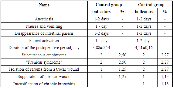
1. Application of our improved methods of simultaneous laparoscopic operations in patients with chronic calculous cholecystitis and combined pathologies of the abdominal organs with a high degree of operational risk due to therapeutic pathologies (coronary artery disease, hypertension, moderate and severe anemia, obesity of III-IV degree), and in patients, who had previously undergone open surgery on the abdominal organs, made it possible to reduce postoperative complications to 3.41% and to avoid deaths.
2. Ease, simplicity, speed and minimally invasiveness of our improved simultaneous laparoscopic operations allows them to be performed even in patients with a high degree of surgical risk due to concomitant therapeutic diseases, who have previously undergone operations on the abdominal organs.
3. The use of our improved simultaneous laparoscopic operations allows to reduce the time of hospital stay, to reduce the cost of treatment and payment of temporary disability certificates, which is of great medical and social importance.

References

[1]  Akilov Kh.A., Saydazimov E.M., Akhmedov A.V. Laparoscopic interventions in elderly and senile people // Surgery of Uzbekistan. - 2004. - No. 1. - P.3-6.
[2]  Aripov U.A., Navruzov S.N., Prokhorova I.P. Combined simultaneous operations in abdominal surgery // Combined simultaneous operations: Abstracts. report Rep. conf. surgeons. - Bukhara, 1992. -- pp. 5-10.
[3]  Bakov V.S. Simultaneous laparoscopic operations for combined pathologies of the abdominal cavity and small pelvis: Dis. ... Cand. honey. sciences. - M., 2000 .-- S. 5-24.
[4]  Balalykin A.S. Past and Present of Abdominal Endoscopic Surgery // Endoscopic Surgery. Materials of the 1st Intern. regional scientific - practical. conf. doctors of the Far East and Siberia. - Yuzhno-Sakhalinsk, 1993 .-- pp. 5-11.
[5]  Baulina N.V., Baulina E.A. Simultaneous operations in surgery and gynecology // Vestn. hir. - No. 2. - 2004. - P.87-91.
[6]  Brekhov E.I. Experience in conducting simultaneous laparoscopic cholecystectomy and gynecological operations / E.I. Brekhov, E.B. Savinova, E.A. Lebedeva // Surgery. Journal them. N.I. Pirogov. - 2010. - No. 12. - pp. 23-26.
[7]  Possibilities of minimally invasive methods of treating acute cholecystitis in patients with high operational risk / Sh. I. Karimov, V.L. Kim, M.Sh. Khakimov et al. // Surgery of Uzbekistan. - 2004. - No. 1. - pp.46-50.
[8]  Gallinger Yu.I. Simultaneous cholecystectomy and appendectomy // Clin. hir. - 1992. - No. 2. - pp.51-53.
[9]  Gallinger Yu.I., Timoshin A.D. Laparoscopic Cholecystectomy: A Practical Guide. - M., - 1992. - P.66.
[10]  Gallinger Yu.I., Karpenkova V.I., Amelina M.A. Ten years of experience in performing laparoscopic cholecystectomy in patients with grade III-IV obesity // Endoscope. hir. - 2004. - No. 3. - pp.10-15.
[11]  Geshelin S.A., Kashtalyan M.A., Gladchuk I.Z. and other Simultaneous operations in patients with cholecystitis // Ukrainian journal of surgery. - Kiev. 2008. -№2. -pp.60-63.
[12]  Duboshina T.B. Geriatric problems in surgery of acute cholecystitis: Author's abstract. dis ... candidate of medical sciences. - Saratov, 1980. -- 16p.
[13]  Karimov Sh.I., Krotov N.F. Complications of laparoscopic cholecystectomy in chronic calculous cholecystitis // Endoscope. hir. - 2000. - No. 4. - pp. 39-41.
[14]  Karimov Sh.I., Nazirov F.N., Khadzhibaev A.M. On the issues of tactics of simultaneous operations for diseases of the abdominal cavity in elderly and senile patients // Endoscope. hir. - 2003. - No. 6. - pp. 25-26.
[15]  Kim V.L. Minimally invasive methods in the treatment of gallstone disease and its complications: Dis. ... Dr. med. sciences. - Tashkent, 2002. -- 5-13p.
[16]  Krotov NF, Nishanov Kh.T., Fuzailov F. Errors and dangers in laparoscopic cholecystectomy // Cholecystitis and its complications: Materials of the Resp. scientific-practical conf. with int. participation. Bukhara, 1997. -- P.67-68.
[17]  Kubyshkin V.A., Strekalovsky V.P., Vukolov A.V. Laparoscopic cholecystectomy: Old problems in a new light // Surgery. - 1997. - No. 1. - pp.65-68.
[18]  Laparoscopic appendectomy / V.N. Sitnikov, V.A. Galin, M.V. Turbin and others / / Endoscope. hir. - 2002. - No. 5. - pp.23-26.
[19]  Laparoscopic cholecystectomy – complications and their prevention / Yu.I. Gallinger, A.A. Movchun, V.I. Karpenkova et al. // Annals of the NCS: Annual scientific publication. - 1997. - Issue. 6. - P.14-18.
[20]  Malinovsky N.N., Savchuk B.D., Ageychev V.A. Combined surgical interventions in surgical practice // Surgery. - 1983. - No. 12. - pp.63-68.
[21]  Minimally invasive methods in the treatment of acute cholecystitis in patients with increased operational risk / Sh.I. Karimov, V.L. Kim, M.Sh. Khakimov et al. // Endoscope. hir. -2003. - No. 6. -pp. 35-37.
[22]  Minimally invasive simultaneous operations in abdominal surgery / B.U. Sabirov, U.A. Sherbekov, Z.B. Kurbanniyazov et al. // Surgery of Uzbekistan. - 2000. - No. 3. - pp.62-64.
[23]  Minimally invasive technologies in the diagnosis and treatment of the painful form of adhesive disease / A.G. Beburishvili, I.V. Mikhin, A.A. Vorobiev et al. // Vestn. hir. - 2004. - T.163, No. 2. - pp.38-40.
[24]  Nazyrov F.G., Kalish Yu.I., Strusky L.P. State and prospects for the development of endoscopic service in the Republic of Uzbekistan // Surgery of Uzbekistan. - 1999. - No. 3. - pp.10-12.
[25]  Rationale for voluntary surgical sterilization as a simultaneous operation in abdominal surgery in women of fertile age / Yu.K. Dzhabbarova, Kh.A. Akilov, K.A. Zhumaniyazov et al. // Surgery of Uzbekistan. - 2000. - No. 2. - pp. 35-37.
[26]  Simultaneous laparoscopic surgical interventions for combined diseases of the abdominal organs of the small pelvis / K.V. Puchkov, V.S. Bakov, V.V. Osipov et al. // Endoscope. hir. - 2004. - No. 6. - P.3-11.
[27]  Ways of preventing complications of laparoscopic cholecystectomy / OV. Galimov, E.I. Senderovich, M.V. Timerbulatov and others / / Endoscope. hir. - 2002. - No. 6. - pp. 45-47.
[28]  Comparative evaluation of the results of video-laparoscopic, mini-laparotomic and traditional cholecystectomy / A.M. Khadzhibaev, F.B. Alidzhanov, A.B. Vakhidov et al. // Surgery of Uzbekistan. - 2004. - No. 2. - pp.51-53.
[29]  Stebunov S.S., Zanko S.N. Simultaneous operations in laparoscopic cholecystectomy // Endoscope. hir. - 1998. - No. 3. - P.18-21.
[30]  Strekalovskiy V.P., Starkov Yu.G. The role of intraoperative diagnostics in laparoscopic treatment of gallstone disease // Endoscope. hir. - 2001. - No. 1. - p.16-20.
[31]  Economic efficiency of the introduction of laparoscopic cholecystectomy in patients with cholelithiasis / RN. Komarov, N.V. Komarov, A.S. Maslagin et al. // Endoscope. hir - 2003. - №4. - P.39-43.
[32]  Andrew M.N., Roty A.R. Incidental Appendectomy with cholecystectomy: Is the Increased Risk justified? // Amer. Surg. -1987. Vol.-53, no. 10. P.553-557.
[33]  Clayman R.V. Comparison of needlescopic appendectomy versus conventional laparoscopic appendectomy. A randomized controlled trial // J Urol. 2005. - Vol. 174 (5), no. 4. - P.120-123.
[34]  Combined abdominal hysterectomy, cholecystectomy and appendicectomy: a study of 25 cases in Abbottabad / S. Griffin, N. Abbassi, Z. Parveen et al. // J. Ayub. Med. Coll. Abbottabad. - 2006. - V. 18, No. 2. - P. 57-59.
[35]  Cuschieri A. Laparoscopic surgery in Europe. Where are we going? // Cir Esp. 2006. - Vol. 79 (1). - P.10-21.
[36]  Jocko J.A. The role of appendectomy in gynaecologic surgery: a canadian retrospective case series // J. Obstet. Gynecol. Can. - 2013. - Vol. 35, No. 1. - P. 44-48.
[37]  Surico D. Laparoendoscopic single-site surgery for treatment of concomitant ovarian cystectomy and cholecystectomy / D. Surico, S. Gentilli, A. Vigone // J. Minim. Invasive Gynecol. - 2010. - Vol. 17, No. 5. - P. 656-659.
[38]  Olivari N., Luerti M., Torzilli G. Simultaneous laparoscopic cholecystectomy and laparoscopically assisted vaginal hysteroadnexectomy. // Dept. of G. Surg., Hospital of Lodi.-Italy.-2007.
[39]  Zaniotto G., Ancona E., Rossi M. Analysis of risk factors of conversion and complication in laparoscopic cholecystectomy. // First European Congress of the European association for Endoscopic Surgery. - Cologne, 1997. - Vol. 412., No. 4.- P.82-84.
[40]  Zieniewicz K., Krawczyk M., Najnigier B. Et al. Complications after laparoscopic cholecystectomy. // First European Congress of the European association for Endoscopic Surgery. - Cologne, 1997. - Vol. 412., no.4.- P.76.