American Journal of Medicine and Medical Sciences

p-ISSN: 2165-901X    e-ISSN: 2165-9036

2020;  10(11): 852-857

doi:10.5923/j.ajmms.20201011.05

Received: Oct. 17, 2020; Accepted: Oct. 29, 2020; Published: Oct. 30, 2020


Diagnostic Accuracy of the Soluble Fms-Like Tyrosine Kinase 1 / Placental Growth Factor Ratio and Polymorphism Agtr 1, Agtr 2 for Preeclampsia

Makhmudova Sevara Erkinovna1, Negmadjanov Bahodur Boltaevich2, Xamraev Xumoyun Xamzaevich1

1Department of Obstetrics and Gynecology, Faculty of Postgraduate Education, Samarkand State Medical Institute, Samarkand, Uzbekistan

2Department of Obstetrics and Gynecology №2, Samarkand State Medical Institute, Samarkand, Uzbekistan

Copyright © 2020 The Author(s). Published by Scientific & Academic Publishing.

This work is licensed under the Creative Commons Attribution International License (CC BY).
http://creativecommons.org/licenses/by/4.0/

Abstract

Preeclampsia (PE) is a disease that continues to be the main cause of maternal and fetal mortality and complications in 5-8% of pregnancies, Preeclampsia develops after 20 weeks of pregnancy and is characterized by hypertension and proteinuria. According to WHO, hypertension during pregnancy is the cause of 9 to 25 per cent of all maternal mortality, but accurate data are difficult to determine. In addition to the fact that PH is one of the leading causes of maternal and perinatal mortality, this disease and its complications cause a range of medical problems. Placental growth factor (PIGF) is relevant for healthy pregnancy, and abnormalities in PIGF functions have been associated with hypertensive disorders of pregnancy.

Keywords: Preeclampsia, Placental growth factor, Pregnancy, Proteinuria, Prognosis, Clinicо-genetic predictors, Polymorphism

Cite this paper: Makhmudova Sevara Erkinovna, Negmadjanov Bahodur Boltaevich, Xamraev Xumoyun Xamzaevich, Diagnostic Accuracy of the Soluble Fms-Like Tyrosine Kinase 1 / Placental Growth Factor Ratio and Polymorphism Agtr 1, Agtr 2 for Preeclampsia, American Journal of Medicine and Medical Sciences, Vol. 10 No. 11, 2020, pp. 852-857. doi: 10.5923/j.ajmms.20201011.05.

1. Introduction

Preeclampsia is one of the most serious complications in obstetrics, which determines high rates of maternal morbidity and mortality. This pathology continues to be a dangerous complication of pregnancy and for the fetus, leading to delayed intrauterine development, premature birth, low birth weight and perinatal mortality. Preeclampsia was described as a “disease of theories” by Chesley in 1978 as the etiology of preeclampsia remained undefined. Recently, it has been believed that the pathogenesis of preeclampsia is associated with endothelium dysfunction, inflammatory response, oxidative stress, and dysfunction of renin-angiotensin system (RAS) [1]. Endothelial cells can regulate the contraction of underlying smooth muscle cells, prevent intravascular coagulation, and maintain the integrity of the intravascular compartment by producing a serials important substances such as nitric oxide (NO), endothelin-1, and tissue plasminogen activator [2], and modifying proliferation patterns of themselves. Some study suggested that proteinuria and cerebral edema present at early stage of preeclampsia might be ascribed to endothelial barrier damage [3]. So, the widespread endothelium dysfunction in matrix may be implicated in the development of preeclampsia. The cause of endothelium dysfunction has yet to be known. It is believed to be multifactorial, including disruption of cytokine function. There are many hypotheses for the occurrence of this pregnancy complication, among which the most relevant is the theory that considers preeclampsia as a multifactorial disease, where many genetic and environmental factors are involved in it's development. The ability to identify risk factors before pregnancy will allow timely assess the likelihood of developing preeclampsia and prescribe preventive treatment. To date, it has been established that more than 100 polymorphic variants of genes are associated with preeclampsia, in particular, genes of metabolism, the main complex of histocompatibility, lipid metabolism, cytokines and growth factors, hemostasis, regulation of endothelial function, vascular system, etc. However, differences in methodology for determining the severity of preeclampsia, the ethnicity of the surveyed, and combination of analyzed allelic variants in different samples determine the ambiguity of the results obtained by different authors. This dictates the necessity for in-depth research to identify the risk group, develop prognostic criteria, and conduct therapeutic and preventive measures to reduce perinatal losses and improve maternal and child health.
It was found out that development of PE is based on a violation of placentation due to the defect in remodeling of myometrial vessels, which leads to incomplete invasion of trophoblast in the early stages of pregnancy. In future, the damaged ischemic placenta begins to secrete an excessive amount of a powerful antiangiogenic factor — a soluble receptor for vasculoendothelial growth factor (VEGF), identified as soluble fms-like tyrosine kinase 1 (sFlt-1). This factor inhibits both VEGF and placental growth factor (PIGF), which ensure normal placental development and function. Circulating in mother's bloodstream, sFlt-1 can contribute to the development of systemic endothelial dysfunction, which is the basis of all clinical manifestations of PE [4,5].
There is reason to believe that the severity of clinical manifestations of PE is due to the period of pregnancy when it first started: the earlier PE debuts, the more severe it is [12].

2. Materials and Methods

We examined 160 pregnant women aged 18 to 40 years with a physiological course of pregnancy observed in the 2,3 –maternity complexes of Samarkand (11-40 weeks). The main and control groups were comparable in age, social characteristics, and obstetric and gynecological history. All women delivered healthy babies at term of 38-40 weeks, with rating on Apgar scale 8-9 scores, with normal weight and growth indicators. The postpartum period was uncomplicated for all of them. The exclusion criteria were multiple pregnancies, hypertension, and a history of preeclampsia. When assessing the reproductive function, it was found that the majority of women in both groups were primaparas (p ≥ 0.05). Pelvic inflammatory processes were equally common (p = 0.05). The group of patients with preeclampsia consisted of 82 pregnant women at 20-40 weeks, including 52 women with moderate preeclampsia and 30 with severe preeclampsia. The diagnosis of preeclampsia was established based on generally accepted criteria – hypertension (blood pressure ≥ 140/90 mmHg) and proteinuria (protein content above 0.3 g in daily urine). The severity of preeclampsia was assessed based on objective indicators and the patient's clinical condition. The group of patients with mild preeclampsia included pregnant women with blood pressure of 140-160/90 mmHg, with proteinuria of more than 0.3 g, but not less than 2 g/day. The group of patients with severe preeclampsia included pregnant women with blood pressure of 160/110 mmHg or more, with proteinuria of more than 2 g/day.
The concentration of PIGF and sFlt-1 in the blood serum of pregnant women was determined using Elecsys PIGF and Elecsys sFlt-1 electrochemiluminescent diagnostic test systems of the Hoffmann La Roche concern (Switzerland) on a Cobas e411 automatic analyzer of the same company. The spectrum of the studied polymorphisms is presented in table 3. Total genomic DNA was isolated from 100 µl of whole venous blood by the sorbent method using the set "Proba-GS Genetics", NP-480-100 (AGTR1_1166 rs5186), NP-476-100 (AGTR2 G1675A rs1403543); for endothelial nitric oxide synthetase, 3 sets were used: NP-554-100 (eNOS_786 rs 2070744), NP-555-100 (eNOS_774 rs 1549758), NP-419-100 (eNOS_298 1799983); single-nucleoid polymorphisms were detected by real-time polymerase chain reaction using the above sets. (DNA technology, Russia).
To obtain the serum, the samples were centrifuged for 15 min at 2000 rates and room temperature. The concentration of markers was determined on the same day, no later than 1.5 hours after blood collection. The concentration of PIGF and sFlt-1 in the blood serum of pregnant women was determined using Elecsys PIGF diagnostic test systems (Ref. no. 05144671190, Roche Diagnostics GmbH, Mannheim, Germany) and Elecsys sFlt-1 (Ref.no. 05109523190, Roche Diagnostics GmbH, Mannheim, Germany) on an automatic Cobas e411 electrochemiluminescence analyzer (Hitachi, Japan). The results were processed using the computer program Statistica.

3. Results

The data obtained indicate that during the physiological course of pregnancy, the concentration of PIGF increases during 11-33 weeks of pregnancy and decreases sharply by the time of delivery. The concentration of sFlt-1 in healthy pregnant women begins to increase significantly from the 34th week of pregnancy and reaches its maximum values at 37-40 weeks of pregnancy, which is probably due to the necessity to reconstruct blood vessels in order to prevent massive bleeding during childbirth. The sFlt-1/PIGF ratio has maximum values in the period of 11-14 and 37-40 weeks of pregnancy, while the minimum values are observed in 24-33 weeks of gestation. (table 1)
Table 1. Medians and reference intervals (5th and 95th percentiles) of PIGF and sFlt-1 concentrations and their correlation values in the dynamics of physiological pregnancy
     
The obtained results allow us to form reference intervals of PIGF and sFlt-1 concentrations and their ratio values in the dynamics of physiological pregnancy from the 11th to the 40th week. Table 1 shows the medians and reference intervals as the 5th and 95th percentiles. It should be noted that these intervals were developed using the Elecsys PIGF and Elecsys sFlt-1 diagnostic test systems (Hoffmann-La Roche, Switzerland) and the Core diagnostic platform (Hitachi, Japan).
In patients with preeclampsia, the concentration of PIGF, sFlt-1 and their ratio values significantly differed from those in patients with the physiological course of pregnancy, and the dependence of the detected changes on the severity of preeclampsia was also observed (table 2).
Table 2. The concentration of PIGF, sFlt-1 and the value of their ratio at 37-40 weeks of physiological pregnancy and in pregnancy complicated by PE
     
It was interesting to study indicators in the group of pregnant women with arterial hypertension (n=14) at 37-40 weeks of pregnancy (table 2). Analysis of table 2 shows that concentration of both factors in patients with PE is about 2 times lower than in healthy pregnant women, while the ratio value is within the obtained reference interval.
Table 3. Polymorphism of the studied genes associated with preeclampsia
     
It was found that in women with preeclampsia, the frequency of low-functional variants in the genes associated with the development of arterial hypertension (type 1 and 2 receptor genes for angiotensin II and nitric oxide synthase) was statistically significantly higher than in women with a physiological course of pregnancy (table 4).
Table 4. Genetic and genotypic frequencies of polymorphisms AGTR1 A1166≥C, AGTR2 G1675A, NOS3-786T/C in women with preeclampsia and in women with physiological pregnancy
     
It is known that hormone angiotensin II causes vasoconstriction and is the main regulator of aldosterone synthesis. The end result of this action is an increase in the volume of circulating blood and an increase in systemic blood pressure. Angiotensin II interacts with two types 1 and 2 angiotensin cell receptors encoded by AGTR1 and AGTR2 genes, respectively. Replacement of adenine (A) with cytosine (C) at position 1166 in the regulatory region of the AGTR1 gene leads to an increase in its expression. The amplification mechanism is caused by the following actions. During synthesis of the receptor protein with non-coding regions of mRNA, translated from the AGTR1 1166A allele according to the complementarity principle, interact with microRNA miR155, and the translation process is inhibited, that leads to a decrease in protein synthesis. MicroRNAs cannot bind to the AGTR1 1166C polymorphic allele, which increases the synthesis of protein product and changes the functional activity of the receptors [10]. The cardiovascular effects of angiotensin II mediated by AT2 receptors are opposite to those caused by AT1 receptors, it means that interaction of angiotensin II with type 2 receptors causes a decrease in blood pressure. An increase in the number of angiotensin II type 2 receptors on the cell surface is determined by AGTR2 1675G allele, since it is associated with activation of gene transcription. The nucleotide replacement of G1675A in the regulatory region of the gene negatively changes the nature of the regulation of gene expression. As a result, carriers of this low-functional polymorphism have a decrease in the number of type 2 receptors and a partial loss of their function (participation in NO production, vascular dilatation), which contributes to an increased risk of hypertension. This study revealed the higher frequency of homozygous carriage of this low-functional polymorphism in women with preeclampsia compared to women with a physiological course of pregnancy. paying attention to the fact that the AGTR2 gene is localized in X chromosome, the phenotypic manifestation of the heterozygous carrier of the 1675A allele can be smoothed due to the phenomenon of allelic exclusion during the process of inactivation in one of sex chromosomes. In homozygotes, the phenotypic effect is not graded by this phenomenon, which probably determines the high frequency of the AGTR2 1675A/A genotype in the group of women with complicated pregnancy.
Endothelial dysfunction is of great importance [9] in the pathogenesis of preeclampsia, it is manifested by an increase in the "sensitivity" of the vascular wall to the pressor effects of mediators with a simultaneous decrease in the production of vasodilators, such as nitric oxide (NO). Nitric oxide is the main endothelial relaxation factor involved in maintaining vascular wall tone and thrombogenesis. Constitutional endothelial NO synthase type 3 (NOS3, synonym еNOS) is involved in synthesis of NO in the endothelium and, consequently, in the regulation of vascular tone, blood flow, and blood pressure [8]. Currently, 3 allelic variants of endothelial NO synthetase (NOS3) gene are most actively studied: 4a/4b in intron 5, structural replacement 894G>T in exon 7, and polymorphism of the promoter region of gene – 786T>C. These polymorphisms are low-functional, it means that if they are present in the genotype, the expression of the NOS3 gene decreases. Reduced production of endothelial NO synthetase causes a decrease in the concentration of nitric oxide in the bloodstream, resulting in reduced vasodilation, which may be an important mechanism for the development of arterial hypertension. There is data in literature about association of low-functional variants of the endothelial NO-synthetase gene with various obstetric pathologies based on changes in vascular tone (PE, placental insufficiency, fetal growth restriction syndrome) [3,6]. According to the results of this study, women with preeclampsia have an increased frequency of allele-786C in NOS3 gene (see table 4).

4. Discussion

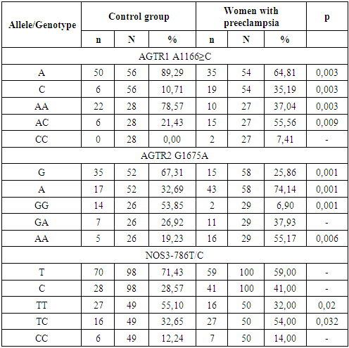
Analysis of the obtained data indicates significant differences in the dynamics of PIGF and sFlt-1 concentrations and their ratio during physiological pregnancy and pregnancy complicated by preeclampsia. The most pronounced changes are in the values of the sFlt-1/PIGF ratio, in addition, the degree of deviation of the listed parameters correlates with the severity of preeclampsia. Apparently, this indicator is the most informative in the early diagnosis of preeclampsia. The results obtained are fully consistent with the published data that the development of preeclampsia is closely associated with an imbalance in synthesis of angiogenic and antiangiogenic factors [5,7,9]. It is known that the process of placenta formation begins with the implantation of cells of fetal origin (cytotrophoblast) into decidual tissue (modified endometrial layer of the pregnant uterus). The cytotrophoblast is not only embedded into endometrium layer (interstitial invasion) and spiral arteries (endovascular invasion), but also reaches the inner third of endometrium. As a result, at the end of the first trimester, several dozen wide, gaping arteries are formed in the utero-placental region and the utero-placental blood flow begins to function actively. Prior to its formation, the function of a powerful stimulus of the first wave of cytotrophoblastic invasion (CTI) is carried out by local tissue hypoxia, which is characteristic of the embryo microenvironment up to 8-10 weeks of development. Hypoxic stimulus increases the expression of specific cell adhesion molecules, stimulates the synthesis of cytokines and vascular growth factors [1].
The presence of polymorphisms of genes that regulate vascular tone (renin-angiotensin system and endothelial nitric oxide synthetase) that predispose to hypertension complications significantly increases the risk of preeclampsia developing. The associations identified in this study can be used as genetic markers of predisposition to the formation of preeclampsia, which will allow timely formation of a risk group and correction of therapeutic and preventive measures.
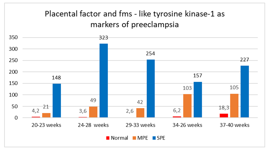
Figure 1. The ratio of sFlt/PIGF in normal pregnancies, moderate (MPE) and severe preeclampsia (SPE)
A. Wang et al. [8] also associate preeclampsia with a violation of the cytotrophoblast invasion process at this stage. The next stage of cytotrophoblast invasion (CTI) deep into the myometrium (the second wave of invasion) occurs at the 16th-18th week of pregnancy. Сytotrophoblast transforms the larger arteries of the lower third of the myometrium, turning them into wide cavities. As a result, the volume of maternal blood entering placenta increases [1]. The separation of two waves of CTI is conditional, since it is an ongoing process, which in its significance is a key mechanism for the development of normal pregnancy or the occurrence of preeclampsia. The statement that preeclampsia begins at the 20th week of pregnancy corresponds only to the time of occurrence of well-known symptoms, while the initial mechanisms are laid and implemented much earlier in the form of defects in the luteal phase of the cycle, violations of implantation and placentation, as well as background diseases of mother. These and other factors are the cause of CTI insufficiency [3]. Endothelial dysfunction plays a central role in the pathogenesis of preeclampsia. Increased synthesis of vasoactive mediators leads to a predominance of vasoconstriction and, as a result, insufficient blood circulation in the placental vessels. Before the appearance of clinical symptoms, the utero-placental blood flow usually decreases and the resistance of the uterine vessels increases, placental ischemia develops [7]. Apparently, in patients predisposed to the development of preeclampsia, decrease in concentration of PIGF and increase in concentration of sFlt-1 indicate abnormal placental development. The reasons of patient’s predisposition to the development of preeclampsia are still not fully established, but it is noted that one of the possible causes is a genetic factor. It has been shown that the development of preeclampsia in mother increases the risk of this pathology in her daughter. It is also believed that the etiology and pathogenesis of preeclampsia are due to the presence of immunopathological mechanisms and a number of environmental factors. Prevention and treatment of preeclampsia is quite a complex task. In this regard, early diagnosis of preeclampsia before its clinical manifestations is one of the tasks in obstetrics, the solution of which will allow to assess the risk and feasibility of preserving the pregnancy [4,9]. The availability of reference intervals of PIGF, sFlt-1 concentrations and their ratio are currently the most informative for the diagnosis of preeclampsia, which allow to evaluate peculiarities of these molecules secretion since the end of the first trimester of pregnancy. Previously, it was shown that already in the first trimester of pregnancy it is possible to diagnose an imbalance in synthesis of PIGF and sFlt-1, as a result of which the ratio of these indicators increases. An increase in sFlt-1 concentration seems to disrupt the intracellular mechanism of PIGF synthesis regulation, which consequently leads to the development of systemic endothelial insufficiency and the progression of clinical signs of preeclampsia (hypertension and proteinuria), as well as to a delay in fetal development [3,9,11].
Thus, based on the performed study, it was found that concentrations of PIGF, sFlt-1 and their ratio values are highly informative indicators of preeclampsia, and the reference intervals of PIGF, sFlt-1 concentrations and their ratio values can be used as "standards". In addition, the determination of the concentration of these markers and the calculation of their ratio should be carried out in the first and second trimesters of pregnancy as part of screening programs for the diagnosis of intrauterine fetal pathology. The determination of preeclampsia markers at the end of the second and third trimester of pregnancy can serve as a basis for the final diagnosis of preeclampsia and the development of tactics for prolonging pregnancy.

References

[1]  Preeclampsia. Guide. Library of a specialist doctor. Ed. Sukhoi G. T., Murashko L. E. M.: GEOTAR-Media. 2010; 576.
[2]  Carty DM, Delles C, Dominiczak AF. Preeclampsia and future maternal health. J Hypertens 2010; 28: 1349–1355.
[3]  Foidart YM, Schaaps YP, Chantraine F. et al. Dysregylation of anti-angiogenic agents (sFlt-1, PIGF and sendoglin) in preeclampsia — a step forward but not the definitive answer. J Reprod Immunol. 2009; 82: 106-111.
[4]  Hammerova L, Chabada J, Drobny J, Batorova A. Lonfi tudinal evaluation of markers of hemostasis in pregnancy. Bratisl Lek Listy 2014; 115 (3): 140–144.
[5]  McElrath T, Lim K-H, Pare E, Rich-Edwards J. et al. Longitudinal evaluation of predictive value for preeclampsia of circulating angiogenic factors through pregnancy. Am J Obst Ginecol. 2010; 11; 407: e1-e7.
[6]  Verlohren S, Herraiz Y, Lapaire O. et al. The sFlt-1/PIGF radio in different types of hypertensive pregnancy disorders and its prognostic potential in peeclamptic patients. Am J of Obstet Gynecol. 2012; 1: 58:e1-e8.
[7]  Lam C, Lim K-H, Karumanchi SA. Circulating angiogenic factors in the pathogenesis and prediction of preeclampsia. Hypertension Res. 2005; 46: 1077-1085.
[8]  Maynard S, Min J, Merchan J, Lim K. Excess placental soluble fms-like tyrosinekinase-1 (sFlt-1) may contribute to endometrial disfunction, hypertension and proteinuria in preeclampsia. J Clin Invest. 2003; 111: 649-658.
[9]  Roberts JM, Bell MJ. If we know so much about preeclampsia, why haven´t we cured the desease? J Reprod Immunol 2013; 99: 1–9.
[10]  Shibata E, Rajakumar A, Roberts RW. et al. Soluble fms-like tyrosine kinase 1 is increased in preeclampsia but not in normotensive pregnancies with small-for-gestational-age neonates: relationship to circulating placental growth factor. J Clin Endocrinol Metab. 2005; 90: 4895-4903.
[11]  Wang A, Rana S, Karumanchi SA. Preeclampsia: the role of angiogenic factors inits pathogenesis. Physiology. 2009; 24: 147-158.
[12]  Yelliffe-Pawlowsky L, Shaw G, Currier R. et al. Association of early preterm birth with abnormal levels of rutinely collected ferstand second-thrimester biomarkers. Am J Obstet and Gynecol. 2013; 208: 128-140.