American Journal of Medicine and Medical Sciences

p-ISSN: 2165-901X    e-ISSN: 2165-9036

2020;  10(10): 803-808

doi:10.5923/j.ajmms.20201010.16

Received: Sep. 2, 2020; Accepted: Sep. 20, 2020; Published: Oct. 26, 2020


Study of Behavioral and Morphological Disorders in Animals with Modeled Pathology of Mild Traumatic Brain Injury

Khaydarov Furqat Ganievich, Khasanova Dilnoza Ahrorova

Anatomy, Clinical Anatomy (OSTA) Department, Bukhara State Medical Institute, Bukhara, Uzbekistan

Correspondence to: Khaydarov Furqat Ganievich, Anatomy, Clinical Anatomy (OSTA) Department, Bukhara State Medical Institute, Bukhara, Uzbekistan.

Email:

Copyright © 2020 The Author(s). Published by Scientific & Academic Publishing.

This work is licensed under the Creative Commons Attribution International License (CC BY).
http://creativecommons.org/licenses/by/4.0/

Abstract

Changes in the brain tissues of animals that have suffered a mild traumatic brain injury correlate with data on neurological deficits and behavioral status of laboratory animals. With a mild traumatic brain injury in experimental animals, circulatory disorders occur in the area of damage to the cerebral cortex, which is expressed in edema of the walls of microvessels, a decrease in their lumen, and changes in the density of the vascular bed. When correcting a mild traumatic brain injury, it is necessary to evaluate the neuropsychiatric disorders by testing, to study the typological features and motor activity of the body.

Keywords: Experimental design turbine, Traumatic brain injury, Morphological changes, Neuropsychiatric disorders

Cite this paper: Khaydarov Furqat Ganievich, Khasanova Dilnoza Ahrorova, Study of Behavioral and Morphological Disorders in Animals with Modeled Pathology of Mild Traumatic Brain Injury, American Journal of Medicine and Medical Sciences, Vol. 10 No. 10, 2020, pp. 803-808. doi: 10.5923/j.ajmms.20201010.16.

1. Introduction

One of the most common brain injuries of medico-social significance is craniocerebral injuries. As the analytical statistics show, Traumatic brain injury (TBI) is a serious problem throughout the world. The prevalence of TBI in the world varies significantly, ranging from 95 to 783 per 100 thousand people per year, and the mortality rate of the population is from 9.5 to 66 per 100 thousand people per year.
The prevalence of TBI in our republic was 692 cases per 100 thousand population, and mortality in different regions of the country varies greatly, from 18.5 to 49 per 100 thousand population [1]. The causes of head injuries are very diverse, but the main mechanism of formation is the impact of an external force factor. Thus, its pathogenesis is characterized by interrelated processes of biochemical, morphological, and immunological disorders [2,3,4,5,6,7].
To date, the attention of scientists is focused on mild craniocerebral trauma, which is due to the complexity of the diagnosis and the scarcity of objective signs of trauma, which could allow differentiation without craniotomy. In the complex of disorders of the central nervous system with mechanical damage, the influence of pathological disorders on the coordination system, behavioral nature, cognitive functions of the body, intracranial pressure, metabolism, etc. is of particular importance [8,9,10,11].
Considering the versatility of disorders in mild traumatic brain injuries, the choice of treatment methods depends primarily on the severity of the injury, the location of the damage in the brain, the age and condition of the victim. So, different experts recommend and use different methods of treatment that contribute to the restoration of the processes of vital organs [12]. According to the latest scientific research to solve the problems associated with the choice of effective treatment methods is the use of experimental models that contribute to a detailed study of damage, as well as the use of corrective drugs that are not always safe for the human body.
In order to further improve treatment methods and develop new ways of correction for mild traumatic brain injury, which is not accompanied by long-term and deep injuries in the brain, it is necessary to develop an experimental model and study the main disorders that allow objectively determining and differentiating the degree of injury.

2. Materials and Research Methods

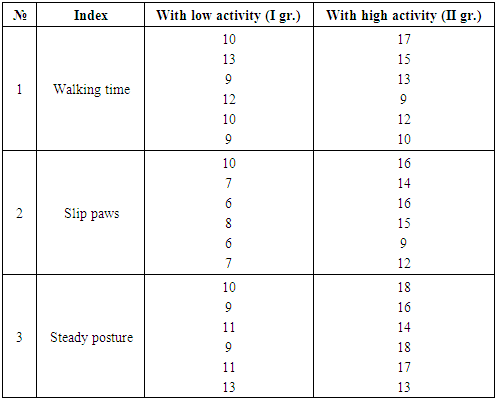
To achieve this goal, experimental studies were conducted on 90 white mongrel rats, males weighing 210 ± 4.8 g, which were kept instandard conditions of the vivarium of the Central Scientific Research Laboratory of Tashkent Pharmaceutical Institute with the provision of a diet in accordance with the daily nutritional standard for animals. Before creating an experimental model of mild traumatic brain injury for the study and comparative analysis of disorders among active and passive animals, the individuals were divided into 2 groups according to the typological status of higher nervous activity using the open field test. Thus, groups of animals with low and high activity were formed.
In the study of neuropsychiatric disorders to identify asymmetric disorders of the brain and substantiate differentiated groups with different emotional and motor activity, the "Cylinder" test was carried out. According to the conditions of the test, the experimental animals were placed in an installation representing a plexiglass cylinder with a height of 30 cm and a diameter of 20 cm with a transparent bottom. The animals, being in an unfamiliar environment, began to exhibit orientation-exploratory behavior, expressed in active movement of the forelimbs on the walls of the cylinder. When climbing on hind legs, animals used one or both forelimbs (normally both legs are used). The results obtained were assessed by the number and quality of movements of the forelimbs with support on the cylinder walls. In addition, in order to determine the coordination of movement after the reproduction of traumatic brain injury on experimental animals on the second and seventh days, the test "Walk on a raised beam" was carried out. This technique was used to assess the state of equilibrium in animals with neurological disorders provoked by an external mechanical shock. So, to study the dynamics of performance indicators, which is one of the most significant clinical signs of the state of the central nervous system, a system was used, which is a raised crossbar of various configurations and thicknesses and a closed safe platform.
According to the method, the experimental animals pass over the crossbar and reach its end, which is a safe area. During the method, the time of passage, slipping of the paws and falling is recorded. The results were evaluated by the time (sec) spent on 3 exponential movements.
The complex of our studies included the study of morphological disorders after the simulation of mild traumatic brain injury in animals. The studies began with euthanasia, chloral hydrate 350 mg / kg was used for anesthesia by puncture of the left ventricle until complete exsanguination. In experimental animals, a biomaterial (brain) was isolated, which was fixed in a 10% formalin solution. For the study, a section of the cerebral cortex in the area of injury was excised. Prepared sections with a thickness of 4-6 micrometers were stained with hematoxylin and eosin. For microscopy and photographing, optical systems were used, consisting of a microscope and an eyepiece camera. On macro-preparations stained with hematoxylin and eosin, the state of capillaries, their blood supply, the state of the meninges, and cellular elements of the brain were studied.

3. Results

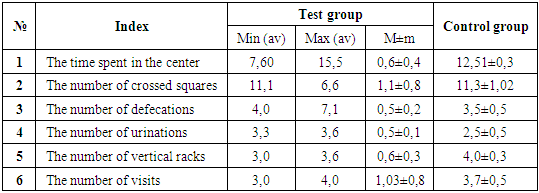
Higher nervous activity, which ensures the continuous operation of all parts of the nervous system, has its own characteristics that characterize the actions of the body under various situations and conditions in the external environment. The behavior of an organism can change dynamically over time, which determines its individual abilities and helps prevent negative consequences as a result of its inadequate actions. The defined typological features of the organism represent a complex process of integration of cortical-subcortical relationships of higher nervous activity [13]. Thus, the features of the higher nervous activity of experimental animals were studied, on the basis of which the main indicators were identified for further breakdown into groups.
To carry out an objective quantitative assessment of the functional state of the central nervous system and assess the effect of mechanical damage on the emotional and mental state, determine the activity and nature of behavior, animals were differentiated into experimental groups with low and high activity using the proven and widespread open field test.
This test made it possible to assess the motor activity, to establish qualitative and quantitative indicators of the behavior of experimental animals under certain conditions. The main indicators for differentiation into groups were the following: the latent period of time spent in the center, the number of crossed squares, the number of defecations, the number of urination, the number of vertical racks, the number of washings. For visual convenience in identifying groups of different activities, animals were marked with different colors (yellow - active, green - low activity).
The test was carried out using a large rectangular chamber (1.50x1.50 m) with walls 0.30 m high, lined with squares. The sides of the central square were 0.30 m, the peripheral squares were 0.15 m. At the intersection of the squares, there were holes 0.02 m in diameter. Lighting was provided by a 150 W shadowless lamp located at a height of 1.00 m above the center of the field. The animals were placed in the center of the field and their activity was recorded every second for 5 minutes. The time spent by the rat in the central square (the duration of the latent period), the time of immobility were recorded, the number of crossed central and peripheral squares (horizontal), the number of racks - lifting on the hind legs (vertical motor activity), the number of washes, the number of defecations and urinations were counted.
According to the experimental studies carried out before the impact of the injury, the following results were obtained: the time spent in the center, measured in seconds, averaged 2.50-2.57 ± 0.6 s, the number of crossed squares - 10.2-12.6 ± 1, 1 pc., The number of defecations - 1.4-1.7 ± 0.5 pcs., The number of urinations - 0.3-0.5 ± 0.5 pcs., The number of vertical racks 4.0-6.5 ± 0, 6, the number of washes 3.8-5.2 ± 1.03 pcs (Tab. 1).
Table 1. Results of the "open field" test before injury
     
According to the results of the test carried out, the experimental animals were divided into 2 groups. Experimental animals with minimum values for all parameters were assigned to the group with low (group I) activity, animals with maximum values in the group with high (group II) activity. Analyzing the test results, it was found that among the experimental experimental animals (90 pcs) the largest number of animals belonged to the active type - 48 pcs and slightly less to the group with low activity - 42 pcs.
After dividing the animals into 2 groups, the next stage of research was the modeling of mild traumatic brain injury. The simulation of such an injury required the most accurate execution of the impact, taking into account its nature and structure of the impact, the duration of its impact and the place of injury. To this end, a number of experimental studies were considered to create a model: fluid-percussion brain injury (FLP), model of TBI using a weight drop method, model of controlled cortical injury (Controlled Cortical Impact Model) [14].
Among the above models for our research, taking into account the set goal and task, the conditions of implementation, we have chosen the model "TBI as a result of a drop in load".
Before the start of the experiment to create a model of mild TBI, the animals were injected intraperitoneally with the drug urethane at a dose of 1 g / kg. This model "TBI as a result of a drop in load" required technical preparedness and appropriate skills. To achieve this goal, we used an experimental constructive turbine for the first time.
The model of mild TBI was carried out in a strict sequence of constructing the experiment. Prepared healthy experimental animals from each group: with low activity, with high activity, were placed on a special surface of a constructive turbine. The animal's head was firmly fixed to the surface using adhesive tape. The animal was laid so that its fixed head, and more specifically its skull, was in the center right under the metal turbine. To reproduce a mild head injury, taking into account all possible violations during its creation, a load with a mass of 24 g was previously prepared using calculations. The role of the load was performed by a metal weight. The turbine is a metal tube 60 m high, with a diameter that allows the free passage of a load with a mass of 24 g (Fig. 1, 2).
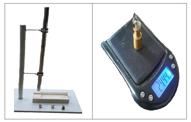
Figure 1. Experimental Structural Turbine and Weight for Modeling Mild TBI
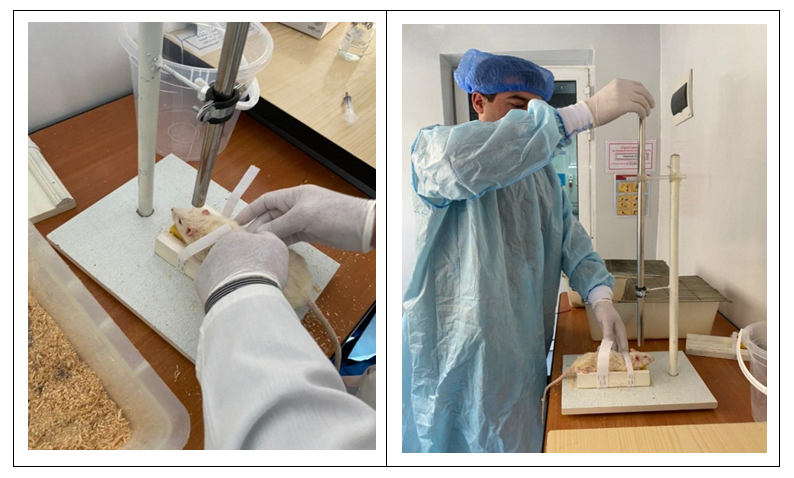
Figure 2. Stages of creating an experimental model of mild TBI
Thus, the animals were inflicted direct mechanical trauma by dropping a load weighing 24 g from a height of 60 m. After modeling, the injured rats were placed in cages (5 individuals in each) for further observation and study of behavior, pathological disorders. The injured animals housed in cages were kept on a standard diet with free access to water and a normal light regime for 14 days.
During the research, characteristic features in the behavior of rats in the early stages of mild TBI were revealed. Numerous clinical studies have revealed that the most / characteristic signs of mild TBI observed in the first hours after the injury are short-term loss of consciousness (up to 30 minutes) or amnesia (up to 24 hours), which subsequently regress and complete recovery occurs [15,16,17]. In this regard, it was necessary to determine in the behavior of animals signs that would indicate the development of the above signs observed in humans. In addition, given that behavioral disorders are among the early violations in this pathology, it was necessary to determine inhibition to determine the depth and direction of behavioral disorders.
So, to identify asymmetric disorders of the brain after modeling mild TBI on the 2nd day, the "Cylinder" test was carried out. The main indicator of the test, as well as asymmetric disorders and movement deficits observed in mild TBI, is the quantitative and qualitative use of the forelimbs by animals. The test consists in examining the horizontal and vertical surfaces of the cylinder walls with the rats' forepaws.
The results obtained showed that in animals, when the forelimbs were supported against the walls of the cylinder, limited movements were observed, and in some individuals only one paw was raised. Thus, in the group of animals with high emotional and motor activity (2nd), independent use of only the left forelimb was observed, compared with the group of low activity (1st), where uniform use of both forelimbs was noted.
According to the test "Cylinder", on models of mild TBI, in the 2nd group of animals with high activity, pronounced pathology develops in the cerebral cortex, as evidenced by the use of one left paw (17%). All of the above indicates a multidirectional course of pathology among a group of animals with high and low activity.
The next stage of the research was to determine the coordination of movements on experimental models of mild TBI. For this purpose, the raised-beam walking test was performed. The test results showed that in animals of the intact group with low and high activity on the second day after modeling a mild degree of TBI, an imbalance occurs, expressed in the lengthening of the latency period of transition from one position to another (Tab. 2). Consequently, due to the received trauma and its consequences, the inhibition of reactions develops in animals, manifested by a slowdown in movements caused by the fear of falling off the bar, which explains his strong grip of the bar with both legs.
Table 2. Test results (raised-beam walking) 2 days after modeling mild head injury, sec
     
The study of morphological changes in the brain cells of animals with simulated mild traumatic brain injury showed the following results. Considering that the clinic of mild TBI is characterized by signs of concussion and contusion of the brain, accompanied by minor focal disorders associated with damage to the brain tissue, it is necessary to highlight the main morphological signs that make it possible to diagnose and predict the course and outcome of the injury in a timely manner.
The study of the patterns of morphofunctional transformations of capillaries, qualitative and quantitative indicators of the structure of the brain in animals with mild traumatic brain injury under experimental conditions showed pronounced structural changes in the substance of the brain, which was determined by the presence of minor areas of local edema of the brain substance in combination with subarachnoid and cortical hemorrhages. With the enlarged image, we identified small areas of brain tissue impregnated with erythrocytes, and a small number of macrophages were also detected.
The study of the cytoarchitectonics of the cerebral cortex revealed no obvious violations in the structure of the arrangement of layers. Despite the absence of violations in the arrangement of layers in the ganglion layer around the pyramidal neurons, pericellular edema was still observed. When comparing neurocytes with a group of intact animals, it was found that their size is somewhat reduced, their cytoplasm is homogeneous and dark-basophilic.
Morphological studies have shown that throughout the surface of the cerebral hemispheres, the choroid is tightly adhered to the brain substance, thickened, its vessels are full-blooded and dilated. Shriveled and hyperchromic neurons were isolated, mainly in the pyramidal layers of the gray matter, a high cell density per unit area was determined. In a comparative aspect, the study of the structure of the cerebral cortex in intact rats did not reveal morphological abnormalities. However, experimental animals with simulated mild TBI showed insignificant morphological abnormalities in the area of injury, which made it possible to give a quantitative and qualitative assessment.
In addition, the study of the reaction of the capillary bed of the cerebral cortex during the period of injury, as shown by the study, begins with a decrease in the diameter of the capillaries and their density, as well as the area of the exchange surface.
Consequently, morphological studies when stained with hematoxylin and eosin indicate changes in the form of minor bruises represented by structureless masses, with hemorrhage engulfing the cortex of the parietotemporal region of the large brain. The revealed intracerebral hemorrhages were small in terms of the volume of damage and the area of distribution - up to 3.0 mm cube. As mentioned above, in the medulla of the perifocal zone, there was edema, hyperemia of small vessels of the cortex and subcortical white matter: precapillaries and capillaries that form networks, pericacapillary edema.
Studies have shown that the revealed morphological changes were more pronounced in the immediate vicinity of the injury site, i.e. in the perifocal zone, and gradually weakened with distance from it. Studies have confirmed that even in cases of mild TBI, where clinical signs are very scarce, there are ultrastructural and microscopically detectable abnormalities in various parts of the brain that can be diagnosed during the issuance of pathological reports.

4. Discussion and Conclusions

To study behavioral and morphological disorders in mild traumatic brain injury, the experimental animal model is recommended to be reproduced by the “TBI as a result of a drop in weight” (free fall) method, which is the simplest and most effective modeling method. In order to further improve the methods of treatment and master new ways of correction in mild traumatic brain injury, it is necessary first of all to assess psychoemotional disorders, including motor activity, which is determined using the informative open field test.
The morphological studies carried out refute the still existing opinion about the absence of morphological changes in mild TBI. Studies have shown that changes in the brain tissues of animals with mild TBI correlate with the data of neurological deficits and behavioral status of laboratory animals. With mild traumatic brain injury, experimental animals develop circulatory disorders in the area of damage to the cerebral cortex, which is expressed in edema of the walls of microvessels, a decrease in their lumen, and a change in the density of the vascular bed. Consequently, when correcting disorders of the brain, in particular with mild TBI, it is necessary to assess by testing neuropsychiatric disorders, to study typological features, motor activity of the body.

References

[1]  Sabirov D.M., Rosstalnaya A.L., Makhmudova M.A. Epidemiological features of craniocerebral trauma // Bulletin of Emergency Medicine - 2019. V. 12. - No. 2. - S. 61-66.
[2]  Gornostaev DV, Lapteva M.I. et al. Epidemiology and pathomorphosis of mechanical trauma // Zh. "Problems of Expertise in Medicine" - 2003. - No. 2. - S. 8-10.
[3]  Zayratyants G.O. Morphofunctional changes in the membranes of the brain during immunomodulatory therapy in patients with traumatic intracranial hematomas // Abstract of the thesis. Cand. honey. sciences. - M., 2006. -- 25 p.
[4]  Korolev I.A. Biochemical changes in craniocerebral trauma // Materials of the All-Russian Scientific Forum of Students "Student Science-2019" - M., 2019. - P. 245-246.
[5]  Myakotnykh VS, Talankina NZ, Borovkova TA Clinical, pathophysiological and morphological aspects of the late period of closed traumatic brain injury // J. neurology and psychiatry them. S. S. Korsakova - 2002. - No. 102 (4). - S. 61-65.
[6]  Nosov A.T., Kajaya N.V. Morphofunctional changes in the brain tissue with repeated mild traumatic brain injury (experimental research) // Ukrainian neurosurgical journal - Kiev, 2006. - №1. - S. 91-98.
[7]  Summarized D.B. and others. Immuno-biochemical monitoring in the acute period of craniocerebral (TBI) and concomitant (CT) injuries // Allergology and Immunology - 2014. – Volume 15. - №2. - S. 147-149.
[8]  Zyablitsev S.V., Khudoley S.A. et al. Influence of modifications of the functional state of the central cholinergic system on the degree of neurological deficit in traumatic brain injury // Neurophysiology - 2015. - T47. - No. 2. - S. 202-205.
[9]  Konovalov A.N., Likhterman LB, Potapov A.A. Clinical guidelines for traumatic brain injury: in 3 volumes. Volume 3: Consequences and complications of traumatic brain injury, standards and recommendations, neurorehabilitation, examination - M., 2002. - 632 p.
[10]  Latysheva V.Ya., Olizarovich M.V., Sachkovsky V.L. Traumatic brain injury: classification, clinical picture, diagnosis and treatment // Textbook - Minsk: Higher School, 2005. -- 158 p.
[11]  Sushkevich G.N., Roshal L.M. et al. The effect of human mesenchymal and neural stem cells on cerebral vessels, vegf and markers of hypoxia in blood serum in traumatic brain injury in rats // Pathogenesis - Dedovsk, 2008. - №3. - S. 24-31.
[12]  Teshaev Sh.J., Khasanova D.A. Topografic-anatomical features of lymphoid structures of the small intestine of rats in norm and against the background of chronic radiation diseases// European science review Vienna, Austria №9-10 2018, Volume 2. Medical science P. 197-198.
[13]  Emelyanov Yu.V. Disorders of psychoneuroimmune relationships in white rats after severe concomitant traumatic brain injury // Abstract of the thesis. Omsk, 2009. -- 24 p.
[14]  Beloshitsky V.V. Principles of modeling traumatic brain injury in the experiment // Ukrainian neurosurgical journal - Kiev, 2008. - №4. - S. 9-14.
[15]  Bofanova N.S. Clinical and functional features of the consequences of traumatic brain injury using stabilometric research as a method for diagnosing motor disorders // News of higher educational institutions - 2013. - No. 2 (26). - S. 66-73.
[16]  Bofanova N.S., Ermolaeva A.I. Clinical and functional features and treatment of the consequences of mild craniocerebral trauma of varying degrees // News of Higher Educational Institutions - Penza, 2014. - №1 (29). - S. 37-45.
[17]  Potapov A.A., Likhterman L.B. et al. Mild traumatic brain injury: clinical guidelines // M.: Association of neurosurgeons of Russia - 2016. - 23 p.