Indiaminov Sayit Indiaminovich1, Kim Antonina Amurovna2
1Doctor of Medical Sciences, Professor, Head of the Department of Forensic Medicine and Pathological Anatomy, Samarkand State Medical Institute, Uzbekistan
2Research Candidate of the Department of Forensic Medicine and Pathological Anatomy of the Samarkand State Medical Institute, Uzbekistan
Correspondence to: Kim Antonina Amurovna, Research Candidate of the Department of Forensic Medicine and Pathological Anatomy of the Samarkand State Medical Institute, Uzbekistan.
| Email: |  |
Copyright © 2020 The Author(s). Published by Scientific & Academic Publishing.
This work is licensed under the Creative Commons Attribution International License (CC BY).
http://creativecommons.org/licenses/by/4.0/

Abstract
For the purpose of identifying the nature and degree of damage to the structures of the brain (В), areas of the cortex with the underlying white matter of the В and the cerebellum were studied in 95 persons who died from carbon monoxide (CO) poisoning. The study was carried out comparatively in two groups: the 1st consisted of persons with only CO poisoning (78), the 2nd - those who died from CO poisoning against the background of acute alcohol intoxication (AAI) - 17 cases. It was found that vascular-tissue changes in the structures of В from direct exposure to CO are manifested when the content of carboxyhemoglobin (HbCO) in the blood is 30% and higher. However, the intensity of vascular and nervous tissue lesions increases sharply when the HbCO content in the blood is 50% or more. In cases of combinations of carbon monoxide poisoning with acute alcohol intoxication, the polymorphism of the nature of vascular tissue changes in the structures of the cortex and subcortical white matter of the В and cerebellum were caused by the toxic effects of both CO and ethanol. At the same time, the intensity of changes also increased at high concentrations of HbCO in the blood (50% and higher) and the presence of more than 3 ppm ethanol in the blood. For the purposes of differential diagnosis of CO poisoning from AAI, the use of modern morphometry methods in studying the structure of the brain is promising.
Keywords:
Carbon monoxide, Poisoning, Alcohol intoxication, Brain, Morphology
Cite this paper: Indiaminov Sayit Indiaminovich, Kim Antonina Amurovna, Morphology of the Brain Structure in Acute Carbon Monoxide Poisoning, American Journal of Medicine and Medical Sciences, Vol. 10 No. 10, 2020, pp. 736-740. doi: 10.5923/j.ajmms.20201010.02.
1. Actuality of the Problem
In forensic practice, the number of studies performed on corpses associated with poisoning occupies one of the leading places in the structure of violent death [1,2]. The causes of poisoning are often hemoglobinotropic poisons, where the leadership belongs to fatal poisoning from carbon monoxide (CO), the relevance of which is preserved throughout the world [3,4,5,6,7]. The diagnosis of CO poisoning is based on clinical and morphological data, the results of a forensic chemical analysis of blood and skeletal muscles. However, these data do not always allow substantiating the thanatogenesis of CO poisoning, especially in cases of the presence of background (competing) conditions in the body. In this regard, microscopic examination of the structure of organs and tissues, due to which the recognition of pathological changes at the cellular level is carried out, is important for establishing the patho- and thanatogenesis of poisoning. The possibilities of morphological methods in forensic medicine have significantly expanded due to the increasingly widespread use of morphometric techniques with the use of computer programs, the use of immunohistochemistry methods [8]. The central nervous system - brain reacts first of all and with significant changes to the effect of the toxic component of CO, which leads to hypoxia of cells and tissues. Despite this, the participation of В in the thanatogenesis of CO poisoning, especially in the presence of competing (background) states, has been insufficiently studied.
2. The Purpose of the Study
To reveal the nature and degree of damage to the structures of В in acute CO poisoning, depending on the presence of signs of acute AAI in the body.
3. Materials and Methods of Research
The material was the brain from 95 corpses of persons who died from CO poisoning. The circumstances of CO poisoning in 90% of cases were heating devices, which influenced the frequency of deaths in winter - 62%, as accidents. In the age aspect, mortality prevailed at a young age (from 18-44 years old) -53%, the middle-age group was (from 45-59 years old) -17%, the elderly (from 60-74 years old) -9% and senile (from 75- 90 years) - 3%. Children under the age of 17 accounted for 18%. Mortality from CO poisoning prevailed in males - 61% of cases. The concentration of HbCO in the blood of the dead was observed in the range of 30% -93%. In the blood of 17 victims, alcohol was found in concentrations from 0.35 ‰ to 3.82 ‰. In most cases, the death of the victims occurred at the site of the poisoning. For microscopic examination, pieces were taken from the areas of the cortex and subcortical white matter, as well as pieces from the cerebellum, which were fixed in a 10% solution of neutral formalin, and embedded in paraffin. Histological sections 7-10 мм thick were stained with hematoxylin and eosin using the Nissl method. Material for histological examination was taken in the earlier postmortem period, during the first day (from 6-8 hours to 16-20 hours). Additionally, the results of macroscopic changes in organs and tissues (conclusions of the examination of corpses), the results of forensic chemical analysis and materials of the circumstances of death were studied and analyzed. The studies were carried out comparatively in 2 groups: 1 - acute CO poisoning and the absence of alcohol in the body - (78 cases); 2- acute CO poisoning against the background of АAI- (17 cases).
4. Research Results and Their Discussion
Microscopic examination of the cortex and subcortical white matter of the В showed that the pia mater is thin in places, thickened in some areas due to the proliferation of loose connective tissue, the fibers of which in the outer layer are collagenized. There is also some thickening of the arachnoendothelium due to moderate cell proliferation. The connective tissue fibers contained single erythrocytes and the usual number of local cells with swollen and pale-colored nuclei, among which there are many large cells of irregular oval shape with an eccentrically located nucleus, granular cytoplasm of pink color, and rounded shapes of these cells were also observed. Vessels of all types and calibers of the shell are full-blooded. In some cases, small areas of fibrosis of the pia mater were noted, in which cells were almost invisible. Adventitia of arteries and less veins of the sheath with phenomena of moderate collagenization. In the substance of the brain, a pronounced blood filling of the vessels of the microvasculature was revealed, the erythrocytes in them were separated by small sections of plasma, foci of erythrocyte diapedesis were noted. As a result of precipitated (precipitated) plasma proteins, hemolyzed erythrocytes, a hyaline thrombus is formed in the vessels of the microvasculature (Fig. 1). Perivascular edema delimited the vessel walls from the brain tissue, forming cracks. Arteries and veins are unevenly filled with blood, the endothelium was characterized somewhere moderately swollen, hyperchromic, weakly pyknomorphic. There were no hemorrhages in the brain tissue.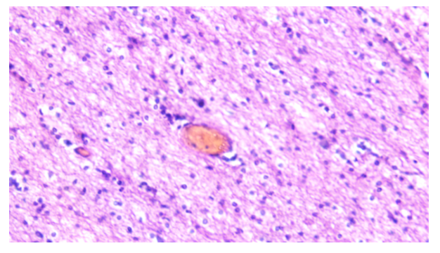 | Figure 1. Woman 22 years old. HbCO in the blood - 54%. Congestion of blood vessels with the presence of hyaline thrombi in small venous vessels in the white matter of the brain. Homogenization of collagen fibers. Staining with hematoxylin and eosin. Lens 40, eyepiece 10 |
The structure of neurocytes underwent dystrophic changes, in the form of swelling of cells, shortening of processes, indistinct contours of nuclei and protoplasms, and the presence of chromatolysis were noted. Quite often there were hyperchromic-wrinkled neurocytes, where the nucleus was almost not contoured, shadow cells, melting neurocytes, neuronophagy. It was also possible to observe the presence of "granular balls". There was a lively reaction from glial cells with the formation of few drainage forms, sattelitosis, pericellular glial edema (Fig. 2). In the cerebellum, the pia mater is congested, as well as in the vessels of the cerebellum stasis, congestion, tissue edema. Dystrophic changes in Purkinje cells, shadows of pear-shaped cells. The intensity of vascular tissue changes in the cortex and in the cerebellar region were most pronounced in those individuals in whose blood a high concentration of HbCO was detected (50% and higher).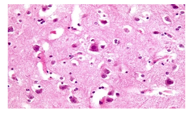 | Figure 2. Woman 22 years old. HbCO in the blood - 54%. Dystrophic changes in neurons in the cortex of the cerebral hemispheres. Satelliteitis. Staining with hematoxylin and eosin. Lens 40, eyepiece 10 |
CO is a highly toxic blood poison and poisoning from it is usually characterized by a slow rate of death, however, a fast brain type of thanatogenesis can also be observed. Our microscopic examination showed that vascular-tissue changes in the structures of В from direct exposure to CO are manifested when the content of HbCO in the blood is 30% and higher. However, the intensity of vascular and nervous tissue lesions increases sharply when the HbCO content in the blood is 50% or more. According to Bogomolova I.N. (2007) in case of CO poisoning, swelling of the В structures and severe edema in the trunk develops. Dystrophic changes with swelling of neurons are revealed. The unevenness of the blood filling of the vessels of the microvasculature, which the author associates with a drop in vascular tone, was noted. In vessels of small caliber, coagulation of plasma proteins with the formation of hyaline thrombi was noted, and with a late onset of death, foci of melting in the В [9]. Some researchers believe that the action of CO affects the maturation of oligodendrocytes, thereby leading to the progression of the process of demyelination of the white matter. This pathology leads to neurological complications, the mechanism of which is not fully understood. According to Sekiya K. (2019), the long-term process of demyelination in CO poisoning is accompanied by a significant decrease in microglial cells. At the same time, their neurotrophic function decreases and as a result, such a phenomenon as encephalopathy develops [10,11]. These data dictate the need for further study of lesions of various structures of В in CO poisoning.Modern advances in methods of radiation diagnostics are widely used in clinical practice to establish the presence and extent of damage, as well as long-term consequences, for example, encephalopathy after acute CO poisoning. Computed tomography (CT), magnetic resonance imaging (MRI), magnetic resonance diffusion tensor imaging (MRDTT) can determine the degree of brain damage at the level of microscopic changes and accurately provide valuable information for the choice of therapy and prognostic assessment. The CT method of В revealed foci of symmetrical lesions of the cerebral pedicles in an elderly patient abusing alcohol in the long-term period after CO poisoning [12,13]. Consequently, modern methods of radiation diagnostics can be used for intravital and postmortem diagnosis of acute CO poisoning, based on lesions of the В structure.In the observations of the 2nd group, the most pronounced vascular-tissue changes were revealed, compared with the results of the studies of the 1st group. At the same time, swelling and edema of the pia mater, pronounced plethora of the vessels of the microvasculature were noted. The walls of large and medium caliber arteries are thickened, their layers are not clearly defined. In their lumen, accumulations of a fine-grained mass are observed, representing plasma coagulants. However, the veins of the membrane are thin-walled in which the aggregation of erythrocytes and leukocytes. Perivascular hemorrhages and erythrocyte diapedesis are noted. In the cortex of the cerebral hemispheres of the GM, there are large arteries, the walls of which are also characterized by swelling, thickening. In this case, the layers of the wall are not determined, the gap in them is narrow or irregular in shape. This characterizes the dystonia of the arteries. Against the background of plethora of blood vessels, stratification of plasma and formed elements of the cell, plasma stasis are noted. Weak to moderate plasmorrhage. Large veins are full-blooded, in them there is an alternation of aggregates of blood and plasma corpuscles. The nuclei are endothelium-hypo- or hyperchromic. The perivascular spaces are enlarged. Medium and small veins contain aggregates of erythrocytes, unevenly full-blooded, blood stasis is determined. Sometimes plasma or discolored erythrocytes are detected in the vessels of the MCR. In the cortex, В neurons are subject to a more pronounced dystrophic process, cytolysis and karyolysis are more common (Fig. 3). Pericellular edema, the space of which is sharply expanded, there are often glia-satellite cells (Fig. 4). In some cases, neuronophagy, moderate proliferation of neuroglial cells, and pericellular edema are observed. Moderate depletion of the cortex by pyramidal cells. A small number of senile amyloid cells are differentiated. In the white matter under the bark, vascular changes are more pronounced. Along with full-blooded vessels, there are also spasmodic ones. The wall of some arteries is disturbed; in the perivascular space, diapedesis of erythrocytes can be found. The structure of the walls of the vessels of the MCB is changed, the layers are not determined, the stasis of erythrocytes or plasma is determined in them. Pericellular edema is determined around neuroglial cells. In the cerebellum - plethora of the membrane. Stasis, tissue edema. Dystrophic changes in Purkinje cells, shadows of pear-shaped cells. Thinning of the granular layer and death of granular cells.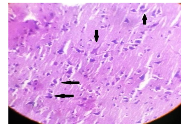 | Figure 3. Male 48 years old, HbCO in blood - 62%, ethyl alcohol - 3.57 ‰. Melting neurocytes and shadow cells in the cortex of the cerebral hemispheres. Staining with hematoxylin and eosin. Lens 20, eyepiece 15 |
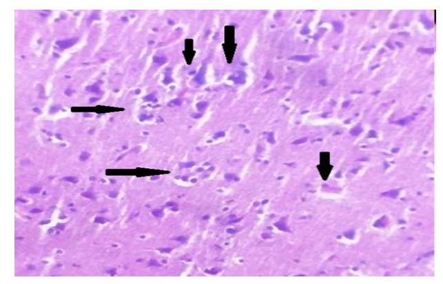 | Figure 4. Male 48 years old, HbCO in blood - 62%, ethyl alcohol - 3.57 ‰. Sattelitosis, pericellular edema in the cortex of the cerebral hemispheres. Staining with hematoxylin and eosin. Lens 20, eyepiece 15 |
Studies have shown that the severity of vascular tissue changes in the structures of the cortex and subcortical white matter of the В and cerebellum were caused by the toxic effects of CO and ethanol, the intensity of the changes increased at high concentrations of HbCO in the blood of 50% and higher and the concentration of ethanol in the blood 3 ppm and higher.It is believed that ethanol suppresses the activity of the respiratory center. Microscopically, in the brain, at the same time, pronounced vascular changes are found in the form of plethora, increased vascular permeability, stasis in the venules, perivascular and pericellular edema, diapedetic hemorrhages. Acute vascular changes are diffusely distributed, but mainly expressed in the molecular layer of the cerebral cortex, white matter and the zone of the dentate nuclei of the cerebellum. According to vascular changes, neurocellular changes are also observed. The neurons are swollen, with the phenomena of chromatolysis. Lysis of nuclei and then of nerve cells is noted, which contributes to an increase in the number of cells - "shadows", an increase in the specific density of neurons [14]. Morphological and morphometric examination of В corpses with acute ethyl alcohol poisoning (ethanol content in blood and urine ranged from 4.1 to 8.9%) revealed signs of damage to the hypothalamus, medulla oblongata and pons with a degree of damage of more than 85-90% and the severity of the lesion more than 20-30% of neurons [14].Most authors believe that metabolic disorders and impaired vascular permeability are characteristic manifestations of ethanol poisoning. In connection with this, with alcohol intoxication, pronounced edema of the В and the pia mater is often observed with the accumulation of a large amount of fluid under it [16]. Experiments show that even with a single acute alcohol intoxication, changes occur in the MCB vessels. In the GM, swelling and dystrophic changes in astrocytes that make up the BBB occur [17]. These data should be taken into account in the postmortem differential diagnosis of CO poisoning from ethanol poisoning. In our opinion, for the purposes of differential diagnosis of these conditions, it is necessary to conduct morphometric studies of the affected structures of the brain in its various departments, since the methods of light microscopy for these purposes turned out to be ineffective.At the same time, microscopic studies are necessary to identify the nature and degree of brain damage in CO poisoning.
5. Conclusions
Our microscopic examination showed that vascular-tissue changes in the structures of В from direct exposure to CO are manifested when the content of HbCO in the blood is 30% and higher. However, the intensity of vascular and nervous tissue lesions increases when the HbCO content in the blood is 50% and higher. In cases of combinations of carbon monoxide poisoning with acute alcohol intoxication, the polymorphism of the nature of vascular tissue changes in the structures of the cortex and subcortical white matter of the В and cerebellum were caused by the toxic effects of CO and ethanol. At the same time, the intensity of changes also increased at high concentrations of HbCO in the blood (50% and higher) and the presence of more than 3 ppm ethanol in the blood. The above data can be taken into account in postmortem diagnostics of CO poisoning conditions. However, for the purposes of differential diagnosis of CO poisoning from acute alcohol intoxication, it is necessary to conduct morphometric studies of the affected structures of the brain in its various parts. The results of morphological and morphometric studies can make it possible to reliably assess the thanatogenesis of this state, especially in cases of combined CO poisoning with possible competing or background conditions, and thereby reveal the pathophysiological basis of lesions of the В structures in these poisonings.Literature data have shown that methods of radiation diagnostics can be used for intravital and postmortem diagnostics of acute CO poisoning, based on lesions of the В structure.
References
| [1] | Indiaminov S.I., Kim A.A. Damage to the structures of the brain in case of poisoning with blood and general functional poisons // Journal of Biomedicine and Practice. 2020. -№3. -74-84. |
| [2] | Kim A.A., Indiaminov S.I., A.Zh. Usarov. Medical and social aspects of carbon monoxide poisoning. // Journal of Biomedicine and Practice. 2020. -№3. -85-92. |
| [3] | Li F, Chan HC, Liu S, et al. Carbon monoxide poisoning as a cause of death in Wuhan, China: A retrospective six-year epidemiological study (2009-2014). Forensic Sci Int. 2015; 253: 112-118. doi:10.1016/j.forsciint.2015.06.007. |
| [4] | Kinoshita H, Türkan H, Vucinic S, et al. Carbon monoxide poisoning. Toxicol Rep. 2020; 7: 169-173. Published 2020 Jan 20. doi:10.1016/j.toxrep.2020.01.005. |
| [5] | Ruas F, Mendonça MC, Real FC, Vieira DN, Teixeira HM. Carbon monoxide poisoning as a cause of death and differential diagnosis in the forensic practice: a retrospective study, 2000-2010. J Forensic Leg Med. 2014; 24: 1-6. doi:10.1016/j.jflm.2014.02.002. |
| [6] | Sircar K, Clower J, Shin MK, Bailey C, King M, Yip F. Carbon monoxide poisoning deaths in the United States, 1999 to 2012. Am J Emerg Med. 2015; 33(9): 1140-1145. doi:10.1016/j.ajem.2015.05.002. |
| [7] | Uysal C, Celik S, Duzgun Altuntas A, et al. Carbon monoxide-related deaths in Ankara between 2001 and 2011. Inhal Toxicol. 2013; 25(2): 102-106. doi:10.3109/08958378.2012.760020. |
| [8] | Bogomolov D.V., Bogomolova I.N., Karavaeva I.E. Prospects for the use of methods of immunohistochemistry in forensic thanatology // Sud.-med.expert. - 2009. - T.52, No. 6. - S. 32-37. |
| [9] | Bogomolova I.N. Pathomorphological changes in internal organs in acute poisoning with carbon monoxide // Problems of expertise in medicine. - 2007. - T.7, No. 1. - S. 26-30. |
| [10] | Parkinson, R. B., Hopkins, R. O., Cleavinger, H. B., Weaver, L. K., Victoroff, J., Foley, J. F., & Bigler, E. D. (2002). White matter hyperintensities and neuropsychological outcome following carbon monoxide poisoning. Neurology, 58(10), 1525–1532. https://doi.org/10.1212/wnl.58.10.1525. |
| [11] | Sekiya, K., Nishihara, T., Abe, N., Konishi, A., Nandate, H., Hamada, T., Ikemune, K., Takasaki, Y., Tanaka, J., Asano, M., & Yorozuya, T. (2019). Carbon monoxide poisoning-induced delayed encephalopathy accompanies decreased microglial cell numbers: Distinctive pathophysiological features from hypoxemia-induced brain damage. Brain research, 1710, 22–32. https://doi.org/10.1016/j.brainres.2018.12.027. |
| [12] | Guo, J., Meng, J., & Han, T. (2014). Zhonghua lao dong wei sheng zhi ye bing za zhi = Zhonghua laodong weisheng zhiyebing zazhi = Chinese journal of industrial hygiene and occupational diseases, 32(7), 533–536. |
| [13] | Shelli R. Kesler, Ramona O. Hopkins, Lindell K. Weaver, Duane D. Blatter. Verbal memory deficits associated with fornix atrophy in carbon monoxide poisoning// Journal of the International Neuropsychological Society. Volume 7, Issue 5. - July 2001, pp. 640-646. |
| [14] | Pigolkin Yu.I., Bogomolov DV, Bogomolova IN. and others. Differential diagnosis of acute poisoning with drugs and ethanol // Sud.- medical expert. - 2003. - T.46, No. 6. - S. 37-43. |
| [15] | Bogomolov D.V., Pavlov A.L., Panchenko L.F .. Bukeshev M.K. Pathology and clinical features of poisoning with alcohol surrogates // Narcology. - 2006. - No. 3 (51). - S. 42-46. |
| [16] | Babakhanyan R.V., Petrov L.V. Principles of Postmortem Diagnosis of Acute Poisoning: A Manual for Physicians / Ed. prof. G.B. Kovalevsky. - St. Petersburg, 2002. -- issue 47. - 48 p. |
| [17] | Spiders V.S., Belyaeva N.Yu., Voronina T.M. Alcoholism and alcoholic illness // Ter. arch. - 2001. - T.73, No. 2. - S. 65-67. |

 Abstract
 Reference
 Full-Text PDF
 Full-text HTML