-
Paper Information
- Next Paper
- Previous Paper
- Paper Submission
-
Journal Information
- About This Journal
- Editorial Board
- Current Issue
- Archive
- Author Guidelines
- Contact Us
American Journal of Medicine and Medical Sciences
p-ISSN: 2165-901X e-ISSN: 2165-9036
2020; 10(9): 669-673
doi:10.5923/j.ajmms.20201009.09
Received: July 13, 2020; Accepted: August 2, 2020; Published: August 26, 2020

Morphometric Characteristics of Goblet Cells of Large Intestine in Various Periods of Postnatal Ontogenesis
 Abstract
 Reference
 Full-Text PDF
 Full-text HTML
E. A. Kharibova, Sh. J. Teshayev
Bukhara State Medical Institute Named after Abu Ali Ibn Sino, Uzbekistan
Copyright © 2020 The Author(s). Published by Scientific & Academic Publishing.
This work is licensed under the Creative Commons Attribution International License (CC BY).
http://creativecommons.org/licenses/by/4.0/

The article presents the study of histophysiological features of different parts of human large intestine (LI) in five age groups: the first adulthood (21 / 22–35 years old), the second adulthood (36–55 / 60 years old), the elderly (56 / 61–75 years old), senile age (76–90 years) and the long-livers (> 90 years). Histological, histochemical and morphometric methods are used. Periods of the first and second adulthood showed an intensive growth of the mucous membrane, submucosa and muscularis in all sections of LI, which is associated with an increase in linear size of the intestine. The muscularis in the elderly and senile age groups and among the long-livers is thicker than in younger age groups due to increase in collagen and elastic fibers. The number of goblet cells (GC) in the epithelial lining in the proximal LI is less than in the distal LI in all age groups. The amount of acid mucins in GC in the proximal section of the LI decreases by puberty, while the amount of neutral mucins decreases by old age.
Keywords: Goblet cells, Age-related changes, Large intestine, Human
Cite this paper: E. A. Kharibova, Sh. J. Teshayev, Morphometric Characteristics of Goblet Cells of Large Intestine in Various Periods of Postnatal Ontogenesis, American Journal of Medicine and Medical Sciences, Vol. 10 No. 9, 2020, pp. 669-673. doi: 10.5923/j.ajmms.20201009.09.
Article Outline
1. Introduction
- In comparison with other organs of the gastrointestinal tract, the colon contains the maximum number of bacteria, so violation of its epithelial barrier leads to translocation of microorganisms and the development of inflammatory bowel diseases, the clinical manifestations of which have age-related features [10,14,15,16]. Information about the structural features of the LI and, in particular, the epithelial-mucosal barrier in postnatal ontogenesis in laboratory animals and humans is fragmentary and not systematized [8,9,12].According to the literature, in the process of postnatal ontogenesis, the LI undergoes deep structural changes: its length and wall thickness increase, crypts, local immune and enteral nervous systems are formed [4,5,6]. It is known that the proximal and distal parts of the LI differ in structural and functional characteristics. During intrauterine development in mammals, the proximal part of the LI is developing from the middle division of primary colon and distal from the rear [6]. Iacopetta B. (2002) found that the crypt depth in the distal more than the proximal colon. The network of capillaries in the proximal part of the LI is multi-layered, and in the distal one-layer, which is associated with greater water absorption and increased transport of electrolytes in the proximal part compared to the distal [17].However, there is no systematic information about the postnatal development of LI, taking into account its departments, in the literature, and the age-related cytophysiological features of goblet cells have not been studied.Thus, the aim of the work was to study the histophysiological changes in the human tissue of different age groups.
2. Materials and Methods
- The work was performed on 100 LI preparations taken from 100 corpses of adults: the first maturity period (21/22–35 years), the second maturity period (36-55/60 years), the elderly (56/61–75 years), senile age (76-90 years) and centenarians. The distribution of objects by age groups was carried out according to periodization and gender ratio.After opening, the LI was extracted from the abdominal cavity, washed from the contents with a phosphate-salt buffer (pH 7.4) at room temperature, divided into two sections – the proximal and distal, each of which was dissected by the mesentery, and the fragments of the intestine were fixed in the Buena fluid. The material was carried out using ascending concentration alcohols and xylenes in the Tissue-Tek VIP5Jr apparatus (Sakura, USA). Dehydrated samples were made in histories on the device Tissue-Tek TEC (Sakura, USA). Sections with a thickness of 4-6 microns were made from paraffin blocks on a microtome model Microm HM340E (ThermoScientific, USA) and stained with hematoxylin and eosin. Using histochemical methods, acidic sulfated (stained with alcian blue pH 1.0 (as) and neutral (detected by SHIK-reaction) Mucins in the BC epithelial lining of the LI were detected. Simultaneous staining of histological sections of human tissue of different age groups was performed. Sections stained with alcian blue and Schiff reagent with iodic acid treatment were photographed using a Leica DM2500 microscope (Germany) at 400 ×400 and under the same lighting conditions. We selected sites with longitudinally oriented crypts. Using the program PhotoM1. 21 (freeware, developed by A. Chernihiv), we determined the thickness of the mucous membrane, submucosal base, circular layer of the muscle membrane, the number of BC on the standard area of 1 mm2 of the epithelial lining, and the optical density of BC stained with alcian blue and with a CHIC reaction. As a result of the chemical reaction, the dye alcian blue specifically binds to the sulfogroups in acidic Mucins, and the SHIK reaction colors the hydroxyl groups that predominate in neutral Mucins. Both colors are selective, which allows us to judge the content of Mucins by the optical density of the bound dye on black-and-white images of histological preparations. The optical density of the studied cells varied significantly in each age group, so it was correlated with the density of the surrounding connective tissue. Statistical processing of the obtained data was performed using STATISTICA 8.0. Due to the fact that the distribution pattern was different from normal, for a pairwise comparison of two groups we used U-Mann–Whitney nonparametric statistics. The differences were considered statistically significant at p≤0.05.
3. Results of the Study
- During histochemical research in BC, we identified neutral CHIC-positive and highly sulfated alcian-positive Mucins, whose role is especially important because they are highly resistant to bacterial glucosidases [5]. At the qualitative level, their content varied.We have shown that the amount of BC in the epithelial lining of the LI did not differ in all the age periods considered by us. In comparison with the proximal part of the LI, the number of BC in the distal part was 2 times greater. The content of acidic and neutral Mucins in both sections of the LI did not differ.During the first maturity period, the thickness of the mucous membrane, the number and depth of crypts in its composition was the smallest. In the composition of the epithelial lining of the LI, a large number of BC was found, which prevailed in the distal part. The thickness of the submucosal base and muscle membrane was also minimal.Compared with the period of the first maturity, the number of BC did not change in the second Mature period, and the content of highly sulfated Mucins in the proximal part decreased by 1.5 times. A comparative study of different parts of the human LI showed that in comparison with the proximal, the number of goblet cells in the distal part was 2 times greater. The content of acidic and neutral Mucins in BC in both departments did not differ. During this age period, the thickness of both the submucosal base and the muscle membrane increased.In the elderly, compared with the senile and long – lived period, the thickness of the mucous membrane was higher in the proximal part, and the depth of the crypts-in the distal. During this period, the content of neutral Mucins in the CD did not change in comparison with the Mature period. the Number of BC and the content of acidic Mucins in them in the distal section were greater than in the proximal. In old age, the thickness of the submucosal base in the proximal part increased, and the thickness of the muscle membrane in both parts of the LI did not change.At morphological research in old age and in centenarians in comparison with old age any changes are not revealed.The content of neutral Mucins in BC in the proximal part of the LI in old age and in centenarians was lower than in old age. In old age and among centenarians, the thickness of the submucosal base in both parts of the LI did not change, and the growth of the muscle membrane was noted only in the distal part (Table 1).
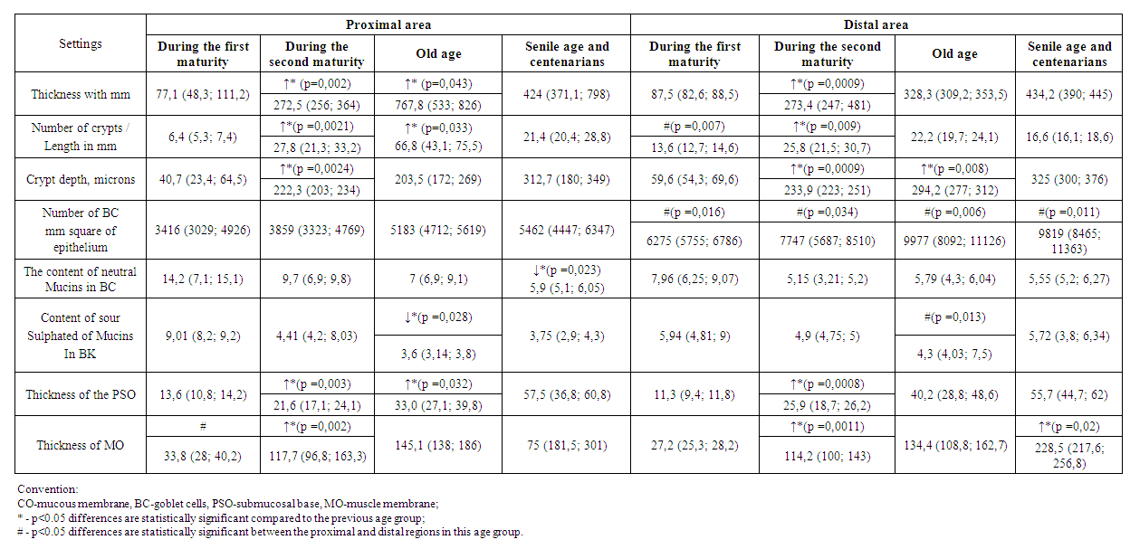 | Table 1. Structural characteristics of human LI of different age groups |
4. Discussion
- BC-single-celled glands that produce mucus, which forms a barrier on the surface of the epithelium, impervious to bacteria and damaging substances, and also serves as a substrate for attachment and nutrition of the commensal microflora. The main component of mucus are Mucins-glycoproteins consisting of a protein axis and a set of oligosaccharide chains associated with it. Peripheral sections of oligosaccharide chains are modified by the remains of sulfuric and sialic acids.We found that from the period of the first maturity to the second Mature age, the thickness of the LIwalls and their structural components – the mucosa, submucosal base and the circular layer of the muscle membrane increases in humans. This is consistent with previously published data from other authors [4,18].In the mucosa of the LI in the period of first maturity, a large number of CD is detected, which prevailed in the distal part. In comparison with the period of the first maturity, the number of crypts and their depth increases in the second Mature period, which corresponds to the data of P. Colony et al. (1989). In the period of second maturity in humans, we have shown an increase in the thickness of the submucosal base, which is associated with a high content of collagen and elastic fibers in its composition. The increase in the thickness of the muscle membrane during this period is due to an increase in the number of smooth myocytes and their size [18].In old age, compared with Mature age, the size of the mucous membrane and submucosal base continues to increase, which is consistent with data from P. Colony (1989) and R. Vigueras et al. (1999). In the proximal LI the above folding in the mucosa, which increases the absolute number of crypts, and in the absence of folds in the distal crypt depth more.We have shown for the first time that the content of acidic Mucins in the CD of the proximal part of the LI decreases by the period of puberty, and neutral ones – by old age. In the distal part, their content does not change. The high content of acidic Mucins in the distal part of the intestine compared to the proximal part is due to their high resistance to bacterial glucosidases, as well as endogenous proteases [19].According to our data, the thickness of the mucous membrane and submucosal base does not change in old age and among centenarians compared to the elderly, which is consistent with the literature data [3]. The thickness of the muscle membrane in the elderly and centenarians is greater than in the elderly, which is consistent with the data of K. Bitar et al. (2003). The increase in the thickness of the muscle membrane is due to an increased content of collagen and elastic fibers, which reduce the extensibility of the intestinal wall. At rest, smooth myocytes in the elderly and centenarians are shorter than in the elderly, and they contract worse [2].In the study of regional differences between different departments of the LI, it was found that in the distal Department in all the age periods considered by us, the number of BC is greater than in the proximal one. According to the literature, the distal section has a greater mucus thickness and the total number of bacteria compared to the proximal section [11].It should be noted that the decrease in the content of acidic Mucins in the elderly and neutral in senile and long-lived reflects age-related histophysiological changes in the epithelial barrier. Obviously, these patterns determine the increase in the incidence of inflammatory diseases in the later periods of postnatal human development. According to J. Ruel et al. (2013), sexually Mature men and the elderly are more likely to develop ulcerative colitis than in other age groups. During puberty, the incidence of cancer is higher in the left (distal) part of the LI, and in older people – in the right (proximal) [10].
5. Conclusions
- During the periods of the first and second maturity in a person in all parts of the LI, there is an intensive growth of the mucous membrane, submucosal base and muscle membrane, which is associated with an increase in the linear size of the intestine.The thickness of the muscle membrane in old age and in centenarians is greater than in sexually Mature people, which is probably due to the increased content of collagen and elastic fibers, which reduce the extensibility of the intestinal wall.The amount of BC in the epithelial lining in the proximal part of the human LI is 2 times less than in the distal one in all age groups. In the proximal part of the LI, the content of acidic Mucins in the BC decreases by 1.5 times by old age, and neutral ones – by senile age.