-
Paper Information
- Previous Paper
- Paper Submission
-
Journal Information
- About This Journal
- Editorial Board
- Current Issue
- Archive
- Author Guidelines
- Contact Us
American Journal of Medicine and Medical Sciences
p-ISSN: 2165-901X e-ISSN: 2165-9036
2020; 10(9): 657-659
doi:10.5923/j.ajmms.20201009.06
Received: July 19, 2020; Accepted: August 10, 2020; Published: August 15, 2020

Evaluation of the Compensatory-Adaptive Mechanisms of Bridge Prosthetics at the Terminal Dentition Defects with the Use of Intraosseous Implants by the Method of Electromyography
 Abstract
 Reference
 Full-Text PDF
 Full-text HTML
Safarov M. T. , Arslanov O. U. , Irisaliev H. I. , Tashpulatova K. M.
Tashkent State Dental Institute, Uzbekistan
Copyright © 2020 The Author(s). Published by Scientific & Academic Publishing.
This work is licensed under the Creative Commons Attribution International License (CC BY).
http://creativecommons.org/licenses/by/4.0/

With partial absence of teeth, patients for various reasons refuse to make removable dentures. Today, dental implantation is widely used for the rehabilitation of such patients. It is known that the adaptation of chewing muscles to new conditions occurs in the first 6 months of using prostheses. In this regard, the method of electromyography allows us to objectively assess the compensatory and adaptive restructuring of the patient's muscle apparatus. Studies have confirmed the restoration of the functional state of the chewing muscles in the orthopedic treatment of patients with various dental defects using dental implants.
Keywords: Teeth, Dental implantation, Adaptation, Bridge prosthetics, Electromyography, Compensatory-adaptive mechanisms, Chewing muscles
Cite this paper: Safarov M. T. , Arslanov O. U. , Irisaliev H. I. , Tashpulatova K. M. , Evaluation of the Compensatory-Adaptive Mechanisms of Bridge Prosthetics at the Terminal Dentition Defects with the Use of Intraosseous Implants by the Method of Electromyography, American Journal of Medicine and Medical Sciences, Vol. 10 No. 9, 2020, pp. 657-659. doi: 10.5923/j.ajmms.20201009.06.
Article Outline
1. Introduction
- In the analysis of literature sources revealed that when the orthopedic treatment of patients with partial absence of teeth up to 26% of patients for various reasons refuse from the manufacture of removable dental prostheses [1,2,5,6]. For rehabilitation of such patients is now widely used dental implant in combination with improved methods of fabrication of fixed dental prostheses [3,4,7,9]. For a reliable functional assessment of orthopedic treatment of patients with the use of dental endosseous implants examined the condition of the masticatory muscles of man [8,11]. The most objective method of assessing the functional state is electromyography (EMG). The use of EMG allows you to explore and find out the functional changes of masticatory muscles during compensatory adjustment of dental apparatus due to loss of teeth [12]. In the process of adaptation to dentures, shorter chewing cycle time by reducing the number of chewing movements and chewing time per act. It is known that the masticatory muscles adaptation to new conditions occurs in the first 6 months of using dentures. In this regard, the technique of electromyography allows us to objectively assess compensatory-adaptive restructuring of the muscular system of the patient, and the influence of masticatory loads on dental implants after prosthetic treatment [13,14].
2. Objectives
- Is to study the functional efficiency of compensatory-adaptive mechanisms of bridge prosthetics at the terminal dentition defects with the use of intraosseous implants by the method of electromyography.
3. Materials and Methods
- Electromyographic studies were carried out by us in the field actually chewing and temporal muscles on the machine, ""Neurotech "" (Russia) at rest and maximum compression of the muscles. As biocriteria in hardware-software complex, used 4-channel electromyograph biosilicon of the firm "Medicor." Plate electrodes used standard. He laid them on the pre-degreased with alcohol skin and secured with adhesive plaster. After the electrodes have started to work with the dialog menu of the software system "of" neurotech"". All patients were divided into 3 groups. I - group consisted of 12 patients with unilateral and bilateral end defect of the dentition, II - group consisted of 14 patients after surgery intraosseous implantation, and III - group consisted of 14 patients who were established bridges with distal based on dental implants.
4. Results
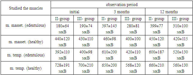
- Comparing the functional activity of the temporal and masticatory muscles to orthopedic treatment of patients of the first group, we identified the following pattern. On the intact party bear the masticatory muscle was 1.5 times higher, and the temporal muscles – in 2,3 times above, than on the side of the defect. Electrophysiological indices of muscle activity in patients with bilateral end defects ranged quite widely and depended on the type of mastication. It should be noted that 80% patients of this group were revealed mainly one-sided, namely, the right type of chewing, and in 20% of patients – even double-sided. In the unilateral type of chewing average values bear on the working side was 1.8 times greater for the masticatory muscles, and 2.1 times for the temporal muscles. In patients with a uniform type of chewing functional activity of the masticatory and temporal muscle were about the same on the right and left. Electromyographic studies in the II group of patients revealed that upon compression of the jaws the maximum amplitude of the BEA was in m. masseter healthy side 440±120 µv; m. masseter on the side of the edentulous - 180±70 µv, and m. temporalis 392±110мкВ; m. temporalis healthy hand - 728 ± 191мкВ. The coefficient of coordination for the masticatory muscles during chewing averaged 2,4±0,13 for the temporal muscle 0,5±0,13; the rest actually masticatory muscles 0,4±0,13 for the temporal 2,1±0,13, indicating incoordination in the work of the masticatory muscles.3 months after the implant procedure with early functional load on the implant was observed a slight decrease in BEA of the muscles at rest. On average, the masticatory muscles, the difference was 20% (m. masseter healthy side is 280±81 mV, m. masseter on the side of the edentulous in the area of the entered implant 190 ± 5,0 µv). From the temporal muscle bear alone decreased on average by 25% and amounted to m.temp. on the side of the edentulous = 450 ± 11мкВ; m. temp. on the healthy side =210 ± 4мкВ. Upon compression of the jaws bear m. masseter ZD. = 460 ± 98, on the side of the edentulous = 397 ± 143 µv; m. temp. ZD. 650 ± 200 µv; m. temp. ad 610 ± 200 µv. The coefficient of coordination for the masticatory muscles under compression was 1.2 ± 0,08; for the temporal muscles 1,07 ± 0,06. The coefficient of coordination for m. masseter alone 0,72 ± 0,05; for m. temporalis 0,5 ± 0,03. This was due to the changes bear the masticatory and temporal muscles, indicating the alignment of the coordination relations work of the masticatory muscles (tab. 1).
|
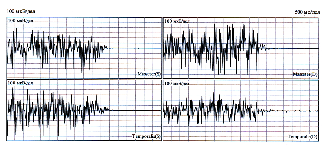 | Figure 1. EMG of the masticatory muscles after the early functional loads on dental implants |
5. Discussion and Сonclusions
- Thus, the findings of electromyography studies confirmed the restoration of the functional state of the masticatory muscles during orthopedic treatment of patients with various defects of the dentition using dental implants. The results of this study are objective evidence of compensatory-adaptive adjustment of the reflex mechanisms of the muscular system in different periods of observations.