-
Paper Information
- Next Paper
- Paper Submission
-
Journal Information
- About This Journal
- Editorial Board
- Current Issue
- Archive
- Author Guidelines
- Contact Us
American Journal of Medicine and Medical Sciences
p-ISSN: 2165-901X e-ISSN: 2165-9036
2020; 10(9): 635-638
doi:10.5923/j.ajmms.20201009.01
Received: April 24, 2020; Accepted: August 8, 2020; Published: August 15, 2020

The Algorithm of Morphological Signs of Thymus During Sepsis in the Unmounded
 Abstract
 Reference
 Full-Text PDF
 Full-text HTML
Sh. Ziyaev
Andijan State Medical Institute, 1, Atabekova Str., Andijan, Republic of Uzbekistan
Correspondence to: Sh. Ziyaev , Andijan State Medical Institute, 1, Atabekova Str., Andijan, Republic of Uzbekistan.
| Email: |  |
Copyright © 2020 The Author(s). Published by Scientific & Academic Publishing.
This work is licensed under the Creative Commons Attribution International License (CC BY).
http://creativecommons.org/licenses/by/4.0/

This article conducted a morphological and morphometric study of the thymus of premature babies who died from various pathologies with the development of an algorithm for morphological signs characteristic of a particular thymus pathology. A morphometric study of the thymus of premature infants who died from various causes showed that with prematurity, the mass and weight coefficient of the thymus are low and they are associated with the degree of prematurity and the variety of causes of death. The compilation of an algorithm for morphological changes to determine a particular pathology in the thymus allowed us to determine the criteria for the normal development and formation of the thymus and lag behind development, hypoplasia, dysplasia and premature atrophy of the thymus against the background of prematurity and development of various diseases. These data can be used as diagnostic criteria in the morphological assessment of the thymus of newborns.
Keywords: Thymus, Sepsis, Perinatal, Histostructure, Newborns, Morphology
Cite this paper: Sh. Ziyaev , The Algorithm of Morphological Signs of Thymus During Sepsis in the Unmounded, American Journal of Medicine and Medical Sciences, Vol. 10 No. 9, 2020, pp. 635-638. doi: 10.5923/j.ajmms.20201009.01.
1. Introduction
- In recent decades, the problem of neonatal sepsis has become relevant again. As you know, in the 80s of the twentieth century there has been a decrease in the number of cases of this formidable disease due to the expansion of the spectrum of antibacterial and immuno-replacement therapy. However, now the frequency of sepsis in newborns has increased and is 0.1-0.2% in full-term and 1-1.5% in premature babies [4-6]. This means that the development of neonatal sepsis is determined primarily by the characteristics of reactivity and mainly non-specific - the ability to form barriers from infection.To date, despite the presence of a significant number of studies, questions of the morphological state of the organs of the immune system in various pathological conditions in children, especially in premature infants, remain poorly understood [1-3]. The central organ of immunogenesis, the thymus, is no exception in this regard. Basic research in recent years has confirmed its key role in immune homeostasis. However, to date, functional studies of this organ prevail over morphological ones. The literature data on the morphological changes of the thymus gland under various pathological conditions are descriptive and characterize mainly qualitative shifts from its histostructure [3,4], whereas in conditions of pathology, in particular during sepsis in premature infants without a quantitative assessment developing in the thymus morphological abnormalities it is impossible to obtain focused information. To date, there is no single unified approach to assessing changes in the thymus histostructure during various pathological processes, as well as ambiguous data on the most common morphological changes in the thymus gland under prematurity. In connection with the above, in this work, the goal was set to conduct a system analysis and develop an algorithm for assessing the morphological signs of differentiation and thymus rearrangement during sepsis, depending on the degree of prematurity.
2. Methodology
- The object of the study was the thymus of 63 premature babies who died in the neonatal period from sepsis. Premature babies by weight were divided into 4 degrees: 2000–2500 g –– I –– grade, 1500–1999 g –– II –– grade, 1000–1499 –– III –– grade and up to 1000 g –– IV –– prematurity. As a control, the thymus of 16 newborns with a body weight of more than 3,000 g born on time and who died from a traumatic brain injury was studied. During autopsy, the thymus was isolated, weighed, and the weight coefficient of the thymus (WCT) was determined. For histological examination, thymus pieces were fixed in a 4% formalin solution on phosphate buffer and, after dehydration in alcohols, embedded in paraffin. Sections with a thickness of 5-8 μm were stained with hematoxylin and eosin, according to Van Gieson, and set up the SCHICK reaction. To unify the accounting for morphological changes in the thymus in terms of prematurity and various pathologies, an algorithm for assessing morphological characters has been developed.
3. Results
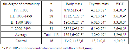
- The results of the study showed that with prematurity not only body mass indices are low, but the mass of internal organs, in particular the central organ of the thymus immunogenesis, also decreases, which leads to the development of an immunodeficiency state in newborns. It was noted that the lower the body mass of premature babies, the lower the mass of the thymus and significantly reduced the weight coefficient of the thymus in comparison with the norm. At I and II degrees of prematurity, although the body weight is slightly increased, the mass of the thymus remains in low numbers and, therefore, the weight coefficient of the thymus also decreases significantly (Table 1).
|
|
4. Discussion
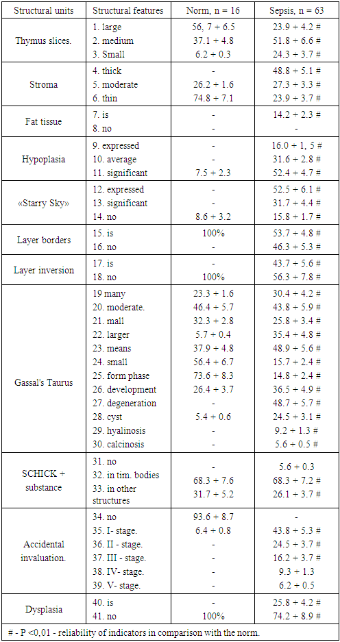
- Studies have established that in most observations, the preservation of the overall histoarchitectonics of the thymus is noted. In these cases, there was a clear division into the cortical and medullar layers, segments of large or medium sizes, the stroma is weakly expressed. There were thymic bodies in the medulla, the number of which is quite variable.The microscopic structure of the thymus in all groups of preterm infants who died from sepsis was associated with weight, weight index and degree of atrophy of the thymus, as well as the severity of the septic phenomenon that caused death. In the control group of newborns, normal values of the thymus histostructure amounted to the following stereotypical sequence of numbering of morphological signs of the thymus: 2, 6, 8, 11, 14, 15, 21, 23, 25, 26, 32, 34. To put it in words, the thymus consists mainly of large and medium lobules, a small amount of stroma, no adipose tissue, lobular hypoplasia and the “starry sky” pattern are not observed, in the segments the layer boundaries are clearly distinguishable, Gassal bodies are medium in size and are found in moderation, they determine PAS positive mass. When studying the thymus, if the following numbers of morphological characters are determined: 3, 6, 11, 16, 21, 24, 25, 33, then this corresponds to a lag in the development and hypoplasia of the thymus. In our observations, these signs were found in premature infants with sepsis on the background of morphological and functional insolvency. At the same time, it is morphologically noted that the thymus lobules are small, the stroma is poorly developed, hypoplastic, in some cases the boundaries of the thymus layers are not determined, the number of Gassal bodies is significantly reduced, their size is small and in a state of formation.It is known that all possible infectious and non-infectious diseases of the mother during pregnancy are risk factors for the development of damage in the organs of the immune system, including the thymus. We have revealed certain morphological abnormalities in the thymus histostructure that are different from the norm and they were accepted as signs of atypical incidence of thymus invasion. Which were indicated by the following numbering: 3, 4, 7, 14, 16, 17, 19, 22, 27, 28, 29, 30, 31, 32, 38, 39, 40. In this case, the thymus was represented morphologically by small segments, with pronounced inversion of the tissue of the lobules, the stroma is thickened due to sclerosis, sometimes with the presence of fat cells. In the lobules, the boundary of the layers is not determined, the Gassal bodies are large, cystically dilated with hyalinosis and calcification. Epithelioreticular cells in a state of hypertrophy and dysplasia, between which the number of lymphocytes is significantly reduced. Such morphological changes characteristic of the atypical incidental thymic invasion in our observations were in preterm infants with perinatal sepsis (Table 2).In the thymus of premature deaths from septic infection in combination with congenital malformations, an increase in the size of epithelioreticular cells with vacuolization of the cytoplasm, the appearance of inclusions of various origins in it and a violation of the formation of nuclei was revealed. These changes are evaluated as epithelioreticular dysplasia and the algorithm of morphological changes was as follows: 3, 4, 7, 9, 16, 17, 21, 24, 25, 38, 39, 40. Dysplasia of the reticuloepithelium and the Gassal body is morphologically detected, uneven the distribution of lymphocytes in both layers of lobules, lobules have different sizes and shapes, the boundaries of the layers are not determined, the stroma is unevenly thickened with the presence of foci of accumulation of adipose tissue, sclerosis and reticulosis.
5. Conclusions
- 1. Morphological and morphometric indicators, as well as the mass and weight coefficient of the thymus of premature infants who die from the septic phenomenon, are significantly lower than the norm and they depend on the degree of prematurity and severity of sepsis.2. The algorithm of morphological signs of the thymus in full-term newborns consists of positive numbering, showing the morphological and functional maturity of the organ in the form of a histotopographic organization of large and medium lobules, slight stroma, without signs of lipomatosis, hypoplasia, dysplasia, with well distinguishable layers in the lobules, and the average size of Gassal bodies.3. The compilation of an algorithm for morphological changes to determine one or another pathology in the thymus allowed us to determine the criteria for the normal development and formation of the thymus, as well as signs of lagging behind the norm, hypoplasia, dysplasia and premature atrophy of the thymus against the background of both prematurity and the onset of a septic phenomenon.4. An increase in the numbers in the thymus algorithm, showing negative morphological changes, corresponded to dysplastic and atrophic processes in the form of: lag formation of histotopography of the organ from the norm, hypoplasia, atypical accidents, dysplasia of the epithelioreticular stroma, which are morphological signs of an immunodeficiency state.