Sanoeva Matlyuba Jonkulovna
PhD, Senior Research Scientist, Bukhara State Medical Institute, Uzbekistan
Correspondence to: Sanoeva Matlyuba Jonkulovna, PhD, Senior Research Scientist, Bukhara State Medical Institute, Uzbekistan.
| Email: |  |
Copyright © 2020 The Author(s). Published by Scientific & Academic Publishing.
This work is licensed under the Creative Commons Attribution International License (CC BY).
http://creativecommons.org/licenses/by/4.0/

Abstract
Because of emerging pathobiochemical reactions in migraine, the phase of episodic and chronic nociceptive reaction occurs, which violates the functional state of the brain. The study of brain dysfunction in migraine is advisable to carry out the most affordable in financial terms, and no less informative method-electroencephalography. The aim of research was to study the correlates of electroencephalography (EEG) of the brain in some complicated forms of migraine. 133 (100%) patients with migraine were under observation. 66 (49.6%) of them were with migraine status, 67 (50.4%) patients with chronic migraine. Quantitative and qualitative comparative analysis of EEG in the study groups showed an increase in the threshold of convulsive readiness, the formation of a disorganized and desynchronous type of bioelectric activity of the brain during migraine attacks with their predominance in migraine with aura. Based on the results of the study, we concluded that neurophysiological parameters of EEG could be objective indicators of complicated forms of migraine, determining the zoning of the pathological process, the presence of exacerbation and aura.
Keywords:
Migraine, Migraine status, Chronic migraine, EEG, Bioelectric activity of the brain in migraine
Cite this paper: Sanoeva Matlyuba Jonkulovna, Electroencephalographic Correlates of Certain Complicated Forms of Migraine, American Journal of Medicine and Medical Sciences, Vol. 10 No. 5, 2020, pp. 314-317. doi: 10.5923/j.ajmms.20201005.08.
1. Relevance
According to different studies, migraine occurs in 5 percent to 38 percent of the population, mainly affects people aged 18-33 years [1], 75-80 percent of people during their lifetime survive at least one migraine attack [2,3]. Brain dysfunction, which is the primary link in migraine pathogenesis, activates the trigemino-vascular system, causing vascular dystonia with pain syndrome [4,5]. Prostaglandins, estrogens, price neutral and peripheral neurotransmitters (serotonin, dopamine, noradrenaline) also be an important role in the pathogenesis of pain in migraine, which causes not only classical hemicrania, but also pronounced vegetative reactions that end in attacks of lethargy and drowsiness [6,7]. The phase of episodic and chronic nociceptive reactions that disturb the functional state of the brain follows the pathobiochemical reactions described above [8]. It is advisable to study brain dysfunction in migraine by the most financially accessible and no less informative method – EEG. Despite years of use, the diagnostic value of the EEG is not disputed in global neurological practice [9,10]. This method is generally recognized and is one of the basic diagnostic methods in modern neurology in both stationary and outpatient conditions, especially in functional brain changes [11].
2. The Aim of Research
Based on the above, the aim was to study the features of electroencephalographic correlates of brainin some complicated forms of migraine.
3. Material and Methods
There were 133 (100,0%) patients with migraine under observation, of them 66 (49,6%) patients with migrainous status, 67 (50,4%) patients with chronic migraine (figure 1).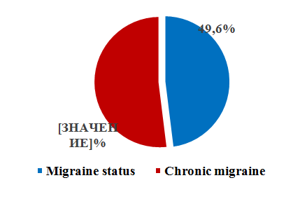 | Figure 1. Distribution of patients |
We performed research with a 16-channel of the EEG. The electrodes were connected to the skull surface in standard background recordings and functional tests: eye opening and closing, hyperventilation test (within 3 minutes). The bioelectrical data were automatically processed according to the program for calculating and mapping spectral-correlation indices of the brain bioelectrical activity. Statistical processing and reliability of differences between the groups of indicators was carried out using Mann-Whitney and Wilcoke-Son methods (Statistica 6.0).
4. The Results of the Research
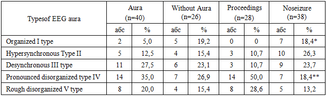
The results of the EEG study for those examined with migrainous status are presented in Table 1. As can be seen from Table 1, based on classification E. A. Zhirmunskaya and B. S. Losev [Gulyaev S.A., 2014, Turdubaeva, G.T., 2015] in migrainous status an organized I-type EEG, with alpha rhythm predominance, characterized by a high degree of regularity, slightly modified amplitude gradient was observed in patients mainly in "Migraine without aura", and in the non-available period, in the anamnesis of these patients rare (2-3 times in 3 months) attacks were observed.Table 1. Characterization of the bioelectric activity of the brain with migraine status (n = 66)
 |
| |
|
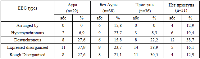
The hypersynchronous type II was characterized by excessive regularity of alpha, beta and theta activity with the loss of regional differences, with the identification of all bipolar leads within the right and left hemispheres, which were 2.5 times more common with "Migraine with aura" in relation to “Migraine without aura” (p> 0.05), while outside the attack period they were detected 2.5 times more often than in the attack period (p> 0.05). For patients with migraine status, the most characteristic was the desynchronous III-type EEG - a complete disappearance or significant decrease in the amplitude of alpha waves, an increase in the amplitude and severity of beta vibrations, the presence of a small number of slow waves, the frequency of which did not differ statistically between patients suffering from " Migraine with aura ”and“ Migraine without aura ”, but outside the attack period, this type of EEG was recorded 2.2 times more often than in the attack period (p> 0.05). Detection of a pronounced disorganized IV type of EEG, characterized by a pronounced, but not sufficiently regular in frequency, disorganized high-amplitude alpha activity, dominant in all areas of the brain, while increasing low-frequency beta activity, the appearance of delta and / or theta waves of a sufficiently high amplitude 1.3 times prevailed in patients with Migraine with aura (p> 0.05), and 2.7 times in the accession period (p <0.01). A coarse disorganized V-type EEG, with a predominance of theta, delta activity, was statistically predominant in patients with Migraine with aura (1.3 times) and in the onset (2.2 times) (p> 0.05).When analyzing the results of background recording, nonspecific changes in the bioelectrical activity of the brain were observed in the form of a decrease in the amplitude and irregularity of the alpha rhythm, the predominance of fast waves, and smoothing of zonal differences. In 14 (21.2%) of the studied indicators, the background EEG was evaluated as an organized type. Paroxysmal activity in the background in the form of single outbreaks of acute waves was determined in 18 (27.3%) cases during attacks of migraine status, with 12 (18.2%) of them suffering from Migraine with aura. When conducting a rhythmic photostimulation test at frequencies of 16–20–24 Hz, the number of patients with photoparoxysmal activity increased 1.3 times, accounting for 24 (36.4%) cases. When carrying out a test with hyper-ventilation (HS), paroxysmal activity was detected in 26 (39.4%) cases, which was mainly represented by bilateral flashes, the appearance of high-amplitude alpha, theta, and delta waves, mainly in the frontal and frontotemporal temporal departments, increased synchronization of bioelectric activity, while a slower recovery of biorhythmics was observed on the side of pain. In 8 (12.1%) patients suffering from Migraine with Aura, bilaterally-synchronized spike-wave discharges were detected. Based on the spectral analysis of the EEG, the predominance of fast-wave activity (beta range), the appearance of paroxysmal activity and outbreaks of spike-wave discharges in the temporal and anterior-frontal leads on the hemicranial side were noted.A comparative analysis of the obtained EEG results of migraine status with chronic migraine was performed (Table 2).Table 2. Characterization of the bioelectric activity of the brain with chronic migraine (n = 67)
 |
| |
|
As can be seen from Table 2, the organized I-type EEG in chronic migraine with aura, as well as in the period of hemicranic attacks, was not detected in any case, in contrast to the group of patients with migrainous status (p>0.05), while in "Migraine without aura" it statistically did not differ between the two comparable groups, and in the non-acute period it prevailed 1.5 times in migrainous status (p>0.05). II-hypersynchronous EEG type in the presence of aura was detected 1.8 times less frequently than in migra-nosed status (p>0.05), while in the onset and interictal periods hyposynchronization was almost 1.5 times inferior to it (p>0.05). Hypersynchronization was 1.5 times more common in chronic migraine without aura than in migrainous status (p>0.05). The frequency of desynchronization with disappearance or significant reduction of alpha amplitude did not statistically differ in both compared groups in the presence of aura, and in the absence of aura it was 1.5 times higher in migrainous status (p>0.05). In the onset and interictal periods desynchronization was 2.2 and (p>0.05) 1.6 times more frequently (p>0.05) respectively in patients with chronic migraine. The IV-expressed disorganized EEG type, with high amplitude alpha activity, amplification of low-frequency beta rhythms, occurrence of pathological waves of high amplitude, was met almost equally in the interictal period - in the presence of aura and without it in patients of both compared groups, whereas in the attack period it exceeded 1.3 times in migraine status (p>0.05). V-coarse disorganized EEG type was 1.4 times more prevalent in the group of patients with chronic migraine both in the presence of aura and without it (p>0.05), however, in the attack and interictal periods it was met almost equally with migrainous status. Results of background EEG recording in chronic migraine show a decrease in alpha rhythm amplitude, predominance of slow waves over fast waves, appearance of single pathological waves without zonal differences. Only 10 (14.9%) patients with baseline EEG indicators had an organized type, which was 1.4 times inferior to the migraine status. Paroxysmal activity on the background EEG in the form of single outbreaks of acute waves was determined 1.7 times less frequently than in migrainous status, which amounted to 11 (16.4%) cases, regardless of the presence of migraine attacks themselves. Of these, 8 (11.9%) suffered from "migraine with aura". In photostimulation, the number of patients with photocouple-similar activity increased 1.2 times, amounting to 13 (19.4%) patients, which statistically did not differ from migraine status. Paroxysmal activity in hyperventilation was observed in 14 (20,9%) subjects in the form of bilateral flares, with their localization in frontal hip and parietal parts. In 11 (16.4%) patients suffering from "Migraine with aura" bilateral-synchronous spike-wave discharges were detected, which exceeded the migrainous status 1.4 times. The EEG spectral analysis was based on proportional activity of fast and slow waves, paroxysmal activity and spike-wave bursts in temporal, antero-frontal and parietal discharges mainly of bilateral localization.
5. Discussion
Quantitative and qualitative comparative analysis of two complex groups of migraine based on the EEG results shows an increase in the threshold of ship-carrying readiness, the formation of disorganized and desynchronous type of brain biolektric activity during the period of migrainous paroxysms, with their predominance in "Migraine with aura". Nonspecific lesions of brain structures with lack of zoning, decreased organized type and pronounced desynchronization of bioelectrical activity in chronic migraine confirms the heavier flow of the brain in comparison with migrainous status. Polymorphic activity, a sign of "perversions of zonal differences" depending on the presence of migraine paroxysms and aura, the predominance of de-synchronous alpha rhythm, excessive appearance of beta rhythm and pathological waves during migraine paroxysms indicates a decrease in the functional activity of neurons that are in the stage of adaptive tension, due to the reactive state of the vascular wall by the mechanism of angiodilatation/ angiospasm during migrenous periods. roxisms, which are the main cause of brain hypoxia with changes in the bioelectric activity of the brain. The prevalence of paroxysmal activity on the background EEG and functional samples, the presence of bila-teral spike-wave bursts in patients predominantly with migrainous status can be considered as the formation of epiactivity during migraine attacks. Obtaining reliable results with prevalence of slow wave bioelectric discharges, pathological waves proves the probability of greater vascular complications in chronic migraine. Thus, the neurophysiological parameters of EEG were objective indicators of complicated forms of migraine, determining the zoning of the pathological process, the presence of exacerbation and aura.
6. Conclusions
1) Neurophysiological correlates are an objective indicator for assessing the bioelectrical activity of the brain, determining the presence of complex forms of migraine with different lateralization of hemicrane.2) The obtained results prove the prevalence of a pronounced disordered EEG type with the appearance of paroxysmal activation and may be the cause of cortical hyperexcitability with the formation of coarser vascular consequences in migraine status and chronic migraine.3) EEG is one of the main methods to assess the functional status and predict vascular brain complications in patients with migraine.
References
| [1] | Gulyaev S.A., Arkhipenko I.V., etc. Electroencephalography in diagnostics of diseases of nervous system. - Vladivostok: FSBU Publishing House, 2012. 200p. |
| [2] | Muzalevskaya D. S., Migraine and perictal headaches in epileptic patients (review) // Saratov Scientific Medical Journal. - – 2016. - Volume 12, No. 2. - P. 278-2811. |
| [3] | Cai S, Hamiwka LD, Wirrell EC. Peri-ictal headache in children: prevalence and characteristic // Pediatr Neurol. – 2008. – Vol. 39. – Р. 91–96. |
| [4] | Vasilenko, A.V.; Migren and epilepsy - "as two sides of the same coin" clinical and diagnostic aspects // Epilepsy and paroxysmal states. - – 2013. - Volume 5, No. 2. - P. 56-68. |
| [5] | Panayiotopoulos CP. “Migralepsy” and the significance of differentiating occipital seizures from migraine // Epilepsia. – 2006. – Vol. 47. – Р. 806–808. |
| [6] | Karaali-Savrun F, Goksan B, Yeni SN, et al. Seizure related headache in patients with epilepsy // Seizure. – 2002. - №11. – Р. 67–69. |
| [7] | Ito M, Adach N, Nakamura F, et al. Characteristics of postictal headache in patients with partial epilepsy // Cephalalgia. – 2004. – Vol. 24. – Р. 23–28. |
| [8] | Gulyaev S.A. Electroencephalographic investigation in clinic: a problem of modern classification // Russian Journal of Child Neurology. - – 2013. - Volume VIII-IX, ¹4. |
| [9] | Gulyaev S.A. Electroencephalographic investigation in clinic: a problem of modern classification // Russian Journal of Child Neurology. - – 2014. - Volume VIII-IX, No. 1. |
| [10] | Turdubaeva, G.T. Classification of electroencephalogram (literature review) // Vestnik KRSU. - 2015. - Vol. 15, ¹ 7. - – P. 156-158. |
| [11] | Isaeva, N.A.; Torubarov, F.S.; Zvereva, Z.F. EEG indicators in persons at risk of ischemic stroke as predictors of the initial cerebral blood circulation insufficiency (in Russian) // Academic notes of St. Petersburg State Medical University. I. P. Pavlov. - – 2014. - Volume XXI, № 3. - P. 55-59. |

 Abstract
 Reference
 Full-Text PDF
 Full-text HTML