-
Paper Information
- Next Paper
- Previous Paper
- Paper Submission
-
Journal Information
- About This Journal
- Editorial Board
- Current Issue
- Archive
- Author Guidelines
- Contact Us
American Journal of Medicine and Medical Sciences
p-ISSN: 2165-901X e-ISSN: 2165-9036
2020; 10(4): 242-245
doi:10.5923/j.ajmms.20201004.16

Body Systemic Response to Optimize Reparative Bone Regeneration
 Abstract
 Reference
 Full-Text PDF
 Full-text HTML
Mahmurov A. M. , Yuldashev A. Y.
Tashkent State Dental Institute, Tashkent, Uzbekistan
Correspondence to: Mahmurov A. M. , Tashkent State Dental Institute, Tashkent, Uzbekistan.
| Email: |  |
Copyright © 2020 The Author(s). Published by Scientific & Academic Publishing.
This work is licensed under the Creative Commons Attribution International License (CC BY).
http://creativecommons.org/licenses/by/4.0/

Reparative regeneration of bone tissue and its optimization is a topical problem in both clinical and fundamental science. It can be performed with the help of local and systemic factors [A.S. Avrunin et al. 2018; A.S. Grioryan et al. 2015; Y.M. Iryanov, 2012; I.V. Mayborodin et al. 2015; G.A. Onoprienko, V.P. Voloshin, 2017; I.DuniS-Cule et al. 2014; W.Li et al. 2015]. They have a significant impact on angiogenesis, differentiation of osteoblasts and osteoclasts, osteogistogenesis [O.L. Grebneva et al., 2012; M.V. Stogov, 2013; A.M.Ali, 2015; Y.H.Kim et al., 2015; W.Li et al., 2015; N.Oxata et al., 2015; V.Shi et al., 2015, etc.]. ] The overwhelming number of works devoted to the improvement of treatment results is based on a mechanical approach based on the need for comparison of fragments and reliable fixation [G.V. Dyachkova et al., 2018, etc.]. However, according to researchers, the restoration of blood supply and metabolism, correction of neuroimmune endocrine relationships is no less critical K.V. Sudakov et al., 2010; L.B. Reznik et al., 2015; W.Li et al., 2015; V.Shi et al., 2015, etc.].
Keywords: Succinate, The surface of a tibial bone, Diaphysis, Index of induction, Glycerophosphate dehydrogenase, Succinate dehydrogenase
Cite this paper: Mahmurov A. M. , Yuldashev A. Y. , Body Systemic Response to Optimize Reparative Bone Regeneration, American Journal of Medicine and Medical Sciences, Vol. 10 No. 4, 2020, pp. 242-245. doi: 10.5923/j.ajmms.20201004.16.
Article Outline
1. The Aim of the Research
- To study the effect of infusion of physiological solution and succinate on the functionally metabolic activity of neutrophils in reparative regeneration of diaphysis bone tissue defect.
2. Research Material and Methods
- Characteristics of the experiment.The experiments were conducted on 42 rabbits of Shinshilla breed weighing 2.0-2.5 kg. After examination of them by the veterinarian, stating the absence of any diseases, under local anesthesia 0.5% solution of novocaine of the middle third of the tibia, a cut of skin and subcutaneous tissue was made. The muscles are bluntly separated from the tibia surface and removed from the middle third of the diaphysis. Then a sharp scalpel made two longitudinal 1.0 cm at a distance of 0.5 cm from each other and two transverse incisions of the periosteum (the scheme of defect formation is shown below), connecting them. The rectangular periosteum strip was removed after separation from the subject bone tissue. Then, per its width (0.5 cm) and length (1.0 cm), boron-machine created the defect of bone tissue to the endoscope. After removing small pieces of bone from the recess, the defect is covered with soft tissue from above. The skin and subcutaneous tissue are sutured with several transverse sutures. The wound is treated with iodinol. The sutures are removed on 8-9 days after the bone defect is created. No complications were observed after the operation.
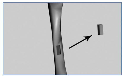 | Scheme of bone defect formation in the tibia diaphysis area |
3. Biochemical Research
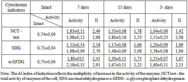
- The functional-metabolic activity of neutrophils (FMAN) is established in the NST-test by the B.N. Park method with co-authors in the modification of M.G. Shubich and V.G. Mednikova [92; p.515-518]. FMAN allows judging about the phagocytic-metabolic activity of granulocytes. The activity of neutrophils in NST-test was taken into account by the semi-quantitative method. The activity of SDG and α-GFDG was determined by N. Novikoff, B. Masek method [140; p.287-302]. The ratio between the activity of SDH and α-HFDG and the NCT-test in the dynamics of the experiment and intact animals is considered as an induction index (AI).
4. Statistical Methods of Reserach
- Morphometric and biochemical results are processed using standard methods of variation statistics using the t-criterion of Student Excel 2010. The arithmetic mean, its average error and reliability (P) were calculated. The difference between the compared values is reliable at P<0.05.
5. Results of Research
- Under physiological conditions circulating in the blood neutrophils are characterized by relatively low activity of SDH (0.71± 0.94 c.u.) and α- GFDG (0.75± 0.06 c.u.); NST-test index is 0.74± 0.6. (Table 1).
6. Discussion
- Blood as the internal environment of the body, as a result of damage to the structure and function of bone tissue of the diaphysis, is adaptively involved in the process of inflammation, whose task is to restore the defect of bone tissue of the diaphysis. Inflammation as a protective and adaptive process limits the damage zone, mobilizes hematopoiesis organs and immunological protection, multiple activates functional and metabolic properties of peripheral blood leukocytes, etc. Regulation of the interaction of cooperating cells (neutrophil-macrophage-lymphocyte, etc.) is carried out due to the feedback of the structures of functional systems. A small number of phagocytes, neutrophils and macrophages are customarily recorded. The impact on the body of a factor that violates the homeostasis of tissues of this or that organ dramatically changes the phagocytic-metabolic activity (PMA), primarily regulatory systems that mobilize all systems to the reactions that correlate homeostasis in the damage zone (inflammation trigger, neutrophil-macrophagal-connective tissue reaction). The stereotype of mobilization of functional-metabolic activity of phagocytic blood and connective tissue cells allows to consider the dynamics of activity of lysosomal and membrane enzymes, energy and oxidative metabolism, processes of synthesis and secretion, migration, proliferation and differentiation. Both in the clinic and the experiment, the most informative, reflecting the reactive shifts in the system of oxidative metabolism, is to determine the ability to spontaneous and induced recovery of nitro blue tetrazol [Nagoyev B.S., M.R. Ivanova Indicators of spontaneous and induced NST-test of leukocytes in patients with hepatitis V. // Epidemiol. and infectious diseases.] From a large number of intracellular enzymes informative, integrally characterizing cell metabolism, we, like other researchers, chose SDG, α-HFDG. They are as informative as possible, characterizing the functional completeness of the cell under study.The SDH transfers hydrogen from succinic acid through ubiquon to cytochrome in the respiratory chain. It's like a mitochondrion marker that reflects the state of oxidation in the cell. α-GFDG is a component of the α-glycerophosphate shunt, which provides continuity and synchrony in the processes of glycolysis and respiration; it transfers hydrogen to the cytochrome C of the respiratory chain. In the study of the functional-metabolic activity of leukocytes attach great importance to HST-test, which detects the induction of peroxidase systems of leukocytes in response to various effects. It is a biochemical marker of cell peroxidase enzymes. Processes occurring in the damage zone after the injury, are presented as follows: when exposed to the polynuclear substrates of cell damage or intercellular substance occurs phagocytosis of the resulting complex of HST-heparin-fibrinogen and its transport to the phagosome. Here, under the influence of enzymes, the recovery of HST and transformation into dark blue formasan. In general, NST-test reflects the degree of activation of oxygen-dependent metabolism, i.e., the function of the hexozomonophosphate shunt and the associated production of free radicals. According to our data, the recovery of NST occurs in neutrophils and macrophages. In the dynamics of inflammation increases the proportion of NST-active neutrophils 4-6 times. Normalization of NST-test parameters occurs only at the stage of completion of inflammation and restoration of damaged tissue. Optimization of the course of inflammation causes both a reliable and significant increase in the rate of NST-test, and return to normal at the stage of stabilization of adaptation and restoration of homeostasis. Our experiments have shown that inflammation (injury) with a seemingly local character is systemic, and the study of enzyme systems of polymorphonuclear leukocytes reflects both its local flow and the body's systemic adaptive response aimed at restoring and preserving homeostasis of the internal environment.The increase in NST-test indexes by 2-3 times indicates the maximum induction of FMAN in the dynamics of inflammation and regeneration, characterizes the rapid and pronounced degree of activation of the phagocytic stage of inflammation in traumatic damage to bone tissue and the formation of blood clots, necrobiotically altered cells and bone particles. A more significant increase in FMAN in the dynamics of reparative regeneration in animals after the administration of succinasol indicates the restoration of oxidative and other processes occurring in mitochondria, normalization of stages of the Kreps cycle.1. Neutrophils are at rest while moving through the system of vessels from bone marrow to tissue or inflammation center. They are active in tissues when performing a specific function, in inflammation (respiratory explosion, phagocytosis, degranulation, extracellular secretion, extracellular lysis of damaged tissues, structures, the decaying neutrophils themselves and other cells) [Serov V.V. Inflammation. -M.: Medina, 1995.-640 p.2. Naasag-Weber M., Hort W.H. Dysfunction of polymorphonuclear leucocytes in uremia.// Semin. Nephrol. -1996. -Vol 16, №3.-192-201.]. From a cell level to an organ violation of structural, functional, metabolic homeostasis, relations of functional systems at tibia fracture (defect) by the formed factors modulates reaction of neutrophils, inflammation. Under conditions of oxygen adequacy, neutrophils realize their functions in full. Ejected proteases open the basal capillary endothelial membrane, provide cleavage of complement, antibodies and other factors with the formation of some mediators that affect both neutrophils and other inflammatory cells. Hydrogen peroxide generated at the same time promotes the activation of collagenase, resulting in a full-fledged enzyme enters the intercellular medium. The splitting of plasma-lemm phospholipids by neutrophil enzymes causes the formation and secretion of some mediators: thromboxanes, prostaglandins, leukotrienes. Neutrophils interact with humoral factors and numerous cells of connective tissue [O.L. Grebneva, M.A. Kovinka, S.N. Luneva et al. The study of humoral components stimulating osteogenesis. // The genius of orthopedics.]. The peculiarity of neutrophils is the presence of phagosome and digestible cellular detritus in the cytoplasm. Neutrophils and macrophages at early stages of inflammation, granulation tissue formation and reparative bone tissue regeneration influence migration, differentiation of fibroblastic and osteoblastic differons, bone tissue remodeling, development of growth factors, vascular formation and development.
7. Conclusions
- The functional-metabolic activity of neutrophils of peripheral blood, according to the activity of SDG, α-GFDG and NST-test in the dynamics of reparative regeneration of bone tissue of diaphyse, infusion of succinasol is significantly more significant than without the administration of the drug.
References
| [1] | R. Avrunin, I. L. Plaksa, M. O. Mavlikeev and others. Early stages of regenerative histogenesis in the periosteum part of human bone callus. // Morphology. -2018. -T.153, №2. -pp.63-69. |
| [2] | O.L. Grebneva, M.A. Kovinka, S.N. Luneva and others. Investigation of humoral components stimulating osteogenesis. // Genius of orthopedics. -2012. -№2. -pp.72-76. |
| [3] | Grigoryan A.S., Orlov A.A., Saburina I.N. and others. Dynamics of the osteogenetic process caused by inoculation of autologous stromal cells extracted from rat fat tissue (experiment. -morphol. research). //Pathol. physiol. and experimentation. -2015. -T.15, №2. -pp.4-11. |
| [4] | G.V. Dyachkova, R.V. Stepanov, K.A. Dyachkov and others. Dynamics of tibia density in patients with a closed fracture of tibia bones at different stages of treatment. Journal of Clinical and Experimental Orthopedics named after G.A. Ilizarov.. -2018. -Т.24, №2.-pp.147-152. |
| [5] | Iryanov Y.M., Iryanova T.Y. Replacement of Bone Defect in Conditions of Osteosynthesis in Application of Titanium Nickel Implant. // Morphology. . -2012. -Т.142, №4. -pp.83-86. |
| [6] | Mayborodin I.V., Morozov V.V., Novikova Y.V. and others. Angiogenesis in tissues after administration of stomal stem cells of bone marrow origin next to thrombin vein in experiment. (in Russian) // Morphology. -2015 -T.148, №4. -pp.12-18.. |
| [7] | Onoprienko G.A., Voloshin V.P. Microcirculation and regeneration of bone tissue: theoretical and clinical aspects. M.: BINOM, 2017.-p.180. |
| [8] | Reznik L.B., Rozhkov K.Yu., Erofeev S.A. and others. Application of physical factors for optimization of bone regeneration (literature review). (in Russian) // Journal of Clinical and Experimental Orthopaedics named after G.A. Ilizarov. -2015. -№ 1.-pp.89-94. |
| [9] | Sudakov K.V., Kuzichev I.A., Nikolaev A.B., Shchelkanov V.I. Evolution of Terminology and Schemes of Functional Systems in the P.K. Anokhin Scientific School - Moscow: European Polygraphic Systems, 2010 –p.238. |
| [10] | Shubich, M.G. and Mednikova, V.G. BT-testudetevnormeihrmeipprignoyi bacterialnykhinfektsii.// Labor. delo.de. -1978, № 9.-pp.515-518. |
| [11] | Ali A.M., El-Alfy B., Amin M. et al. Can platteled-rich plasma shorten the consolidation phase of distal of distraction osteogenesis? An experimental study. // Eur. J. Orthop. Surg Traumatol -2015. -Vol.25, N3. -pp.543-548. |
| [12] | Dunic-Cule I., Brkljacic J., Rogic D. et al. Systemically avallable bone morphogenetic protein twoand seven affect bone metabolism.// Int. Orthop. -2014. -Vol.38, №9. -pp. 1979-1985. |
| [13] | Kim Y.H., Tabata Y. Dual-controlled release system of drugs for bone regeneration.// Adv. Drug Deliv Rev. -2015. -Vol.45, №12. -pp.19-175. |
| [14] | Li W., Zhu S., Hu J. Bone regeneration is promoted by orally administered bowine lactoferrin in a rabbit tibial distraction ostegenesis model.// Clin. Orthop. Relat. Res. -2015. -Vol.43, №7. -pp.2383-2393. |
| [15] | Marion N.W., Mao J.J. Bone reconstruction with bone marrow stromal cells.//Methods in Embriology. -2006. -2006. -Vol.420, №1. -pp.287-302. |
| [16] | Novikoff N., Masek B. A citochemical method of the ultrastructural localization оf NADF-diaphorase activity using tetrazolium salt in bone mаrrow.// J. Histochem Cytochem. -1976. -Vol.24, №4.-pp.612-613. |
| [17] | Ohata T., Maruno H., Ishimura S. Changes over time in callus formation caused by intermittently administering pth in rabbit distraction osteogenesis models.// J.Orthop. Surg. Res. -2015. -Vol. 130, №10. -pp.88-98. |
| [18] | Shi M., Zhou Y., Shao J. et al. Stimulation of osteogenesis and angiogenesis of hBMSCs by delivering Si ions and functional drug from mesoporous silica nanospheres. // Acta Biomater. -2015. -№21. -pp.178-189. |
| [19] | Stogov M.V. Growth factors in human serum during operative tibial lengthening with the Ilizarov vethod. // J.Orthop. Res., 2013 -Vol.31, №12. -pp.1966-1970. |