-
Paper Information
- Next Paper
- Paper Submission
-
Journal Information
- About This Journal
- Editorial Board
- Current Issue
- Archive
- Author Guidelines
- Contact Us
American Journal of Medicine and Medical Sciences
p-ISSN: 2165-901X e-ISSN: 2165-9036
2020; 10(4): 209-211
doi:10.5923/j.ajmms.20201004.07

Experience with the Visual Evoked Potential Method in Children with Cognitive Impairment
 Abstract
 Reference
 Full-Text PDF
 Full-text HTML
Mirdjurayeva Nargiza 1, Shamansurov Shaanvar 2, Gulyamova Maktuba 3
1A Researcher at the Tashkent Institute of Postgraduate Medical Education
2Professor, Doctor of Medical Sciences, Head of the of Children's Neurology Department of the Tashkent Institute of Postgraduate Medical Education
3Associate Professor of Neurology Department at the Tashkent Institute of Postgraduate Medical Education
Correspondence to: Mirdjurayeva Nargiza , A Researcher at the Tashkent Institute of Postgraduate Medical Education.
| Email: |  |
Copyright © 2020 The Author(s). Published by Scientific & Academic Publishing.
This work is licensed under the Creative Commons Attribution International License (CC BY).
http://creativecommons.org/licenses/by/4.0/

The article is devoted to the study of cognitive functions in preschool children with cognitive disabilities. The study examined the anamnesis and neurophysiological characteristics of preschool children with attention deficit hyperactivity disorder, mental development disorders and mental retardation. We analysed 108 children aged 5-7 years. The data obtained in the study make it possible to focus the attention of children's neurologists, pediatricians and psychiatrists on the diagnostic value of the VEP method.
Keywords: Visual evoked potentials, Attention deficit hyperactivity disorder, Mental development disorder, Mental retardation
Cite this paper: Mirdjurayeva Nargiza , Shamansurov Shaanvar , Gulyamova Maktuba , Experience with the Visual Evoked Potential Method in Children with Cognitive Impairment, American Journal of Medicine and Medical Sciences, Vol. 10 No. 4, 2020, pp. 209-211. doi: 10.5923/j.ajmms.20201004.07.
Article Outline
1. Relevance
- Cognitive impairment is an actual problem in pediatric neurology and is found in 20% of children and adolescents [1]. Cognitive functions are understood as the most complex functions of the brain, with the help of which the process of rational cognition of the world is carried out and purposeful interaction with it is provided [2,3]. The results of numerous studies on the assessment of mental health in preschool and school-age children are very worrying [4,5]. On a background of adverse changes of an ecological situation, social and economic problems, narrow specialization of medicine, dynamics of increase in receipt in preschool and school establishments of children with difficulty of adaptation to educational activity, development of school significant functions, concentration of attention and performance of educational tasks is brightly traced [6].Evoked Potentials (EP) is a method of detecting weak and superficial changes in brain electrical activity in response to a stimulus of different modality. The method allows obtaining objective information about the state of peripheral and central links of various sensory systems such as vision, hearing, etc. It is a non-invasive and objective method of testing CNS functions, for which there are no contraindications [7]. The International Society of Clinical Neurophysiologists recommends conducting electrophysiological studies (the technique of visual evoked potentials) to assess the functional state of the central nervous system. [7,8]. However, to date, there are no clear criteria based on experimental material sufficient for the diagnosis and localization of pathological processes in the visual sensory system.
2. Materials and Methods of Research
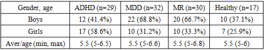
- We examined 108 children whose parents combined complaints about not being ready for school, not paying attention, not assiduous and not remembering information. According to the degree of exposure to etio-pathogenetic factors, different clinical picture and results of paraclinical methods of study, and psychological testing assessments, the children examined were divided into four groups:The 1st group included 29 children with hyperactivity and attention deficit syndrome. In the study group, 12 (41.4% of this group) boys and 17 (58.6% of this group) girls were studied. In accordance with clinical and neuroimaging signs, the group was formed:The 2nd group included 32 patients with psychological disorders. In the study group, 22 (68.8% of this group) were boys and 10 (31.2%) were girls.The 3rd group included 29 children with a preliminary diagnosis of "mental retardation, mild degree", including 20 (66.7%) boys and 10 (33.3%) girls. The control group 4 consisted of 17 healthy children. Of these, 10 (37.1%) were boys and 7 (25.9%) were girls. The group characteristics and age subgroups of patients included in the study are presented in Table 1.1.
|
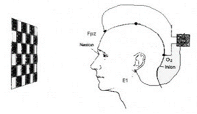 We measured the peak latency of waves (PL) - the time from the moment the stimulus was applied to the top of the wave, which characterizes the speed of the pulse through the visual analyzer.
 3. Results of the Study and Their Discussion
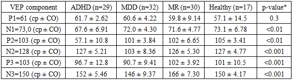
- Based on the results of numerous studies previously carried out using the visual evoked potential technique, the appearance of peak P1 is the result of yellow spot stimulation. Component N1 is detected by the striatum. The peak latency of component P2 is generated in the cortex at 18-19 Brodman's field. Negative peaks of N2 and N3.Thus, during the examination of VEP in children, we identified the following deviations. In children of the 1st group, the component N1 67.6 ± 6.91 and the successive peak P2 57.1 ± 10.8 were decreased, which indicated the disturbance of subcortical system connections with the cortex. A decrease in the latency of component N2 103 ± 8.36 and the following P3 90.7 ± 9.41 generated in the cortex of the striatum was observed in children of the 2nd group with NPR, which characterized the disturbance of the primary processing of the incoming signal at the cortical level (17-18 Broadman fields). In children of the 3rd group with mild mental retardation, an increase in latency N3 166 ± 7.30 was noted, which is the result of associative cortical activity - fields 18-19 for Brodman, which reflects the stage of visual information analysis.
|
4. Conclusions
- 1. The analysis of the data obtained during the VEP methodology revealed reliable results, which should be taken into account in cognitive disorders in pre-school children.2. Changes in the above parameters were observed in the examined children in the form of lengthening the reaction time (response delay).3. The increase in the latency of visual evoked potentials indicates a slowdown of the nerve impulse conduction, at different stages of the visual analyzer, which may serve as an additional diagnostic criterion in assessing cognitive functions in preschool children.