-
Paper Information
- Next Paper
- Previous Paper
- Paper Submission
-
Journal Information
- About This Journal
- Editorial Board
- Current Issue
- Archive
- Author Guidelines
- Contact Us
American Journal of Medicine and Medical Sciences
p-ISSN: 2165-901X e-ISSN: 2165-9036
2020; 10(1): 55-58
doi:10.5923/j.ajmms.20201001.12

Gross and Microscopic Anatomy of the Vater Papilla (Hepatopancreatic Ampulla) in Animals with and without Gall Bladder
 Abstract
 Reference
 Full-Text PDF
 Full-text HTML
Rakhmonov Zafarjon Mamadievich1, Oripov Firdavs Suratovich2, Dekhkanov Tashpulat Dekhkanovich3
1Assistant Professor of the Department of Human Anatomy, Operative Surgery and Topographic Anatomy of Samarkand State Medical Institute, Republic of Uzbekistan
2Chief of the Department of Histology, Embryology and Cytology of Samarkand State Medical Institute, Associate Professor, Republic of Uzbekistan
3Professor of the Department of Histology, Embryology and Cytology of Samarkand State Medical Institute, Republic of Uzbekistan
Correspondence to: Rakhmonov Zafarjon Mamadievich, Assistant Professor of the Department of Human Anatomy, Operative Surgery and Topographic Anatomy of Samarkand State Medical Institute, Republic of Uzbekistan.
| Email: |  |
Copyright © 2020 The Author(s). Published by Scientific & Academic Publishing.
This work is licensed under the Creative Commons Attribution International License (CC BY).
http://creativecommons.org/licenses/by/4.0/

The relevance of research problem. Congenital absence of any organ is severe anomaly of development and often leads to the serious consequences. Objective. To study comparable macroscopic and microscopic features of Vater papilla in mammals those have and do not have a gall bladder. Materials and methods. Materials for our investigations were Vater papillae of 10 rats and 6 horses (they don’t have gall bladder), 12 rabbits and 11 representatives of cattle (they have gall bladder). Results of the study. The comparative morphology of the Vater papilla of animals with and without gall bladder was studied. It was established that those animals that have a gall bladder larger than the Vater papilla have an ampoule with a complex network of folds, which is not the case for those who do not have a gall bladder. Conclusions: in rabbits and cattle that have a gall bladder, the Vater papilla forms a pronounced ampule (Vater ampule) with a complex network of folds, and in rats and horses that do not have a gall bladder, such an ampule was not found.
Keywords: Vater papilla, Comparative morphology, Gall bladder
Cite this paper: Rakhmonov Zafarjon Mamadievich, Oripov Firdavs Suratovich, Dekhkanov Tashpulat Dekhkanovich, Gross and Microscopic Anatomy of the Vater Papilla (Hepatopancreatic Ampulla) in Animals with and without Gall Bladder, American Journal of Medicine and Medical Sciences, Vol. 10 No. 1, 2020, pp. 55-58. doi: 10.5923/j.ajmms.20201001.12.
Article Outline
1. The Relevance of Research Problem
- Congenital absence of any organ is severe anomaly of development and often leads to the serious consequences. Perhaps there are organs which have not all representatives of vertebrate animals closer to the living conditions and nutrition character. Gall bladder belongs to such organs. Some animals (rats, horses, camels, elephants, rhinoceros and etc.) and birds (pigeons, turtle-doves, ostriches and etc.) do not have it. This is a big biological problem which attracts the attention of investigators and current century far from its final decision. At the same time, literature data regarding functional importance of gall bladder testifies that it takes part in regulation of digestive activity of the small intestine and influences to the functional activity of Oddy’s sphincter (Vinnik Yu.S. et al., 2012, Osipenko M.F. et al. 2013). There are scientific works devoting to the features of structural organization of the major duodenal papilla (Kostirkina V.V. 2006, Horiguchi S.I., Kamisawa N. 2010), in which the presence of the sphincters’ complex have been noted. There are research works regarding the role of gall bladder (Tyuryumin Ya.L, Shanturova V.А., Tyuryumina Е.E., 2011). It is natural that we have a question how does compensation of these functions happenin those animals that don’t have gall bladder? Gall bladder by its construction (in general with common biliary duct) makes a pressure which promotes the opening of Oddy’s sphincter. Therefore we can consider that in vertebrate animals which don’t have gall bladder sphincter apparatus of the terminal part of biliary duct also should have its features. According to the data of literature (Pokhobova Е.Yu., Belova G.V., 2012) the ampoule contains 3-5 layers of semilunar folds which form valves. There are comparative-morphologic works devoting to this problem (Dundarov Z.А., Gavrilenko V.I., Golubeva N.N. 2007, Zimatkin S.М., Markovets N.I. 2016). The above mentioned data could allow us to consider that the causes of the absence of gall bladder in some vertebrate animals is not fully established and it is a big medical-biological problem that far from its final decision.
2. Objective
- To study comparable macroscopic and microscopic features of Vater papilla in mammals those have and do not have a gall bladder.
3. Materials and Methods
- Materials for our investigations were Vater papillae of 10 rats and 6 horses (they don’t have gall bladder), 12 rabbits and 11 representatives of Bull ‘cattle’ (they have gall bladder). The structure of Vater papilla in horses and representatives of cattle has been studied by macroscopic method under binocular loupe. Vater papilla of rabbits and rats has been studied by macroscopic method by receiving a series of consecutive cuts. Materials were fixedin 12% solution of indifferent formalin neutralized by saturated solution of borax. Histological processing of the material and embedding in paraffin have been carried out by general accepted scheme. Serial consecutive cuts of the material were stained by hematoxylin-eosine with the use of Van Gizon method and impregnated by using of Grimelius method.
4. Results of the Study
- The ampulla of Vater, also known as the hepatopancreatic ampulla or the hepatopancreatic duct, is formed by the union of the pancreatic duct and the common bile duct. The ampulla is specifically located at the major duodenal papilla (figure 1, 2).
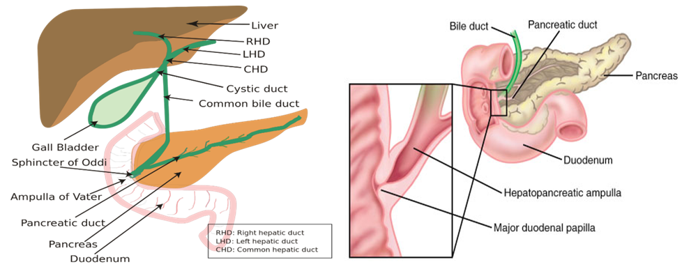 | Figure 1. Structure ampulla of Vater |
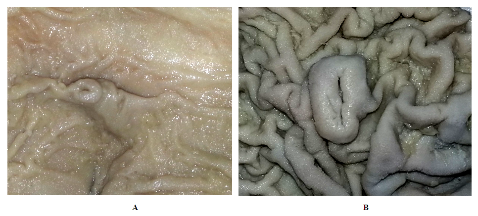 | Figure 2. Macroscopic structure of Vater papilla of bull (А) and horse (B). Macropreparation. MBS.Оb.4, ок.6 |
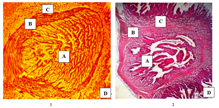 | Figure 3. Cryostat (1) and paraffin (2) sections of the ampulla of the Vater papilla of an adult rabbit. 1-impregnation according to Grimelius. 2-staining with hematoxylin-eosin. Ob. 20, Ok. 10. A–cavityampoules with folds. B–muscleshell of the ampoule. B–submucous membrane of the duodenum with Brunner's glands, D –cavityof the duodenum |