-
Paper Information
- Next Paper
- Previous Paper
- Paper Submission
-
Journal Information
- About This Journal
- Editorial Board
- Current Issue
- Archive
- Author Guidelines
- Contact Us
American Journal of Medicine and Medical Sciences
p-ISSN: 2165-901X e-ISSN: 2165-9036
2020; 10(1): 44-48
doi:10.5923/j.ajmms.20201001.10

Activity of Pathomorphologic Changes in Intestinal Lymphoid Tissue Caused by Sharp Intestinal Infections
 Abstract
 Reference
 Full-Text PDF
 Full-text HTML
Zhurayeva G. B.
Candidate of Medical Sciences, Senior Lecturer, Head of the Pathological Anatomy Bukhara State Medical Institute, Uzbekistan
Correspondence to: Zhurayeva G. B., Candidate of Medical Sciences, Senior Lecturer, Head of the Pathological Anatomy Bukhara State Medical Institute, Uzbekistan.
| Email: |  |
Copyright © 2020 The Author(s). Published by Scientific & Academic Publishing.
This work is licensed under the Creative Commons Attribution International License (CC BY).
http://creativecommons.org/licenses/by/4.0/

Acute intestinal infection is common in children of all ages and takes a special place in infant mortality. Newborns and young children are more susceptible to the development of AII (acute intestinal infection) caused by opportunistic microorganisms if they have primorbid conditions such as premature birth, malnutrition, rickets, anemia and congenital anomalies. In case of artificial and biased vaccination in children, the pathogenic micro florae often causes dysentery, salmonellosis and staphylococcal enterocolit, and other acute intestine infection (AII) [1,2,3]. In the development of AII, especially in a child, an important role is played by general and local factors of the immune system. Several factors play a special role in the local system of protection of the intestinal mucosa, which prevents intestinal infections. Results of morphometric researches showed that babies who died from acute intestinal infections had initially the grown number of stomal cells in lymphoid tissue of both iliac and large intestines of reticular and macrofagal origin with development of destructive changes in them. Parallel with these changes increase of the number of average lymphocytes are noted as the reaction of the immune system of intestines to an infection of active average lymphocytes. In addition to the changes above mentioned, during the subsequent age periods of babies there is growth in the quantity of small lymphocytes in composition of lymphoid follicles, which increases on average in 10-15 times as compared to the norm. Prevalence of acute intestinal infections at children's age and high rates of mortality have caused to consider them as one of actual problems.
Keywords: Acute intestine infection, Iliac and large intestines, Lymphoid tissue, Primorbid condition
Cite this paper: Zhurayeva G. B., Activity of Pathomorphologic Changes in Intestinal Lymphoid Tissue Caused by Sharp Intestinal Infections, American Journal of Medicine and Medical Sciences, Vol. 10 No. 1, 2020, pp. 44-48. doi: 10.5923/j.ajmms.20201001.10.
Article Outline
1. The Aim of the Research
- Identification of morphological criteria, morphometric parameters of mucous membrane and lymphoid tissue of intestines of nursing infants, as well as pathomorphological changes which developed as a result of acute intestinal infections.
2. Materials and Methods
- The basis of this research was formed by data of autopsies which are carried out at nursing infants who died of different types of acute intestinal infections during five years –from 2003 to 2007 in the Bukhara regional patholologic bureau and the Republican pathologic center. When calculating weight of children, ratio of physical development the 81 2 (50) 2017 criteria of the European bureau of the World Health Organization (WHO) (2000) were applied, presented in the methodical proceedings "Feeding of newborns and nursing infants". For the purpose of simplification of the calculation of a level of constant weight increase of children in mathematical formulas and in numbers, the below-brought coefficient of physical development – Q = real increase in weight / ideal increase in weight has been established. The criterion of weight of a child includes a condition of development of all internals, including a condition of the proceeding development of bodies of the digestive tract. In our materials, lag of development of digestive tract organs was noted in 84,6% of cases, as even in the period of nonutility a child could die due to any sharp intestinal infections. In all our cases, autopsy at children was carried out in 0,5-1 hour after death. At first 5,0-10,0 ml of blood was taken from heart and vascular system, plasma was emitted from it in the centrifuge, immunological researches were conducted with the help of a radial immune diffusive method of Mancini and with the use of nonspecific serum (The Moscow NIIVS to them I.I. Mechnikova). Immunoglobulin E is defined by radio immune methods. The number of T - and V-lymphocytes is counted on sensitivity to theophylline. For bacteriological researches the distal part of small intestine, and also the part of large intestine were used. When dissecting children in the course of the research the small intestine was excreted completely and the same length of one part of the large intestine, the excreted interiors were packed in a thin paper, rolled up by a parcel, fixed for 3 days in 10 percentage solution of neutral formalin. Each parcel including both bowels of intestines was dehydrated in alcohol, in chloroform and was filled in paraffin. Then received histological cuts of 5-8 microns thick were painted with hematoxylin-eozine according to Van-Gizona and CHIC reaction. Histological and histochemical painted cuts were studied under a microscope; informative sites of the cut were photographed. For the purpose of specification of quantitative indices of lymphoid tissue of intestines in walls of thin and thick intestines, morphometric researches were conducted by the method of Avtandilov G.G. with calculation of quantity of lymphoid follicles and their cellular structure. The calculation was carried out under the microscope lens 40 with imposing on lymphoid follicles of intestines of 10 times Avtandilov's grid, consisting of 100 points, only 1000 points. The reticular of cells, lymphoblast, large, average and small lymphocytes, and also the degenerating cells and macrophages are counted as wee as the quantity in lymphoid follicles and their relative percent. Quantitative indices were processed by statistical methods, size arithmetic averages, square errors of averages and indicators of reliability were calculated. 82 2 (50) 2015.
3. Results and Discussion
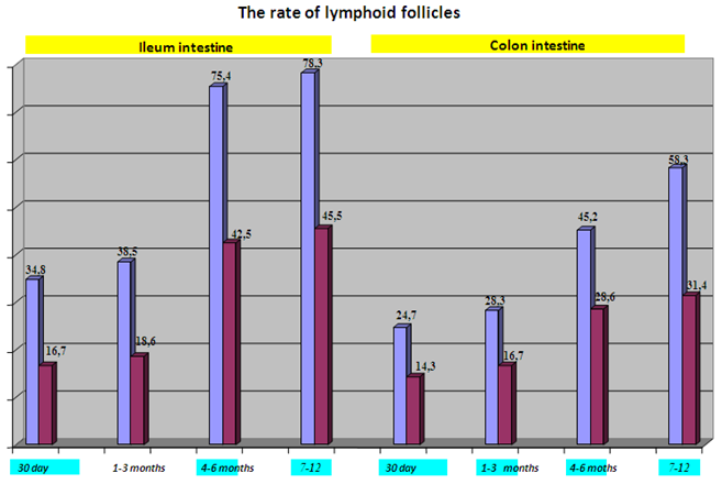
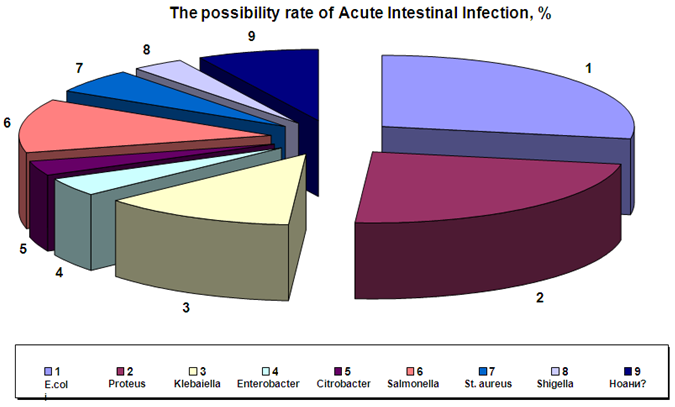
- Results of the microscopic researches showed, at autopsies of children with pnevmopatiya and craniocereberal injuries the mucous membrane of intestines had a uniform hystotophografic structure, but their thickness and length are various. It is revealed that in the first half of one-year-old life of newborns the mucous membrane of both thin and thick gut isn’t completely undeformed. Thus mucous fibers are short, of different form and size, the quantity of scaphoid cells isn't enough and they in the process of vacuolization. Especially, hypoplasia is noted in its connecting tissue mucous membrane, but not in its lymphoid cells, there are not enough vessels, and connecting tissue fibers are located randomly. Such rare lymphoid congestions consist of small and average size of lymphocytes. In Strom, being in their basis, seldom reticular cells are also randomly scattered. At the edges of lymphoid cellular congestions, post-capillary venial in which wall migration of lymphocytes is observed. At this appearing lymphoid follicles the mucous membrane is equal without tissues. Lymphoid follicles are located in a sub mucous layer. Newborns at the age of 7-12 months in the iliac gut wall have normal morphofunctional conditions of mucous membrane and lymphoid tissues. The tissues are extended, some are branched out, in its membrane the quantity of lymphoid cells is increased. Integumentary cylindrical epithelium is equal, nearly of one form and size among them there are enough scaphoid cells. Together with it, in this age period lymphoid follicles are located lonely and at distance from each other. In some of them in the center there are germinate centers and they turn on secondary lymphoid follicles. At this age in the wall of the small intestine lymphoid follicles are observed which are located in proximity of a muscular layer. They are rather smaller in size, their reticular cells are located randomly, the lymphocytes are on the periphery of the follicle infiltration in surrounding tissues. In the thick gut wall in this time of the research there are enough primary and secondary lymphoid follicles. If the primary lymphoid follicles are located in its connecting tissue membrane, the secondary have places in a submucous layer of the gut wall. The basis of secondary follicles is wide, the top is truncated and ridges on a surface of the mucous membrane. In which there are centers of reproduction and they consist of reticular cells and lymphoblast. Thus, babies during the first 6 months, had lymphoid follicles of different size and form, distance between follicles is wide; the germinate center is defined only in 65% of cases. During the next 6 months almost all lymphoid follicles have identical cellular structure and structure. Morphometric research of the iliac gut wall showed that the quantity of lymphoid follicles changes depending on age. It means that newborns have on average 34,8±5,2%, 1-3 months - 38,5±6,3%, 4-6 months – 75,4±9,4%, 7-12 months – 78,3±8,5%. It is revealed that in 83 2 (50) 2015 the thick gut wall the quantity of lymphoid follicles is even less in comparison with thin: newborns have 24,7±4,2%, 1-3 months – 28,3±4,3%, 4-6 months – 45,2±6,4%, 7-12 months – 58,3±7,5%. So it is possible to judge that in the wall of both thin, and large intestine in dynamics of age till one year the quantity of lymphoid follicles gradually increases and at the end of one-year-old life their number becomes twice more than at newborns. The carried-out calculation of morphometric indicators of relative percent of the organization of cellular structure of lymphoid follicles of children aged one year showed considerable differences depending on the lived months. At newborns the most part of lymphoid follicles comprise of small lymphocytes which is 87,8±8,6% in average. The reticular cells (2,1±0,2%) as the basis of lymphoid follicles and lymphoblast (0,9±0,2%) in functionally passive state also makes 3% of the total of cells. On the square of the germinate center meets in a small amount large (1,2±0,3%) and the average size (1,2±0,3%) of lymphocytes. As a part of lymphoid follicles of the thick gut the quantity of small lymphocytes is even less 84,5±6,6%, the reticular cells of 3,1±0,2% and lymphoblast 1,2±0,3%. In the germinate center big (2,2±0,5%) and average lymphocytes (6,6±0,7%) are noted. On the periphery of lymphoid follicles destructive cells (1,8±0,4%) and fagociting macrophages (0,9±0,2%) are noted. The quantity and structure of the cells of lymphoid follicles of the small intestine of newborns testifies to the lack of antigen influence and shows a condition of rest of immune system. On morphometric indicators the organization of structure of cells of lymphoid follicles of the mucous membrane of the large intestine, sharply differs from monthly relative percent of cells at children about one year. The small lymphocytes comprise the most part of lymphoid follicles at newborns 84,5±6,6%. The reticular cells (3,1±0,2%) and lymphoblast (3,1±0,2%) comprise the stroma of lymphoid follicles which also are functionally passive, the rest cells rated at 4,3%. As a part of lymphoid follicles destructive cells (1,8±0,4%) and phagocyte macrophages (0,9±0,2%) are noted. The development of high-quality and quantitative changes in the structure of cells of lymphoid knots of the large intestine of children after six months of life is evident. According to the microscopic researches, the number of lymphoid knots sharply increases; the set of their separate congestions of big sizes is revealed. It is known that such condition of local immune system is noted in histotopographic and the morphofunctional relation, generally in an organism of nursing infants in the second half of the first year of life. In congestions of lymphoid follicles of mucous membrane of the large intestine the volume of the germinate center extends, the number the reticular cells in it increases up to 4 times, lymphoblast – twice, and large lymphocytes – by 3 times, the same is observed and in quality as well. On the germinate center small lymphocytes 84 2 (50) 2015 form the dense ring which is thickened and condensed in its structure number of small lymphocytes, in comparison with newborns, decreases by 17%, their morph functional activity comes to light. On the surface of mucous membrane, including in a layer of integumentary epithelium infiltration the degree of small lymphocytes in symbioses and epithelium of mucous membrane is very high. Results of the clinical-anamnestic analysis of these children with AII showed that from the total (94 cases), the first age group included 19,1% of cases, the second – 27,7%, the third – 28,8%, the fourth – 24,4%. These children often had premorbid diseases: it is revealed that 28,4% of children had prematurity, at 23,6% - a hypotrophy, at 18,5% - rickets, at 21,7% - anemia and at 7,8% - congenital malformations. At children died from AII in all age groups there are low indicators of body weight, noted as at the birth as at death. Low weight indicators of a body are proved by the ratio calculation of physical development of the child which was as follows: in the first group it is 25% lower than the norm, in the second – 17%, in the third – 12% , in the fourth – 19% lower than the norms. It is established that in etiologic structure of activators of AII opportunistic microorganisms, such as koliinfection (28,4%), protease (23,7%), klebsiyell (12,6%), enterobakter (4,3%), sitrobakter (3,6%) are prevailing ones. Sometimes AII were caused by pathogenic microbes: in 3,5% of shield, 12,4% - salmonellosis, 5,6% - staphylococcus. In 7,9% of cases the etiology of AII isn't established. Studying of immunological indicators of the children who died of AII showed that at newborn IgA averaged 0,27±0,06 g/l, IgG – 5,4±0,8 g/l. At the age of 1-3 months is even lower, IgA – 0,11±0,02, IgG – 1,86±0,4 g/l. Thus in blood the quantity of a cortisol (5487,62±34,5 nmol/l) and immunoglobulin E considerably increases (143,65±12,34 ¬./l). In the senior age groups some increase in indicators of immunoglobulin’s was noted that apparently is connected with formation of immune system of an organism. At AII IgM caused by pathogenic microorganisms indicators and IgG were high and sometimes reached IgM – 5,6, IgG – 15,8 g/l. In 7-12 months the period when artificial feeding prevailed and AII were caused by gramm negative microorganisms, there were lower indicators of immunoglobulin. At this IgG decreased to 2,81 g/l, IgM raised to 1,94 g/l. Results of the pathomorphologic of researches showed that at children of AII caused by opportunistic microorganisms were exposed by mucous desquamative, mucous and necrotic and necrotic-ulcer inflammatory enterocolit. Such severe course of the infectious process and profound pathomorphologic change of mucous membrane of the intestines were caused by premorbid diseases as prematurity, hypotrophy, anemia, rickets and congenital malformations. Thus, morphologically in the intestines there was desquamation of integumentary epithelium, necrosis and ulceration of mucous membrane, diffusion of 85 2 (50) 2015 inflammatory infiltration of its membrane generally with mononuclear and single polynuclear cells. Studying of lymphoid tissue of the intestines showed that in connection with the infections at the died newborns lymphoid follicles is undeformed, atypically and unevenly hyperplastic. Thus, the main morphofunctional zones of lymphoid follicles are not revealed, the active lymphocytes are located round the post-capillary venules. Their borders aren't defined, lymphocytes infiltrate its own cover and submucous layer diffusively, among them there is a large number of macrophages and destructive cells. In more senior age periods of babies it is noted that if lymphoid follicles of the intestines is initially atrophic and hypolastic, under the influence of AII in them there is chaotic and uneven proliferation of cells and secondary changes in a type of hemorrhage and necrosis can be noted. Thus, in one cases the diffusion proliferation of all cellular elements of lymphoid follicles is noted, in other cases the hyperplasia of the germinative center with reticulosis is defined, in the third cases their main morphofunctional zones are not revealed. Apparently, these various patomorphologic changes of mucous membrane and lymphoid follicles of the intestines depend on the initial condition of lymphoid tissue of the intestines and on pathogenicity of AII activator. Results of the morphometric research of the cellular structure of lymphoid follicles of the gut wall of the babies who died of AII showed that since early months of life quantity the reticular cells, macrophages, degenerate cells and average lymphocytes considerably increases. Such increase of the quantity of cells of lymphoid follicles proceeds in the next months. In the age period of 7-12 months, in comparison with norm, increases their quantities, on average, on 10-15 times. If to interpret these morphometric changes depending on the clinic-anamnestic data, it is possible to assume that babies in initial stages of the life, the formation of lymphoid system of intestines falls off the norm and in development of response reaction the prevailing proliferation of reticular cells and macrophages with destructive changes in some of them is noted. The morphometric indicators of lymphoid follicles of the thick gut wall at AII change in almost identical dynamics as well as in lymphoid follicles of the iliac gut wall and 10/7 parts of them include small lymphocytes, 1,6 part to average lymphocytes, out of 100 1,5 part refer to reticular cells, 1 part to macrophages. These indicators proceed to accrue also during the subsequent age periods. As a part of lymphoid follicles the following proportion of cells is noted: small lymphocytes make 10/3 parts, reticular cells of 10/1,6 parts, macrophages of 100/5, degenerate cells of 100/3 parts. Thus, the results of morphometric studies have shown that at children who have died of AII, since the beginning in the lymphoid tissue of both iliac gut and the colon there is growth of the number of stromal cells of as reticular as macrophageal origin with the development of 86 2 (50) 2015 destructive changes. Simultaneously with these changes the higher number of lymphocytes in the medium is observed as an indicator in the intestinal immune response on the infection of medium active lymphocytes. In the next age periods babies in the content of lymphoid follicles, there is increase of the number of small lymphocytes, which as compared with the norm increases by 10-15 times in average attached to the above changes.
4. Conclusions
- At the nursing infants the number of lymphoid follicles in the gut wall and colon gradually increases and at the end of one year the parameters become twice higher. In the cellular structure qualitative and quantitative changes were observed, usually in the second half of the year. In the newborns the undeformed lymphoid tissue of the intestine is revealed simultaneously with atopic hyperplasia in response to the infection. When lymphoid follicles are initially in the condition of hypoplasia and atrophy, in response to the infection the uneven and random proliferation of the cells is observed. Thus, the main macrofunctional zones of lymphoid follicles are not revealed morphologically, and the secondary changes in the type of necrosis and hemorrhage are detected. The development of AII with conventionally pathogenic microorganisms, is primarily dependent on the existence premorbidial diseases and initial morphological and morphometric failure of lymphoid tissue of the intestine. Thus, catarrhal desquamative, catarrhal and purulent necrotic-ulcerative enterocolitis is revealed pathomorphologically. The AII in the lymphoid follicles in the intestinal wall, morphometrically since the first months of baby's life, the number of reticular, macrophageal, degenerative cells and medium lymphocytes, in the period of 7-12 months increased in 10 -15 times. Thus, the dynamics of reticular cells was noted: 1-3 months in 5 times, 4-6 months in 10, 7-12 months in 15 times. The number of macrophages increased in 7 times, and on average lymphocytes in 5 times.√ The number of lymphoid follicles in the ileum wall increases dramatically from birth to infancy, and their number more than doubled in infants√ In the first 6 months of infancy, lymphoid follicles have different sizes, and their intervals are not the same, only 65% of the germination area. By the second 6 moths, lymphoid follicles have almost the same structure and content√ Lymphoid nodes of the intestine in children who died from injuries from one month to three months do not undergo significant changes in the structure of lymphoid cells√ Significant qualitative and quantitative changes in the structure of cells of the intestinal lymph nodes appear after the sixth month of life
|
|