-
Paper Information
- Next Paper
- Previous Paper
- Paper Submission
-
Journal Information
- About This Journal
- Editorial Board
- Current Issue
- Archive
- Author Guidelines
- Contact Us
American Journal of Medicine and Medical Sciences
p-ISSN: 2165-901X e-ISSN: 2165-9036
2020; 10(1): 9-12
doi:10.5923/j.ajmms.20201001.03

Morphological Parameters of Rat Testes in Normal and Under the Influence of Chronic Radiation Disease
 Abstract
 Reference
 Full-Text PDF
 Full-text HTML
R. R. Baymuradov, Sh. J. Teshaev
Anatomy, Clinical Anatomy (OSTA) Department, Bukhara State Medical Institute, Bukhara, Uzbekistan
Correspondence to: R. R. Baymuradov, Anatomy, Clinical Anatomy (OSTA) Department, Bukhara State Medical Institute, Bukhara, Uzbekistan.
| Email: |  |
Copyright © 2020 The Author(s). Published by Scientific & Academic Publishing.
This work is licensed under the Creative Commons Attribution International License (CC BY).
http://creativecommons.org/licenses/by/4.0/

In this work objective was to study the morphological parameters of the testes of rats of 3 months of age in normal condition and under the influence of chronic radiation disease. The study was conducted on 40 white randomly bred male rats, weighing from 95 to 120 g, divided into 2 groups. Chronic radiation sickness was induced by irradiation of rats (4 Gy). Animals were removed from the experiment at 90 days of age, 20 days after its inception. Mathematical processing was also performed in which the standard deviation and representativeness errors were determined. An analysis of the results showed that in rats of the experimental group exposed to radiation (chronic radiation sickness), at the sexually mature (90 day) age, the diameter of convoluted seminiferous tubules is 8% less than the control, and their cross-sectional area is 13% behind. Other anatomical parameters, such as the mass of the testes lag behind by 8%, the length of the testes by 6%, the thickness of the testes by 5% and the volume of the testes by 10%, respectively. It has been established that after exposure to chronic radiation, puberty is delayed and all the morphological parameters of the testes lag behind.
Keywords: Testes, Convoluted tubules, Morphological parameters, Chronic radiation sickness
Cite this paper: R. R. Baymuradov, Sh. J. Teshaev, Morphological Parameters of Rat Testes in Normal and Under the Influence of Chronic Radiation Disease, American Journal of Medicine and Medical Sciences, Vol. 10 No. 1, 2020, pp. 9-12. doi: 10.5923/j.ajmms.20201001.03.
Article Outline
1. Introduction
1.1. The Relevance of the Problem
- In the 20th century, the role of chemical factors was enormous, but now physical factors come first. Among these factors, radiation requires special attention and pathogenically affects our body in one way or another.At the moment, among the problems caused by the environment, male infertility is very important. The male factor in infertility in marriage is 30-50% [1], so reproductive dysfunction in men has acquired special medical and social significance. While chemical factors enter our body mainly through contaminated food [2-7] changing sperm parameters and decreasing their production [8,9], radiation cannot be directly perceived by the human senses and therefore, they often cause more serious consequences, thereby increasing the decrease in fertility in men.The effect of ionizing radiation significantly upsets the balance of metabolism, which maintains the integrity of structures and homeostasis in the cells of various body tissues [10]. In addition, it is well known that gonads are highly sensitive to the effects of ionizing radiation. Therefore, the testes along with the bone marrow are assigned to the 1st group of critical radiation organs [11].The effect of radiation is gradually amplified, despite the fact that the testes are an immune protected part of the body in which cells are protected from harmful pathogenic factors. The hemato-testicular barrier, which is formed by the boundaries of Sertoli cells, restricts the access of germinal antigens to interstitial immune cells and the passage of antibodies from interstitium into the tubular lumen is considered the most recognized mechanism of immunological protection. Macrophages, peritubular cells, and Leydig cells are also involved in this protective mechanism [12,13].In addition, in a number of experiments it was proved that irradiation reduces spermatogenesis, changes the production of various hormones and causes infertility. In both rodents and humans, the degree of damage to the testes is directly related to the dose of radiation [14,15], and the germinal epithelium is very sensitive to radiation damage [16], while changes in spermatogonia begin even after low doses, like 0.1 Gy and permanent infertility can occur after fractional doses of 2 Gy or higher [17].
1.2. The Aim of the Study
- The goal of the study was to study the morphological parameters of the testes of rats of 3 months of age, normal and against a background of chronic radiation sickness.
2. Materials and Research Methods
- The study was conducted on 40 white random-bred albino male rats, weighing from 95 to 120 g, which were kept under vivarium conditions with a 12-hour light regime, with a standard diet and free access to water. They were divided into 2 groups: 1- control group (n = 20), 2- experimental group (n = 20). To simulate chronic radiation sickness, rats were irradiated. Irradiation began when the rats were 70-days old. The manipulation lasted for 20 days and ended at the age of 90-days old. Every day dose was 0.2 Gy (the total dose - 4.0 Gy). For irradiation we used DGTT device "AGAT R1" (Baltiets plant, Narva, Estonia, 1991, production, operation since 1994, reloading of 2007, with a capacity of 25.006 cGy/min.). The slaughter of rats was carried out by instant decapitation under ether anesthesia. Our study was fully did in accordance with the document "Rules of work with experimental animals", approved by the ethics committee of the Bukhara State Medical Institute named after Abu Ali ibn Sino (№. 18 dated 16.01.2018). It was based on the provisions of the Helsinki Declaration of the World Medical Association of 1964, supplemented in 1975, 1983, 1989, 1996, 2000, 2002, 2004, 2008, 2013.To conduct further research after opening the abdominal cavity, the testes were removed and the anatomical parameters of the testes, such as mass, length, width, volume were studied. Then extracted testes were fixed in Bouin's solution and poured into paraffin blocks according to generally accepted methods. Then, paraffin sections were prepared with a thickness of 6-7 μm, which were stained with hematoxylin-eosin. Histological and morphometric studies of testis tissue were performed using a microscope NLCD-307B.According to the data obtained, the cross-sectional area of the convoluted seminiferous tubules was calculated by the formula:
 where Sn is the area of a single cut of the tubule, d is the diameter of a separate cut of the tubule,
  Mathematical processing was carried out directly from the general data matrix “Excel 7.0” on a Pentium-IV personal computer; the standard deviation and representativeness errors were determined.
3. The Results of the Study
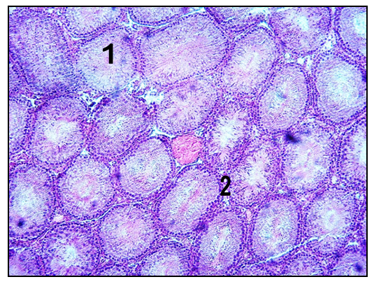
- Analysis of the morphological parameters of the testes of rats showed that in the experimental group the parameters are lagging behind in comparison with the control.In male rats of 90 days of age in the control group, the body weight varied between 95,6 ± 120,3 g and in average 106,8 ± 1,53 g. Testes are oval and are mainly found in the scrotum, less often in the inguinal-scrotal canal. The mass of the testes varies individually from 0.62 g to 0.88 g, on average 0.78 ± 0.016 g. The relative weight of testes was 1,46%. The length of the testes is 1.23 - 1.78 cm, on average 1.42 ± 0.034 cm. The width of the testes ranges from 0.9 to 1.3 cm, an average of 1.11 ± 0.025 cm. The volume of testes individually ranges from 0.61 to 0.83 cm3, an average of 0.69 ± 0.014 cm3. Microscopic examination of testicular tissue sections showed that, at this age, the diameter of the convoluted seminiferous tubules increases, a free lumen appears to promote mature sperm, so the density of the testis tissue decreases sharply.In male rats of 90 days old, the testes are coated with capsule. The thickness of the capsule is in the range from 13.2 to 29.6 μm, an average of 23.1 ± 1.02 μm. The capsule is represented by connective tissue of a fibrous structure consisting of thick bundles of collagen fibers. Bunches of underdeveloped elastic fibers become apparent. Outside, the membrane is covered with 3-4 layers of longitudinally oriented flat, mesothelial cells.The structural and functional unit of the testes are convoluted seminiferous tubules.The diameter of the convoluted seminiferous tubules in 90-day-old rats varies from 142.1 to 196.3 μm, an average of 171.3 ± 3.36 μm. The cross-sectional area of the convoluted seminiferous tubules ranges from 15318.3 to 26832.6 μm2, an average of 22191.4 ± 713.88 μm2. The walls of the convoluted seminiferous tubules are formed by connective tissue, a fibrous structure consisting of bundles of collagen fibers, reticular fibers, as well as thin bundles of elastic fibers (Fig. 1).
 | Figure 1. Testis of a 90-day-old rat in the control group. 1 - convoluted seminiferous tubules. 2 - inter-tubular spaces. Hematoxylin - eosin stain |
 | Figure 2. Testis of a 90-day-old experimental group rat. 1 - convoluted seminiferous tubules. 2 - inter-tubular spaces. Hematoxylin - eosin stain |
4. Discussion and Conclusions
- After studying the results of the studies, it was found that the mass of testes in rats of the control group from 1 day of birth to 90 days increases 39 times. And the absolute growth is 0.76 g. At the same time, the length of the testes increases 4.17 times, the thickness of the testes is 5.28 times and the volume of the testes is 49.3 times.It was detected that in rats of the experimental group, starting from the newborn to 3 months, the weight of the testes increases 36 times. The absolute growth is 0.70 g. Other parameters, such as the length of the testes, the thickness of the testes, the volume of the testes increase 3.94, 5.05 and 45 times. In this case, the absolute growth is 1.0 cm3, 0.85 cm3 and 0.616 cm3, respectively.It was found out that in rats of the intact group, the wall thickness of the capsule from the moment of birth to 3 months increases 2.2 times. And the diameter of the convoluted tubules increases 3.58 times, the cross-sectional area 12.1 times. The same parameters in rats of the experimental group increased 2.11 times, 3.31 times and 10.7 times.An analysis of the results showed that in rats of the experimental group exposed to radiation (chronic radiation sickness), at the sexually mature (90 day) age, the diameter of convoluted seminiferous tubules is 8% less than the control, and their cross-sectional area is 13% behind. Koruji M et al. proved that the number of normal tubules, the thickness of the epithelium, the diameter of the tubes and the diameter of the lumen were significantly reduced when irradiated with a high dose compared to control testes [18]. These data are similar to ours.As for the other anatomical parameters, the mass of the testes lags by 8%, the length of the testes by 6%, the thickness of the testes by 5% and the volume of the testes by 10%, respectively. Vereshchako GG et al. Had a similar result, when fractional irradiation at a total dose of 2.0 Gy in combination with anabolic drugs (e.g. phenobolin at a dose of 2.5 mg / kg) led to a significant decrease in the relative weight of the testicles and, in particular, epididymis [19]. In the works of Klepko AV et al. it was found that at the age of 2.5 months, irradiation in doses of 0.1, 0.3, 0.6 and 1.0 Gy does not affect the overall dynamics of the weight of animals, however, all the anatomical parameters of the testes still decrease [20].In addition relative weight of testes also redused under the influence of radiation which is close to results of other experiment [21]. In the testes of rats exposed to fractional radiation, which resulted in the development of chronic radiation sickness, there was a change in the microstructure of the gonads. Negative effects were manifested by a decrease in the mass, length, width and volume of the testes. It has also been proved that the capsule wall thickness decreases, the diameter of the convoluted seminiferous tubules and their cross-sectional area.