-
Paper Information
- Previous Paper
- Paper Submission
-
Journal Information
- About This Journal
- Editorial Board
- Current Issue
- Archive
- Author Guidelines
- Contact Us
American Journal of Medicine and Medical Sciences
p-ISSN: 2165-901X e-ISSN: 2165-9036
2019; 9(2): 515-518
doi:10.5923/j.ajmms.20190912.14

Features of Clinical and Laboratory Diagnostics of Enteroviral Serous Meningitis
Ergasheva Munisa Yakubovna
Applicant for the degree PhD Scientific, Research Institute of Virology of the Ministry of Health of the Republic of Uzbekistan
Correspondence to: Ergasheva Munisa Yakubovna, Applicant for the degree PhD Scientific, Research Institute of Virology of the Ministry of Health of the Republic of Uzbekistan.
| Email: |  |
Copyright © 2019 The Author(s). Published by Scientific & Academic Publishing.
This work is licensed under the Creative Commons Attribution International License (CC BY).
http://creativecommons.org/licenses/by/4.0/

Relevance of the research problem: The presence of the circulation of many serotypes of enteroviruses can affect the initial manifestations of the disease, the course of meningitis, which leads to the study of the clinical and laboratory features of this disease. Objective: To determine the frequency of detection of enterovirus infection in patients with serous meningitis, with following aspectual feature and development algorithm of clinical and laboratory diagnosis of Enteroviral meningitis. Materials and methods: 122 patients with serous meningitis were studied, cultural study feces and polymerase chain reaction liquor of patients with serous meningitis to identify enteroviruses. Diagnostic value of clinical symptom was determined. Results of the study: it was revealed that enterovirus infection was detected in 1/3 of all patients with serous meningitis, while the most common causative agents were ECHO 30 and ECHO 6, as well as Cox B 1-6. A number of clinical and diagnostic signs characteristic of Enteroviral serous meningitis were identified. Conclusions: the polymerase chain reaction method had a greater diagnostic importance compared to the cultural method. The disease appeared acutely, with the characteristic triad of symptoms for meningitis, passed with an average degree of gravity and moderately expressed meningeal syndrome on the background of pharyngitis and herpetic tonsillitis.
Keywords: Enteroviral serous meningitis, Polymerase chain reaction, Cultural research, Informational content of a clinical sign
Cite this paper: Ergasheva Munisa Yakubovna, Features of Clinical and Laboratory Diagnostics of Enteroviral Serous Meningitis, American Journal of Medicine and Medical Sciences, Vol. 9 No. 2, 2019, pp. 515-518. doi: 10.5923/j.ajmms.20190912.14.
Article Outline
1. The Relevance of the Research Problem
- Significant polymorphism of clinical manifestations with the absence of a clear dependence by the serological type of pathogen, the high frequency of asymptomatic forms of Enteroviral infection (EVI), prolonged virus carriage, the absence of specific methods of prevention, make EVI an uncontrolled disease [5,7].In turn, the epidemiological significance of enterovirus infection is determined by: high contagiousness, broad spreading, outbreak of morbidity, a variety of causative agents of EVI, the possibility of severe consequences, up to fatal outcomes [1].In spite of the monitoring and numerous studies EVI worldwide, nowadays, the epidemiological situation of EVI remains tense, there is a rise in the incidence, including Enteroviral meningitis is the most common clinical form of EVI and particularly not prosperous situation in neighboring countries. According to the authors, in recent years 85-90% of cases of viral serous meningitis in children and (less commonly) in young people are caused by enteroviruses [8,9].The presence of circulation of many types / serotypes of enteroviruses, their evolutionary development can affect the initial manifestations of the disease, the course of meningitis [3,6], which leads to the study of clinical and laboratory features of serous meningitis of Enteroviral etiology at the present stage.
2. Objective
- To determine the frequency of detection of enterovirus infection as a causative agent in patients with serous meningitis, followed by characterization of the typical distribution of non-polio enteroviruses and the development of the clinical and laboratory diagnostic algorithm of EVM.
3. Material and Methods
- 122 patients of various ages with serous meningitis, hospitalized in the Regional Infectious Hospitals of Tashkent and Samarkand were examined. The diagnosis of serous meningitis was based on a combination of complaints, anamnesis, clinical picture of the disease, a neurological examination and laboratory tests.To confirm the presence of enterovirus infections in patients with serous meningitis method was used cultural studies of feces, and cerebrospinal fluid by PCR, followed by neutralization to determine the distribution of species of Enterobacteriaceae.Separation of enteroviruses from the feces of patients with serous meningitis was carried out in the virology department on a nutrient medium "Needle DMEM with L- glutamine". Molecular biological research method (PCR) was carried out in the reference laboratory of the Research Institute of Virology of the Ministry of Health of the Republic of Uzbekistan. Analysis was performed by using the test-system "The amplitude Sense Enterovirus » (Russian Federation, Moscow).Features of clinical and laboratory manifestations of EVMs were analyzed with the calculation of the information content of the diagnostic coefficient by A. Kulbak [2].
4. Results of the Study
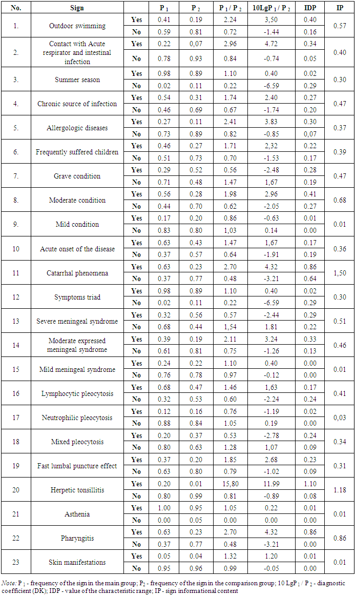
- A comparative analysis of the frequency of emitting EV from various clinical materials by various methods showed that 92 samples of feces from patients with serous meningitis in 32 (34.7%) cases and 30 samples of cerebrospinal fluid from patients with serous meningitis in 9 (30%) cases detection of EV were positive.Preliminary results of a classical virological study did not affect the treatment tactics, as the attending physicians received them only at the 3-4th week of the disease, when the patients were already discharged from the hospital.The detection of the pathogen in the cerebrospinal fluid during PCR made it possible to say about the role of enterovirus in the development of serous meningitis. The results were obtained during the first days after the patient’s admission to the hospital (2-3 days of hospitalization), which made it possible to make a final diagnosis of “Enteroviral etiology meningitis”, prescribe appropriate antiviral therapy and discontinue antibiotic treatment in unclear diagnostic cases in the first days since hospitalization.When comparing different diagnostic methods, all examined patients were divided into groups according to the time of admission to the hospital: patients who arrived on the first day after the disease, within 2 to 3 days from the onset of the disease and later than 3 days after the onset of symptoms of the disease.Comparison of the positive results obtained in the cultural and molecular genetic studies depending on the timing of the patient’s admission to the hospital revealed that in patients admitted to the hospital on days 1–2 of the disease, the positive results recorded an advantage with the cultural method of research (59.4 % ± 8.7 and 22.2% ± 13.9; p> 0.05). When the patient was hospitalized on the 3-4th day of the disease, the results were almost the same both the molecular genetic method and the cultural investigation (31.3% ± 8.2 and 33.3% ± 15.7; p > 0.05). When conducting studies of patients hospitalized later than 4 days of illness, positive results in a cultural investigation lowest frequency (9.4%±5.1) were noted. During PCR, data on the causative agent of meningitis were obtained in 44.4% ± 16.6 (p <0.05) samples of cerebrospinal fluid, which was explained by higher stability of the nucleic acid than the intact viral particles.When conducting virological testing of CSF and feces of patients with a positive result on the EVI, followed by neutralization with a specific set of ECHO and virus antigens Coxs, it was revealed that the most frequent strain in patients with serous meningitis of Enteroviral etiology was ECHO 30 - 18 cases of 41 (43.90% ± 7.75). Coxs B strain was detected in 4 (9.76% ± 4.63) cases, ECHO 6 strain was detected in 2 cases out of 41 (4.88% ± 3.36), and in just one case, the serotype ECHO 7 and ECHO 12 was determined (2.44% ± 2.41 and 2.44% ± 2.41). During the neutralization reaction in the remaining samples, the exact pathogen was not determined (NTEV) in 11 cases (26.83% ± 6.92). Thus, in most cases, the results of the investigation in the neutralization reaction gave positive results - 37 cases (90.24% ± 4.63). Of these, results in the neutralization reaction with feces of patients with serous meningitis and a positive result of a culture study on EVI were detected in all 32 cases (100%).The cultural method for the isolation of EV from CSF followed by a neutralization reaction was positive in 5 out of 9 cases (55.56% ± 16.56). It should be noted that the cultural method for the detection of EV in cerebrospinal fluid, gives a very low frequency. Works of Shtanberg A.V., 2009 [8] shows the frequency of a positive result in cerebrospinal fluid samples for the cultural detection of the virus was only 9%, with a positive result of PCR and corresponding clinical picture of the EVM. In this regard, the data of 9 cases of a positive PCR reaction to EVI, despite a negative cultural response, are indicators of the incidence of this infection, since the result was obtained from sterile body fluids.Thus, our data on the identification of EVM serotypes confirmed the data of world literature, suggesting that the most common strain causing serous meningitis of enteroviral etiology is ECHO 30 and ECHO 6 [7]. At the same time, one of the features of serous meningitis of Enteroviral etiology was the fact that in both Tashkent and Samarkand there were cases of meningitis caused by the Coxs B 1-6 strain, which at the present stage are detected with serous meningitis [8] , but an indication on them as EVM pathogens is extremely rare. When comparing our data with the results of domestic scientists conducted in Samarkand and Tashkent regions at the end of the 20th century , we can say that in our region and 30-40 years ago, Coxs B1 -6 and ECHO 6 were identified, and were considered as one of the reasons for sporadic cases aseptic meningitis [4].On discharge of the objectives of this work was to develop an algorithm of clinical and laboratory diagnostics Enteroviral meningitis, based on the list of the most informative anamnestic, clinical and laboratory features.In the framework of this investigation, an approach was chosen based on the determination of the information content of signs according to Kulbak. This method, in comparison with other methods of minimizing informative redundancy, is the most simple and accessible for algorithmization.The methodology for calculating the information content of signs according to Kulbak is based on the determination of diagnostic coefficients calculated for the main and control groups of patients. Using the proposed methodology, on the basis of the generated database, diagnostic coefficients and information values were calculated for each of the signs and the most informative signs were selected for the diagnosis of EVM (table 1).
|
5. Conclusions
- 1. Of all cases of serous meningitis, the detection of enterovirus as a pathogen was observed in 1/3 of cases, while the polymerase chain reaction method was of greater diagnostic value compared to the cultural method.2. The most common causative agents of EVM were ECHO 30 and ECHO 6; cases of Coxs B1 - 6 due to serous meningitis were also detected.3. Coxs B1 - 6 and ECHO 6 can be considered a permanent inhabitant of the range and causing sporadic cases of EVM incidence, while ECHO 30 is an imported strain.4. The most significant clinical and laboratory signs EVM were: swimming in the open non flow water, contact with patients with acute respiratory infections or acute intestinal infection, the presence of chronic infection source, frequent incidence of children as well as allergic pathology. The disease occurs acute, with the characteristic triad of meningitis symptoms, moderate expression of the meningeal syndrome with background of pharyngitis and accompanied gerpetic tonsillitis.
 Abstract
 Reference
 Full-Text PDF
 Full-text HTML