-
Paper Information
- Previous Paper
- Paper Submission
-
Journal Information
- About This Journal
- Editorial Board
- Current Issue
- Archive
- Author Guidelines
- Contact Us
American Journal of Medicine and Medical Sciences
p-ISSN: 2165-901X e-ISSN: 2165-9036
2019; 9(2): 467-470
doi:10.5923/j.ajmms.20190912.04

Quantitative Relationship of Lymphocytes in the Lymphoid Nodules of the Small Intestine of Rats in Normal and Under the Influence of Kotoran
 Abstract
 Reference
 Full-Text PDF
 Full-text HTML
N. E. Tukhsanova
Anatomy, Clinical Anatomy (OSTA) Department, Bukhara State Medical Institute, Bukhara, Uzbekistan
Correspondence to: N. E. Tukhsanova, Anatomy, Clinical Anatomy (OSTA) Department, Bukhara State Medical Institute, Bukhara, Uzbekistan.
| Email: |  |
Copyright © 2019 The Author(s). Published by Scientific & Academic Publishing.
This work is licensed under the Creative Commons Attribution International License (CC BY).
http://creativecommons.org/licenses/by/4.0/

In our work, we determined the number of lymphocytes in the lymphoid nodules in normal condition and when exposed to the herbicide which passed through the mother’s milk. The results of the study showed that in the control group in all age categories, the number of large lymphocytes increases in the distal direction throughout the small intestine, and in the experimental group, the number of small lymphocytes decreases. An increase in the number of large (not mature) lymphocytes in the lymphoid nodules of the small intestine of the experimental group indicates a response of the body to a chemical effect.
Keywords: Lymphocyte, Kotoran, Herbicide
Cite this paper: N. E. Tukhsanova, Quantitative Relationship of Lymphocytes in the Lymphoid Nodules of the Small Intestine of Rats in Normal and Under the Influence of Kotoran, American Journal of Medicine and Medical Sciences, Vol. 9 No. 2, 2019, pp. 467-470. doi: 10.5923/j.ajmms.20190912.04.
Article Outline
1. Introduction
- The results of our studies provide in-depth knowledge about the change in the morphological parameters of the structures of the small intestine of one, two, three-month-old rats, normal and under the influence of which. The course of changes occurring for the first time in the immune status of rats under the inhibitory effect of a pesticide, which is age-related, was studied for the first time. The dynamics of the established changes allows us to predict the development of gastrointestinal diseases in the early stages. For the first time, the change in the cellular composition of the lymphoid structures of the small intestine under the influence of which in the age aspect was studied. The results of the study can be used when giving lectures on gastroenterology and immunology.
1.1. The Relevance of the Problem
- Human health is the main criterion for assessing the quality of life indicator. One of the important factors that play a role for human health is the environment. Because it creates the conditions for the full and harmonious development of man.The rapid development of the chemical, pharmaceutical, engineering industries, and the widespread use of a large arsenal of pesticides in agriculture and households pose a real threat to public health. Currently, the environment is contaminated with pesticides, since modern agricultural production cannot be imagined without pesticides [11].Currently, one of the widely used pesticides used in agriculture of the Republic of Uzbekistan is the herbicide kotoran. Kotoran (Urea,N,N-dimethyl-N'-[3- (trifluoromethyl)phenyl]) C10H11F3N2O is a white crystalline substance. The herbicide is used to control weeds in cotton crops by spraying the soil. Solubility in water at 25°C mg / l, soluble in organic solvents. LD50 for rats is 1515 mg / kg. It is stored in the soil for a rather long time (Melnikov N.N. 1995).Chemical environmental factors enter the human body primarily through the gastrointestinal tract, it represents the most extensive habitat of microflora in the human body, since its surface area is more than 300 m2 [3].The intestine has a big importance in the immune status of the body [1,3,4,6,11]. It has been established that, about 80% of all immunocompetent cells of the body are located in the intestinal mucosa, about 25% of the intestinal mucosa consists of immunologically active tissue and cells. Each meter of the intestines of an adult contains about 1010 lymphocytes [2,3,7,8,10].In the literature available to us there are a number of works devoted to the cellular composition of the lymphoid structures of the intestine. But they do not show the dynamics of the development of these structures in the age aspect and under the influence of kotoran. All of the above dictates the need for this study.
1.2. The Aim of the Study
- The goal of the study was to study the quantitative content of lymphocytes in the lymphoid structures of the small intestine of rat pups up to 1 month in the norm and when exposed to the herbicide kotoran.
2. Materials and Research Methods
- The study was conducted on 56 rats. They were divided into 2 groups: control (20 rats) and experimental (36 rats) group. All animals were kept in the same conditions and on a regular diet. As an experimental model, 20% of the concentrate of cotoran was taken. Poisoning of rats occurred through the milk of the mother (female rats) who were administered kotoran through a thin metal probe intragastrically at a dose of 0.05 mg (1 ml) in the morning before the day of slaughter. Female rats of the control group were administered 1 ml of distilled water through a probe. Slaughter of rats was performed under ether anesthesia on the 1st, 16th, 30th day of postnatal ontogenesis. After opening the abdominal cavity, the small intestine was removed and pieces from the initial, middle and lower segment were taken. The materials were fixed in Buena solution, carried out on alcohols of increasing concentration and poured into paraffin. Sections 5-10 microns thick were made from the blocks on the microtome, and stained with hematoxilin - eosin. On the microscope slide morphometric measurement was carried out using the ocular ruler. Throughout the small intestine, the depth of the crypts, the height of the villi and the wall thickness of each layer were measured separately. To study the cell composition of lymphoid nodules, a grid with 69 nodal points was inserted into the eyepiece of the microscope. When the eyepiece 10 lens 40 (under oil immersion) counted the number of points that fell on small, medium and large lymphocytes.The obtained digital data were processed by the method of variation statistics using R. B. Strelkov's tables (1986). The reliability of the results was estimated at p≤0.05. The studies were carried out in compliance with the rules of humane treatment of animals, which are governed by the “Rules for conducting work using experimental animals”, approved by the ethical committee of the Bukhara State Medical Institute (2018).
3. The Results of the Study
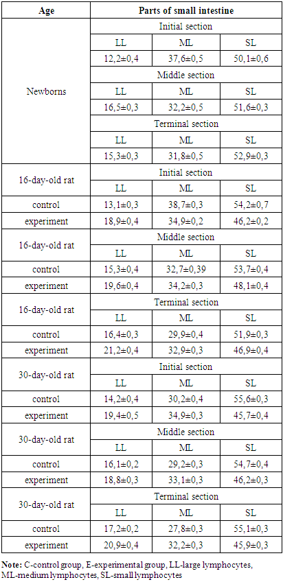
- In newborns, rat lymphoid formations of the small intestine are represented by diffuse lymphoid tissue. In the submucosa, diffuse accumulation of small and medium lymphocytes in the form of single-row chains is observed. In diffuse clusters of the initial segment of the small intestine, the content of large lymphocytes ranges from 11 to 15 cells per 100 lymphocytes, an average of 12.2 ± 0.4; medium lymphocytes - from 34 to 39, on average-37.6 ± 0.5; and small lymphocytes - from 47 to 53, an average of -50.1 ± 0.6.In newborn rat pups in the lymphoid accumulations of the middle part of the small intestine, the number of large lymphocytes per 100 lymphocytes was in the range from 15 to 18 cells, an average of 16.5 ± 0.3; medium lymphocytes from 30 to 35, on average 32.2 ± 0.5; small lymphocytes from 51 to 54, on average 51.6 ± 0.3.A study of the cellular composition of the lower segment of the small intestine showed that the number of large lymphocytes per 100 lymphocytes ranged from 14 to 17, an average of 15.3 ± 0.3, medium lymphocytes from 29 to 34, an average of -31.8 ± 0.5, small lymphocytes from 50 to 56, an average of 52.9 ± 0.3 cells.In a 16-day-old rat of the control group, in the lymphoid nodules of the initial section of the small intestine, the content of large lymphocytes per 100 lymphocytes ranges from 12 to 15, an average of 13.1 ± 0.3, medium lymphocytes from 36 to 40, an average of 38.7 ± 0.3, small lymphocytes - 52 to 59, on average - 53.7 ± 0.4 cells.In lymphoid nodules of the middle part of the small intestine of 16-day-old rats, the number of large lymphocytes ranged from 14 to 17, an average of 15.3 ± 0.4; secondary lymphocytes 30-35, on average-32.7 ± 0.39; small lymphocytes - 52-56, on average - 53.7 ± 0.4 cells.The number of lymphocytes in the lymphoid nodules of the lower part of the small intestine per 100 lymphocytes: large lymphocytes - from 15 to 18, an average of 16.4 ± 0.3 cells, medium lymphocytes - from 28 to 32, an average of 29.9 ± 0.4, small lymphocytes - from 52 to 54, on average - 51.9 ± 0.3 cells.In a 30-day-old rat of the control group, lymphoid clusters located at different distances from each other are determined at the border between the mucous membrane and the submucosa of the small intestine.In lymphoid nodules of the initial section of the small intestine per 100 cells, there are from 12 to 16 large lymphocytes, an average of 14.2 ± 0.4; medium lymphocytes - from 28 to 32, an average of 30.2 ± 0.4; small lymphocytes - from 54 to 58, on average - 55.6 ± 0.3 per 100 lymphocytes.The number of large lymphocytes in the lymphoid nodules of the middle section of the small intestine of rats of 30 days of age per 100 lymphocytes ranged from 14 to 18, an average of 16.1 ± 0.2, medium lymphocytes from 28 to 31, an average of 29.2 ± 0, 3, small lymphocytes - 52 to 56, an average of -54.7 ± 0.4 cells (Figure 1).
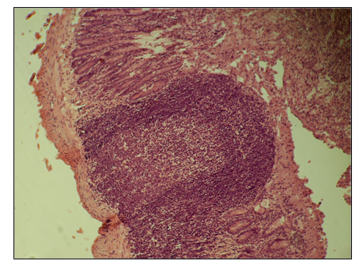 | Figure 1. The cellular composition of the small intestine lymphoid node of the one-month-old rats (control group) |
|
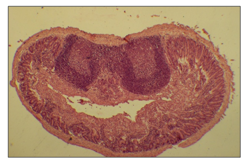 | Figure 2. The cellular composition of the small intestine lymphoid node of the one-month-old rats (experimental group) |
4. Discussion and Conclusions
- The results of the study showed that in the control group in all age categories throughout the small intestine in the distal direction the number of large lymphocytes increases, the number of medium and small lymphocytes decreases. Our data on the number of large lymphocytes is consistent with the data of G.G. Aminova and M.R. Sapin (2007), where they indicate that the cellular composition of the lymphoid nodules of the cecum in humans varies little with age, especially large lymphocytes (fibroblasts and reticulocytes) [2].In 16 day old rats, the experiment shows an increase in the proportion of large lymphocytes, compared with medium and small lymphocytes. In 30 day old experimental group rats, the number of large lymphocytes decreases in the lymphoid nodules, and the number of small lymphocytes increases.Studies have shown that the number of large lymphocytes of the small intestine in rats of the experimental group from the first birthday to 30 days of age increases at the following rates: in the upper part 1.59 times, in the middle part 1.14 times and in the terminal part 1.37 times. It was revealed that the number of medium lymphocytes in 30 day rats (experimental group) from the moment of birth in the middle and lower part of the small intestine increases 1.03 and 1.01 times, respectively. Whereas in the initial part is reduced by 1.08 times.The rate of increase in the number of small lymphocytes in general is significantly reduced in relation to the first day of life. It is 1.09 times in the initial part of the small intestine, 1.12 times in its middle part and 1.15 times in the terminal part of the small intestine.In an experiment in the lymphoid nodules of the small intestine, an increase in the number of large lymphocytes is observed with age. Against the background of a decrease in small, the number of medium lymphocytes almost does not change. The number of small lymphocytes in lymphoid nodules in all age groups and in all segments of the small intestine in experimental rats is less than the control.When a chemical substance (kotoran) enters the body, the lymphoid tissue reacts to it with qualitative changes. The same reaction is observed on the part of lymphoid formations in various inflammatory processes [9].An increase in the number of large (not mature) lymphocytes in the lymphoid nodules of the small intestine of the experimental group indicates a response of the body to a chemical effect.Kotoran, which passed through mother’s milk, causes an increase in lymphocytic infiltration of the epithelium, contributes to an increase in the proportion of large lymphocytes in lymphoid nodules.