-
Paper Information
- Previous Paper
- Paper Submission
-
Journal Information
- About This Journal
- Editorial Board
- Current Issue
- Archive
- Author Guidelines
- Contact Us
American Journal of Medicine and Medical Sciences
p-ISSN: 2165-901X e-ISSN: 2165-9036
2019; 9(1): 453-456
doi:10.5923/j.ajmms.20190911.10

Thyroid Cells Development under Thyreostatic Burdon
 Abstract
 Reference
 Full-Text PDF
 Full-text HTML
Rasulov Khamidulla Abdullaevich
Tashkent Pediatric Medical Institute, Ph.D., associate professor, Tashkent, Uzbekistan
Correspondence to: Rasulov Khamidulla Abdullaevich, Tashkent Pediatric Medical Institute, Ph.D., associate professor, Tashkent, Uzbekistan.
| Email: |  |
Copyright © 2019 The Author(s). Published by Scientific & Academic Publishing.
This work is licensed under the Creative Commons Attribution International License (CC BY).
http://creativecommons.org/licenses/by/4.0/

Today thyroid gland pathologies take one of the leading places among endocrine disorders. Factors such as unfavorable ecology, change of environmental micro element composition, hereditary predisposition play a certain role in the development of thyroid pathology. Morphology of thyroid tissue, which is well known reliably reflects the degree of thyroid functional activity in various physiological and pathological conditions of an organism, in fact is not studied in various stages of experimental hypothyroids early postnatal ontogenesis. Merkazolil is one of the thyreostatics that block pyroxide oxidase, participating in the synthesis of thyroid hormones of thyroid gland, which prevents the iodization process. The active part of the drug enters the thyroid gland tissue which formed a stable compound with hydrogen peroxide, as a result of which the enzymatic function catalyzes the iodine compound. The synthesis of thyroid hormones in the beginning decreases, and follicular contents increase in the future. A-cells-thyrocytesthat are lined on the wall of the follicle of the gland, B-cells are found in the parenchyma of the gland were first described in 1898 year were found in nodular goiter, adenomas and some thyroid diseases. C-cells are calcitoninocytes that produce the parathyroid hormone antagonist of calcitonin. It is necessary to find out the effect of mercazolil on the functional morphology of the thyroid gland in laboratory rats undergoing hypothyroidism, and also to establish the significance of the changes undergoing in the growing organism in the formation of structural damage to the organ.
Keywords: Thyroid gland, Ashkenazi cells, Oxyphilic epithelium, Merkasolil
Cite this paper: Rasulov Khamidulla Abdullaevich, Thyroid Cells Development under Thyreostatic Burdon, American Journal of Medicine and Medical Sciences, Vol. 9 No. 1, 2019, pp. 453-456. doi: 10.5923/j.ajmms.20190911.10.
Article Outline
1. Introduction
- Thyroid gland- a butterfly-shaped organ located in the base of the neck.Ashkenazi cells- B-cells are found in the parenchyma of the gland.Oxyphilic epithelium-they are found throughout the body as the thyroid gland.Merkasolil- one of the thyreostatics that block pyroxide oxidase, participating in the synthesis of thyroid hormones of thyroid gland.Today thyroid gland pathologies take one of the leading places among endocrine disorders. Factors such as unfavorable ecology, change of environmental micro element composition, hereditary predisposition play a certain role in the development of thyroid pathology. Morphology of thyroid tissue, which is well known reliably reflects the degree of thyroid functional activity in various physiological and pathological conditions of an organism [4], in fact is not studied in various stages of experimental hypothyroids early postnatal ontogenesis.At the same time certain morphological functional shifts in thyroid gland are observed in the process of physiological aging, which are testified in multiple publications of modern scientists [1,3-5].The aforesaid is relevant to the basic cell population of thyroid gland, represented by A-cells (thyreocytes) forming follicular compartment of the gland and secreting iodine-containing thyroid hormones. Besides that, thyroid gland is a multicomponent organ including two more populations of cells such as B-cell (Ashkenazi cells, Hurthle cells, oncocytes, oxyphilic cells) and C-cells (parafollicular cells, calcitoninocytes) [6,7,9].A-cells-thyrocytesthat are lined on the wall of the follicle of the gland,B-cells are found in the parenchyma of the gland were first described in 1898 year were found in nodular goiter, adenomas and some thyroid diseases.C-cells are calcitoninocytes that produce the parathyroid hormone antagonist of calcitonin.Products of B and C-cells’ secretion play a significant part both in the activity of an organism and functioning of Thyroid Gland producing thyroid hormones [2,10].It is difficult to imagine, that in the decrease of thyroid functional activity and various unfavorable impacts on body, including iodine deficiency, populations of these cells stay non-sensitive and intact. Taking into account all the aforesaid, it is interesting to clarify the effect of merkasolil on the functional morphology of TG in laboratory rats with hypothyroids, and to determine the importance of each of these factors in the formation of structural lesions in the organ.No special researches were performed in this field.
2. The Objective
- To study morphological functional condition of Thyroid Gland in the aspect of early ontogenesis with its hypofunction.
3. Materials and Methods
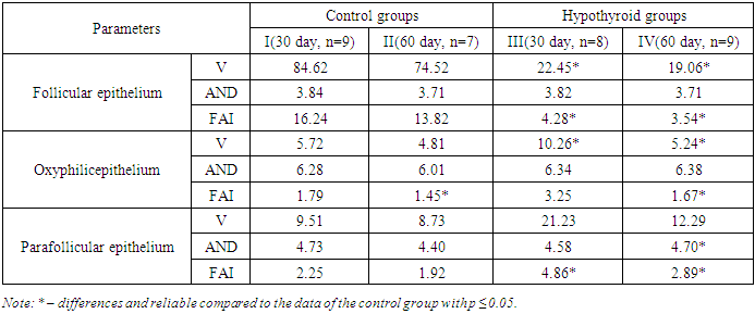
- We used 33 not thoroughbred infant rats being 150-180g. In the first set (I and III groups — 9 rats at the 30th day and 7 rats at the 60th day, respectively) we studied morphological structure of thyroid gland in control animals. In the second set (II and IV groups — 8 rats at the 30th day and 9 rats at the 60th day respectively) we studied morphological alterations in anatomical structures of thyroid gland with hypothyroids. In the blood serum of the experimental group, the content of total thyroxine was 58.6 ± 1.17 nmol/L, significantly lower than the control (<0.05). The study included 2 control groups (9 and 7 individuals) to compare the obtained results of morphological studies of the experimental groups. Hypothyroid was caused by administration of one of thyreostatics – Merkasolil in the dosage 3 mg/kg of animals’ body mass.All rats were held in similar conditions of a vivarium and received usual food. Animals were excluded from the experiment by means of ether overdose. An experimental model of hypothyroidism was carried out according to the method of V.Y. Khryshchanovich 2012 year. During the experiment, cases of overdose and side effects were not detected.Samples of thyroid tissue from both lobules of TG of control and experimental rats with hypothyroid were fixed in 12% solution of neutral formalin, dehydrated in alcohol with growing concentration, and coated by paraffin. Serial histological slices were stained by hematoxillin and eosine, and using Van-Gizon’s method.Morphometric analysis of thyroid epithelial cells was performed in compliance with V.P. Volkov’s recommendations (2015). Density of each kind of cells of thyroid parenchyma (V) was determined by calculation in the microscope field of vision 300W Pixel CCD Electronic Eyepiece Dual Mechanical Mobile platform, attached 150x140 mm flat platform, range of mobile devices: 75x55 mm, KOHLER reflective backlight, 6 V / 20 W halogen lamp with adjustable brightness.That parameter characterizes expression of hyper and/or hypoplasia of certain cells pool as a whole.Average diameter of the nuclei of the studied cells (AND), indicating the degree of hyper or hypotrophy of each cell, was determined by means of measurement of the greatest (a) and the smallest (b) sizes of nuclear and consequent calculation using the following formulae [4,11]:AND=√abAccording to the results of morphometric measurements, we calculated integral parameter such as functional activity index (FAI) for each cell population using the formulae:FAI=V*AND/20Statistical processing of the obtained data was performed by means of non-parametric statistic methods differing by sufficient capacity, simplicity, reliability, and self-descriptiveness [8].FAI- functional activity index makes it possible to mathematically assess the state of the glandular parenchyma.V- the volume cells glandular parenchyma the field of view determines the number of cellular elementsAND- the average diameter of the nuclei.
4. Results and Discussion
- That research demonstrates that morphological functional condition of TG is significantly affected by both age and mercasolil administration (Table 1). Dynamics of structural alterations in various cell populations was remarkably different in its expression and direction.
|
5. Conclusions
- As a result of experimental research we revealed different directions of alterations in functional and morphological status of various TG cells’ populations associated with age and conditioned by thyreostatic effect of merkasolil.Morphological shifts in TG tissues cause more severe structural cellular disorders. Impact of the thyreostatic (merkasolil) is dominating pathogenic agent determining both degree and severity, and the terms of development of morphological functional alterations in TG, which serve to be material basis of its hypofunction.