American Journal of Medicine and Medical Sciences

p-ISSN: 2165-901X    e-ISSN: 2165-9036

2019;  9(1): 409-416

doi:10.5923/j.ajmms.20190911.01


Immune System of Normal Endometry and Its Effect on the Development of Endometrial Cancer

S. V. Kamishov, M. N. Tillyashaykhov

Republican Specialized Scientific and Practical Medical Center of Oncology and Radiology of the Ministry of Health of the Republic of Uzbekistan, Tashkent, Uzbekistan

Copyright © 2019 The Author(s). Published by Scientific & Academic Publishing.

This work is licensed under the Creative Commons Attribution International License (CC BY).
http://creativecommons.org/licenses/by/4.0/

Abstract

The contribution of the immune system to the formation of tumors has not been studied for a long time, although it was already described for the first time several decades ago. However, more and more data have been accumulating for the past decade on the role that the immune system plays in the development and progression of a tumor and its possible role in the patient's prognosis. In addition, interest is growing for preclinical and clinical research concerning the use of the immune system in the treatment of cancer.Immunotherapy for gynecological cancers in general, and especially for endometrial cancer is still in its early days. Only a small number of studies, with varying success rates, have been published. We currently provide a concise overview of the available literature on the role of the immune system in the normal endometrium and in endometrial cancer as well as the possible implication for future immunotherapeutic studies.

Keywords: Immune system, Endometrium, Endometrial cancer

Cite this paper: S. V. Kamishov, M. N. Tillyashaykhov, Immune System of Normal Endometry and Its Effect on the Development of Endometrial Cancer, American Journal of Medicine and Medical Sciences, Vol. 9 No. 1, 2019, pp. 409-416. doi: 10.5923/j.ajmms.20190911.01.

1. Introduction

Many of the risk factors associated with the etiology of endometrial cancer have been repeatedly described in the world literature. Obesity and lack of physical activity are two important risk factors for the development of uterine tumors, along with elevated blood pressure, high energy intake, high serum glucose and elevated exposure to estrogen [1]. Hormonal fluctuations during the menstrual cycle, as described, modulate the immune functions considered by Wira et al. [2]. Hormonal fluctuations and interactions with immune cells create a protective environment against the penetration of pathogenic microorganisms, creating a favorable environment for the implantation of embryos and the development of the fetus. Obesity which is associated with an increased risk of endometrial cancer, is considered a chronic inflammatory condition that causes an increased release of pro-inflammatory cytokines, such as IL-6 and CRP [3].
In addition to the effects of the described risk factors on the immune system, the vast majority of endometrial cancers are diagnosed in postmenopausal women and often in elderly patients. Age has an important influence on the immune system, the so-called immune-senescence, which parallels hormonal changes occurring with increasing age [4]. Aging causes a general decline in immune functions and leads to a latent pro-inflammatory state.
These data taken together indicate that risk factors associated with the onset of endometrial cancer have an important effect on the immune system. In the current review, we give an overview of the role that the immune system plays in a normal non-pregnant uterus, and how changes in the immune system can play a role in the development of uterine tumors and possible clinical outcomes. This knowledge is important for the further successful development of immunotherapeutic strategies for uterine cancer.

2. The Uterine Immune System under Physiological Conditions and in Cancer

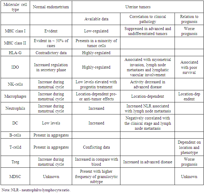
The immune system in the normal uterus serves a dual purpose. On the one hand, it plays a role in protecting against pathogens, and on the other hand, it is able to adapt to the immunosuppressive state in order to create a feto-maternal tolerance to a semi-allogeneic fetus. These individual functions include the complex interaction of the hormonal fluctuations of the menstrual cycle and the immune system. Normal endometrium is naturally under strict hormonal control. He is under constant control of changes in estradiol and progesterone during the menstrual cycle. Both the innate and adaptive arm of the immune system are influenced by these hormonal changes. Several risk factors have been described for endometrial cancer, which may be linked with increased inflammation of the endometrial tissue, reviewed by Modugno et al. [5].
It has been shown that increased exposure of estrogen is associated with the development of endometrial cancer due to the mitogenic effect of estrogen [6-8]. Therefore, estrogen-related carcinogenesis may be associated with inflammatory events. Chronic inflammation has been associated with cancer [9]. Several pathways of inflammation are involved in carcinogenesis. Many of these pathways are initialized, among other things, by activation of STAT 3 or NF-κB [10].
Immune functions of normal and malignant endometrial cells
The endometrial epithelium serves as the primary line of defense against viruses and other pathogens entering the uterus. The epithelial cells form an integral part of the mucosal immune system. Along with the formation of a physical barrier, epithelial cells perform several direct functions associated with the immune system, one of which is the secretion of defensins [11]. Defensins form a part of the innate immune system, considering their immediate antimicrobial function and their ability to activate the adaptive immune system. For example, it has been shown that defensins attract T-cells and immature dendritic cells (DC) in response to binding to the C-C chemokine receptor of type 6 (CCR6) [12]. Other secreted molecules include macrophage inflammatory protein (MIP) 3α, also a ligand for CCR6, and secretory leukocyte protease inhibitor (SLPI) [13-14]. Contradictory, uterine epithelial cells secrete unidentified, soluble immune mediators that confer a tolerogenic phenotype to DC [15].
Obesity and diabetes have also been shown to be associated with increased release of pro-inflammatory molecules, such as IL-6, TNF-α, CRP, leptin and macrophage migration inhibitory factor [3, 16]. Two studies evaluating serum levels of IL-6, TNF-α and CRP and the risk of developing endometrial cancer showed that increased levels of CRP are associated with the risk of endometrial cancer [17-18]. Wang et al. found this correlation after correcting of BMI and age [18]. Friedenreich et al., in addition, found that the risk of CRP and endometrial cancer was associated with high BMI and serum IL-6, and the risk of endometrial cancer was associated with low BMI [17].
Indolamine 2,3-dioxygenase (IDO) which is responsible for suppressing T-cells by depriving T-cells of the essential nutrient tryptophan, is activated in the secretory and proliferative endometrium. The presence of the enzyme can fulfill a dual protective role: it acts as an antibacterial agent, while on the other hand it causes T-cell inhibition. The latter will create an immunosuppressive state to allow embryonal implantation [19]. IDO is also expressed by endometrial carcinoma cells [20–22], and it has been proven to be associated with myometrial invasion, lymph node metastases, and lymphatic vascular involvement [21]. In addition, the high expression of IDO correlated with a decrease in the involvement of CD8 + TIL and NK-cells and was associated with poor survival [20]. Thus, both in normal and malignant endometrium, IDO’s primary function seems to be induction of immunosuppression in order to allow embryonal implantation or tumor growth. Endometrial epithelial cells are also powerful antigen-presenting cells. Ferguson et al. found expression of MHC class I in endometrial glands as well as stromal cells and endothelial cells. MHC class II, on the contrary, was found to be expressed in the endometrial glands in approximately 50% of normal endometrium samples [23]. Fahey et al. have shown that cultured epithelial cells express CD40 and CD1d and that epithelial cells as well as stromal endometrial cells can elicit tetanus toxoid specific T-cell responses [24-25]. In endometrial tumors classic MHC of class I was suppressed in 48.5% of 520 tumors, which is associated with a worse prognosis of the disease [26]. In addition, the non-classical MHC class I molecule, HLA-G, was increased in 39.8% of samples, corroborating results of Barrier et al., who found expression of HLA-G in 55% of samples [27]. Despite the need for further study, the increased regulation of HLA-G molecules in endometrial tumors may be a protective mechanism to avoid NK-lysis of cells with reduced regulation of MHC class I molecules, as shown in other tumors [28-29]. MHC class II was found to be present in only a small portion of malignant endometrial cells. The low presence of both classical MHC I and II molecules and the activation of non-classical HLA-G indicate a weak antigen-presenting ability of endometrial tumor cells [30-31]. Cells in the underlying stroma, however, do show MHC II positivity [30]. Taken together, normal endometrial cells can present antigens in the context of MHC molecules, probably as a defense mechanism against pathogenic microorganisms. Endometrial tumor cells, however, inhibit the expression of MHC molecules to provide an immune output. Several members of the B7-H family have been described lately. We recently described the presence of these molecules both in normal endometrium and uterine tumors [22]. The expression of PD-L1 (B7-H1) and B7-H4 was detected in the vast majority of normal endometrium, while PD-L2 (B7-DK) was present in about half of the normal endometrium, albeit at low levels. All of these molecules were also present in endometrial cancer [22]. Comparing the expression levels of all molecules, no up-regulation in endometrial tumors was found. In spite of the fact that the studied population was rather small for a thorough analysis, there was a tendency to a decrease in survival in PD-L1 + tumors [22]. The results on B7-H4 are contradictory to a previously published study in which B7-H4 was reported to be significantly up-regulated in endometrial tumors [32,33]. The expression pattern of this molecule was mainly cytoplasmic in combination with strong membrane staining and, as it has been shown, negatively correlates with the number of TIL, both the T-cell population as a whole (CD3 +) and the individual CTL population (CD8 +) [32]. For the latter, this correlation was also found for B7-H3 [34]. These data indicate that, since for most of these mediators no up-regulation was found in endometrial tumors, these molecules may also exert their immunosuppressive functions in both the normal and cancerous situation, as described above for IDO. Table 1 gives a concise overview of the immunological players in endometrium and endometrial tumors and their implication in tumor biology.
Table 1. Overview of immunological mediators for uterine tumors
     
Innate immune cells
Macrophages represent approximately 10% of the total cellular population of the endometrium. They are mostly present in the endometrial stroma and myometrial connective tissue [35]. Their frequency is the highest prior to menstruation, along with the frequency of neutrophils. The latter play a role in the breakdown of the endometrial tissue at menstruation as well as in the elevation of immune protection during the disruption of the endometrial epithelium protective barrier [36]. Macrophages play a paradoxical role in cancer, in a sense that they can have both a pro- and anti-tumorigenic function [37]. Tumor-associated macrophages (TAM) located in the focal necrotic center of the tumor and TAM at the tumor margin correlated with disease progression and with clinic-pathological features of the tumor [37-38]. TAM at the tumor margin were associated with the formation of lymph node metastases indicating tumor progression, whereas macrophages in the tumor nest, the bulky area of the tumor surrounding the tumor center, were associated with a better relapse-free survival. This may be explained by local factors within the tumor, exerting different functions on macrophages. The tumor center, for example, is known to be hypoxic. This is suggested to trigger the angiogenic capacities of macrophages, leading to renewed oxygen supply and tumor progression [37]. Another cell type, like macrophages derived from the myeloid lineage, are myeloid derived suppressor cells (MDSC). To date, to our knowledge, MDSC have only been described in endometrial cancer by Vanderstraeten A. et al. [22]. MDSC analysis was subdivided into the presence of monocytic MDSC (lin-HLA-DR-/loCD11b+CD14+) and granulocytic MDSC (lin-HLA-DR-/loCD11b+CD14-).
Both MDSC of the monocytic and granulocytic type were found, although the majority of the identified population was of the granulocytic type. This subtype has been described to have the strongest suppressive capacity compared to the monocytic subtype [39] providing evidence of increased immunosuppression in endometrial tumors.
The largest representative of the innate immune system however, is that of natural killer cells (NK-cells). Like for the cells described above, their numbers in normal endometrium vary depending on the phase of the cycle. The highest number of NK cells is found in the secretory phase of the cycle. At this point NK-cells represent about 70% of the total leukocyte population [35]. This is probably the result of both an increase in the level of IL-15 in the endometrium in the secretory phase, and an increase in the influx of NK-cells from peripheral blood [19]. However, the research by Manaster et al. showed that the percentage of NK cells remains relatively constant at approximately 30% of the total lymphocyte population [40]. Male et al found the presence of precursor NK-cells, so-called stage 3 NK-cells in uterine mucosa as well as mature, stage 4, NK-cells. [41]. The authors postulate that stage 3 NK-cells (CD34-CD117+CD94+) migrate into the uterus where they mature to obtain their distinct phenotype (CD34-CD117-/+CD94+) [41]. Uterine NK-cells are different from their counterparts in blood [42]. Like NK-cells in blood they express CD94, CD56 and CD9, but do not express CD16, CD8 or CD57. In addition, CD56 is expressed at about ten-fold higher levels in uterine NK-cells than in blood NK-cells [42]. It has been little described about the functional differences of peripheral blood NK-cells and uterine NK-cells. NK cells in both the proliferative and secretory phases of the endometrium can be inert cells that do not possess both their cytotoxic ability and the ability to secrete cytokines. However, it can be corrected when cells are cultured in the presence of IL-15 [40]. Stimulation with IL-15 resulted in the up-regulation of the activating NK-cell receptors NKp30 and NKp44, but no difference was found in expression of NKp46 and NKG2D. In addition, IL-15 activated endometrial NK-cells showed increased in vitro cytotoxic capability and secreted IP-10 (CXCL-10) and IFN-γ [40]. Uterine NK-cells are thus suggested to be inert lymphocytes without the cytotoxic capabilities of peripheral NK-cells. These NK-cells are inactive during the normal menstrual cycle and are suggested to mature to fully functional NK- cells during pregnancy [40]. There are only few studies focusing on NK-cells in endometrial carcinoma patients. NK-cell activity in peripheral blood against K562 cells was found to decrease with an increase in histological differentiation grade and myometrial invasion in early stage (stage I) endometrial carcinoma [43]. A study published by Timonen et al. in 1987 in 8 endometrial cancer patients and 1 endometrial stromal sarcoma patient showed that unstimulated peripheral blood lymphocytes had shown cytotoxic responses against autologous tumor and against HeLa cells in 7/9 patients [44]. This activity was increased upon addition of recombinant IL-2. The IL-2 activated lytic precursor cells belonging to the subpopulation of lymphocytes which includes NK-cells [44]. Ferguson et al. found that NK-cells were virtually absent in endometrial tumors [23].
Intratumoral NK-cells were analyzed immunohistochemically in endometrial carcinoma patients after progestin treatment [45].
The total cytotoxic (granzyme B+) lymphocyte population in the tumors increased in 6.5 times after treatment with progestin. While CD56 + NK cells were low or absent before treatment, the frequency of NK cells increased to 76% of the total population of cytotoxic (granzyme B +) cells in the endometrial foci, which showed signs of regression. In contrast, in the case of lesions of a stable or progressive nature, an increase in the number of NK-cells was not observed. CD8+ CTL showed a mild increase in regressing lesions, while they remained approximately constant in stable or progressive lesions. Thus, progestin treatment can attract NK-cells into uterine tumors which is associated with disease improvement. In addition, these findings can explain the increased level of NK-cells in the secretory phase of normal endometrium, when progesterone levels are the highest one.
Dendritic cells (DCs) have also been described in the human endometrium at relatively low levels compared with other immune cells [46]. Throughout the cycle, these cells are located in the functional and basal layers. The frequency of immature CD1a + DK increases during the cycle, while the mature CD83 + DK population remains relatively constant. It indicates that, according to their natural function, mature dendritic cells migrate from their resident tissue. HLA-DR + DK has been shown to be present in uterine tumors in both glandular cells and interstitial tissue [47].
The functional capacity of tumor-infiltrating DC, however, has been shown to be compromised in uterine tumors, due to significantly reduced expression of the CD86, CD80 and CD40 costimulatory molecules compared with DC in normal endometrium [48].
Adaptive immune cells
T and B cells, both members of the adaptive immune system, can also be detected in the normal endometrium. They are present in the uterine mucosa in the form of unique aggregates consisting of the nucleus of B-cells surrounded by T-cells. Besides, these structures are surrounded by a capsule of macrophages and monocytes [49]. These structures have been suggested to be similar to the mucosa-associated lymphoid tissue (MALT), which can be found in the gastrointestinal system [50]. The T-cells present in these aggregates are almost exclusively CD8+ CD45RO+, indicating that they are memory type effector cells [51]. These aggregates have been shown to increase in size from the proliferative phase, at this point without the B-cell nucleus, up to the secretory phase of the menstrual cycle. In addition, they are absent in the menopause, indicating that their expansion is hormone-driven [49]. This is further exemplified by the observation that T- cells within the aggregate express estrogen receptors [52].
In addition, Yeaman et al. showed that the accumulation of T-cells was the result of the T-cells migration towards the endometrium and not the proliferation of single resident T-cell clones [51]. The function of these aggregates is largely unknown. However, they may serve a purpose in both the creation of an immunosuppressive environment to allow feto-maternal tolerance on the one hand and on the other hand to create a protective environment against pathogens during menstruation when the epithelial barrier is disrupted. The former is exemplified by the observation that the cytotoxic T-lymphocytes in the proliferative phase of the cycle have cytotoxic capacity, while this function is severely dampened in the secretory phase during which a conception can occur [53]. Besides, CD8 + T-cells are still able to show their full cytotoxic function, which further indicates that during the secretory phase of the menstrual cycle, a temporary state of immunosuppression occurs for possible implantation of the embryo. The difference in cytotoxic capacity of T-cells during the different phases of the menstrual cycle is subject to hormonal control in order to maintain the balance between immune protection and tolerance [53]. The latter function of the lymphoid aggregates is supported by the location from which they originate. During the proliferative phase, the aggregates expand from inside of the basal stroma, the inner third of the endometrium that is not shed during menstruation. Consequently, the lymphoid aggregates may provide immune protection against pathogens during menstruation. Alternatively, the presence of these aggregates in the basal stroma may be a means to prevent loss of T- and B-cells during menstruation [49]. This type of lymphoid structures, recently termed tertiary lymphoid structures, resemble the MALT found in the gastrointestinal system, as it was mentioned above. These structures have been described in several tumor types, such as colorectal cancer, lung cancer, melanoma, ovarian cancer, renal cell cancer and breast cancer [54]. The co-localization of both T- and B-lymphocytes in these aggregates has been shown to correlate with improved patient survival [55]. MALT-like structures in uterine tumors have not been described to date, but tumor infiltrating lymphocytes (TIL) have been shown in different studies.
Chang et al. found that CD8+ (TIL) showed less expression of granzyme B and perforin than their blood counterparts, indicating possible functional defects or tumor-induced suppression [56]. However, in vitro activation of TIL resulted in adequate activation of TIL and induction to the same polarization profile as found in peripheral blood (i.e. main polarization to Th1 type cells). TIL have been associated with prognosis in endometrial cancer with contradictory reports. The prognostic value of this infiltrate depends on the location within the tumor. Increased numbers of TIL, of unspecified composition, at the invasive margin of the tumor (i.e. the tumor-myometrial junction) did not have a beneficial effect on patient survival according to a study by Silverberg et al. [57]. These results were contradicted by Kondratiev et al., who found that, although confirming the presence of CD8+ TIL at the tumor invasive margin, the presence of those TIL was associated with improved prognosis [58]. However, the latter study only considered CD8+ TIL at the invasive border while Silverberg et al. considered the total lymphocyte population which may clarify these different findings. Two additional investigations studied the total lymphocyte population at the invasive margin [59-60]. Deligdisch et al observed that the presence of an infiltrate consisting of lymphocytes and plasma cells, potentially indicating the described tertiary lymphoid structures, appeared to be related to low-grade endometrial tumors, and suggested that TIL are associated with a favorable prognosis [60]. A later study by Ambros et al. refuted this suggested association [59]. Intratumoral CD8+ TIL have been associated with improved disease-free survival in both type I and type II of endometrial cancer [61]. These intratumoral TIL were found more frequently in low grade tumors than in high grade tumors.
The presence of CD45RO+ T-cells, indicating memory T-cells was also shown. Moreover, the presence of memory T-cells was associated with increased overall survival and with reduced events of recurrence [61]. Chang et al. described that the majority of tumor infiltrating CD8+ T cells are CD28-CD45RA-CD45RO+ defining terminally differentiated T-cells. In addition, the T-cells appeared to be in an activated state, exemplified by the expression of CD69, CD103 and CD152 [56]. In the proximal tumor draining lymph nodes (TDLN), the CD4/CD8 ratio is increased [62]. In addition, Yamamoto et al. found that clonally expanded T-cells are absent from TDLN in patients with local endometrial tumors, while clonally expanded T-cells could be retrieved from TDLN and peripheral blood in patients suffering from metastatic cancer, supporting the role of immune responses to solid tumors [63]. This specific appearance of T-cell clones in TDLN of metastatic tumors may be a consequence of direct T-cell priming by (metastasized) tumor cells present in the TDLN in metastatic tumors. It leads to the expansion of T-cell clones in affected lymph nodes. This direct priming does not occur in unaffected lymph nodes, as is the case in early-stage disease [64]. The results of Yamamoto et al. expand earlier findings of Garzetti et al. [65] who did not find any clinical significance in the lymphocyte distribution in lymph nodes in patients with early stage disease. In addition, Garzetti et al. showed that myometrial invasion with or without lymphovascular space involvement was associated with increased CD16+ and CD56+ cells defining NK-cells in pelvic nodes [65]. Regulatory T-cells (Treg) have a natural function to suppress ongoing immune responses when they are no longer necessary. However, this suppressive property of these cells may also cause suppression of an antitumor immune response. Treg have been shown to be increased in peripheral blood of normal controls in the late follicular phase [66]. Analysis of the Treg frequency in the endometrium showed that Treg were present in only low frequencies in the endometrium and that the frequency was higher in the proliferative phase compared to the secretory phase [67]. Collectively, these data indicate that the frequency of Treg cells appears to increase during the proliferative phase and is reduced after ovulation.
Several studies have reported the presence of regulatory T-cells (Treg) in endometrial carcinoma.
Intratumoral CD4+CD25+ Treg expressing higher levels of FoxP3, CD103 and GITR are increased compared to peripheral blood [56]. In this particular study it was also shown that, like CTL, intratumoral Treg also expressed granzyme B indicating the capacity to lyse effector cells. However, Treg in stromal tissue were found to be significantly lower in tumor samples compared to normal endometrium [68]. Although lower, high Treg counts in tumor samples were shown to correlate with increased vascularity [68], tumor grade, stage, the extent of lymph node metastases and myometrial invasion [56] as well as worse disease-free survival [69]. The latter has also been shown to result from the presence of high Treg/CD8 and Treg/CD4 ratios [69]. In distal TDLN, the proportion of functional regulatory T-cells is increased [62].
Taken together, the fluctuations of the different cell types during the normal menstrual cycle are a further indication of the dual role the immune system plays in the uterus as it was described earlier. The immunosuppressive ability that certain types of cells, such as Treg, have within the framework of the feto-maternal tolerance also contributes to the salvation of immunity. Some cell types, such as macrophages and T-cells, appear to have different effects on the outcome of the tumor, depending on the location in which they are located. This probably indicates that in different places of the tumor or in the microenvironment of the tumor, the immune system can be influenced differently in such a way that the functionality of the immune cells affects either the anti-tumor or pro-tumor profile.

3. Conclusions

The data currently presented clearly show that the immune system is present and active in both normal endometrium and endometrial tumors. In the normal endometrium, the immune system plays a central role in protecting against pathogens and in providing tolerance to the feto-maternal line. Like this dual role in the healthy situation, it also has both a pro- and anti-tumorigenic function. In our opinion, the interplay between positive and negative players and mechanisms in tumor development and progression provides possible intervention options in the treatment of endometrial cancer, which deserves further attention in future research.

References

[1]  Amant F, Moerman P, Neven P, Timmerman D, Van Limbergen E, Vergote I. Endometrial cancer. Lancet. 2005; 366(9484): 491-505.
[2]  Wira CR, Fahey JV, Ghosh M, Patel MV, Hickey DK, Ochiel DO. Sex hormone regulation of innate immunity in the female reproductive tract: the role of epithelial cells in balancing reproductive potential with protection against sexually transmitted pathogens. American journal of reproductive immunology. 2010; 63(6): 544-65.
[3]  Visser M, Bouter LM, McQuillan GM, Wener MH, Harris TB. Elevated C-reactive protein levels in overweight and obese adults. JAMA: the journal of the American Medical Association. 1999; 282(22): 2131-5.
[4]  Pfister G, Savino W. Can the immune system still be efficient in the elderly? An immunological and immunoendocrine therapeutic perspective. Neuroimmunomodulation. 2008; 15(4-6): 351-64.
[5]  Modugno F, Ness RB, Chen C, Weiss NS. Inflammation and endometrial cancer: a hypothesis. Cancer epidemiology, biomarkers & prevention: a publication of the American Association for Cancer Research, cosponsored by the American Society of Preventive Oncology. 2005; 14(12): 2840-7.
[6]  Austin H, Austin JM, Jr., Partridge EE, Hatch KD, Shingleton HM. Endometrial cancer, obesity, and body fat distribution. Cancer research. 1991; 51(2): 568-72.
[7]  Potischman N, Hoover RN, Brinton LA, Siiteri P, Dorgan JF, Swanson CA, et al. Case-control study of endogenous steroid hormones and endometrial cancer. Journal of the National Cancer Institute. 1996; 88(16): 1127-35.
[8]  Zeleniuch-Jacquotte A, Akhmedkhanov A, Kato I, Koenig KL, Shore RE, Kim MY, et al. Postmenopausal endogenous oestrogens and risk of endometrial cancer: results of a prospective study. British journal of cancer. 2001; 84(7): 975-81.
[9]  Hanahan D, Weinberg RA. Hallmarks of cancer: the next generation. Cell. 2011; 144(5): 646-74.
[10]  Elinav E, Nowarski R, Thaiss CA, Hu B, Jin C, Flavell RA. Inflammation-induced cancer: crosstalk between tumours, immune cells and microorganisms. Nature reviews Cancer.
[11]  Wira CR, Grant-Tschudy KS, Crane-Godreau MA. Epithelial cells in the female reproductive tract: a central role as sentinels of immune protection. American journal of reproductive immunology. 2005; 53(2): 65-76.
[12]  Yang D, Chertov O, Bykovskaia SN, Chen Q, Buffo MJ, Shogan J, et al. Beta-defensins: linking innate and adaptive immunity through dendritic and T cell CCR6. Science. 1999; 286(5439): 525-8.
[13]  Fahey JV, Wira CR. Effect of menstrual status on antibacterial activity and secretory leukocyte protease inhibitor production by human uterine epithelial cells in culture. The Journal of infectious diseases. 2002; 185(11): 1606-13.
[14]  Fahey JV, Schaefer TM, Wira CR. Sex hormone modulation of human uterine epithelial cell immune responses. Integrative and comparative biology. 2006; 46(6): 1082-7. 2013; 13(11): 759-71.
[15]  Ochiel DO, Ghosh M, Fahey JV, Guyre PM, Wira CR. Human uterine epithelial cell secretions regulate dendritic cell differentiation and responses to TLR ligands. Journal of leukocyte biology. 2010; 88(3): 435-44.
[16]  Dandona P, Aljada A, Ghanim H, Mohanty P, Tripathy C, Hofmeyer D, et al. Increased plasma concentration of macrophage migration inhibitory factor (MIF) and MIF mRNA in mononuclear cells in the obese and the suppressive action of metformin. The Journal of clinical endocrinology and metabolism. 2004; 89(10): 5043-7.
[17]  Friedenreich CM, Langley AR, Speidel TP, Lau DC, Courneya KS, Csizmadi I, et al. Case-control study of inflammatory markers and the risk of endometrial cancer. European journal of cancer prevention: the official journal of the European Cancer Prevention Organisation. 2013; 22(4): 374-9.
[18]  Wang T, Rohan TE, Gunter MJ, Xue X, Wactawski-Wende J, Rajpathak SN, et al. A prospective study of inflammation markers and endometrial cancer risk in postmenopausal hormone nonusers. Cancer epidemiology, biomarkers & prevention: a publication of the American Association for Cancer Research, cosponsored by the American Society of Preventive Oncology. 2011; 20(5): 971-7.
[19]  Lobo SC, Huang ST, Germeyer A, Dosiou C, Vo KC, Tulac S, et al. The immune environment in human endometrium during the window of implantation. American journal of reproductive immunology. 2004; 52(4): 244-51.
[20]  de Jong RA, Kema IP, Boerma A, Boezen HM, van der Want JJ, Gooden MJ, et al. Prognostic role of indoleamine 2,3-dioxygenase in endometrial carcinoma. Gynecologic oncology. 2012; 126(3): 474-80.
[21]  Ino K, Yamamoto E, Shibata K, Kajiyama H, Yoshida N, Terauchi M, et al. Inverse correlation between tumoral indoleamine 2,3-dioxygenase expression and tumor-infiltrating lymphocytes in endometrial cancer: its association with disease progression and survival. Clinical cancer research: an official journal of the American Association for Cancer Research. 2008; 14(8): 2310-7.
[22]  Vanderstraeten A, Luyten C, Verbist G, Tuyaerts S, Amant F. Mapping the immunosuppressive environment in uterine tumors: implications for immunotherapy. Cancer immunology, immunotherapy: CII. 2014; 63(6): 545-57.
[23]  Ferguson A, Moore M, Fox H. Expression of MHC products and leucocyte differentiation antigens in gynaecological neoplasms: an immunohistological analysis of the tumour cells and infiltrating leucocytes. British journal of cancer. 1985; 52(4): 551-63.
[24]  Fahey JV, Wallace PK, Johnson K, Guyre PM, Wira CR. Antigen presentation by human uterine epithelial cells to autologous T cells. American journal of reproductive immunology. 2006; 55(1): 1-11.
[25]  Wallace PK, Yeaman GR, Johnson K, Collins JE, Guyre PM, Wira CR. MHC class II expression and antigen presentation by human endometrial cells. The Journal of steroid biochemistry and molecular biology. 2001; 76(1-5): 203-11.
[26]  Bijen CB, Bantema-Joppe EJ, de Jong RA, Leffers N, Mourits MJ, Eggink HF, et al. The prognostic role of classical and nonclassical MHC class I expression in endometrial cancer. International journal of cancer Journal international du cancer. 2010; 126(6): 1417-27.
[27]  Barrier BF, Kendall BS, Sharpe-Timms KL, Kost ER. Characterization of human leukocyte antigen-G (HLA-G) expression in endometrial adenocarcinoma. Gynecologic oncology. 2006; 103(1): 25-30.
[28]  Ibrahim EC, Guerra N, Lacombe MJ, Angevin E, Chouaib S, Carosella ED, et al. Tumor-specific up-regulation of the nonclassical class I HLA-G antigen expression in renal carcinoma. Cancer research. 2001; 61(18): 6838-45.
[29]  Paul P, Rouas-Freiss N, Khalil-Daher I, Moreau P, Riteau B, Le Gal FA, et al. HLA-G expression in melanoma: a way for tumor cells to escape from immunosurveillance. Proceedings of the National Academy of Sciences of the United States of America. 1998; 95(8): 4510-5.
[30]  Lazaris A, Chatzigianni EB, Xidias G, Panoskaltsis TA, Thomopoulou GC, Eftychiadis CA, et al. Tissue evaluation of immune markers in endometrial and cervical carcinomas. Journal of experimental & clinical cancer research: CR. 2004; 23(2): 269-75.
[31]  Tamiolakis D, Venizelos J, Lambropoulou M, Nikolaidou S, Tsikouras P, Jivannakis T, et al. Local immune response in serous papillary carcinoma of the endometrium. Histology and histopathology. 2005; 20(2): 403-8.
[32]  Miyatake T, Tringler B, Liu W, Liu SH, Papkoff J, Enomoto T, et al. B7-H4 (DD-O110) is overexpressed in high risk uterine endometrioid adenocarcinomas and inversely correlated with tumor T-cell infiltration. Gynecologic oncology. 2007; 106(1): 119-27.
[33]  Qian Y, Shen L, Cheng L, Wu Z, Yao H. B7-H4 expression in various tumors determined using a novel developed monoclonal antibody. Clinical and experimental medicine. 2011; 11(3): 163-70.
[34]  Brunner A, Hinterholzer S, Riss P, Heinze G, Brustmann H. Immunoexpression of B7-H3 in endometrial cancer: relation to tumor T-cell infiltration and prognosis. Gynecologic oncology. 2012; 124(1): 105-11.
[35]  Wira CR, Fahey JV, Sentman CL, Pioli PA, Shen L. Innate and adaptive immunity in female genital tract: cellular responses and interactions. Immunological reviews. 2005; 206: 306-35.
[36]  Hickey DK, Patel MV, Fahey JV, Wira CR. Innate and adaptive immunity at mucosal surfaces of the female reproductive tract: stratification and integration of immune protection against the transmission of sexually transmitted infections. Journal of reproductive immunology. 2011; 88(2): 185-94.
[37]  Ohno S, Ohno Y, Suzuki N, Kamei T, Koike K, Inagawa H, et al. Correlation of histological localization of tumor-associated macrophages with clinicopathological features in endometrial.
[38]  Tamiolakis D, Venizelos J, Lambropoulou M, Nikolaidou S, Tsikouras P, Jivannakis T, et al. Local immune response in serous papillary carcinoma of the endometrium. Histology and histopathology. 2005; 20(2): 403-8.
[39]  Miyatake T, Tringler B, Liu W, Liu SH, Papkoff J, Enomoto T, et al. B7-H4 (DD-O110) is overexpressed in high risk uterine endometrioid adenocarcinomas and inversely correlated with tumor T-cell infiltration. Gynecologic oncology. 2007; 106(1): 119-27.
[40]  Qian Y, Shen L, Cheng L, Wu Z, Yao H. B7-H4 expression in various tumors determined using a novel developed monoclonal antibody. Clinical and experimental medicine. 2011; 11(3): 163-70.
[41]  Brunner A, Hinterholzer S, Riss P, Heinze G, Brustmann H. Immunoexpression of B7-H3 in endometrial cancer: relation to tumor T-cell infiltration and prognosis. Gynecologic oncology. 2012; 124(1): 105-11.
[42]  Wira CR, Fahey JV, Sentman CL, Pioli PA, Shen L. Innate and adaptive immunity in female genital tract: cellular responses and interactions. Immunological reviews. 2005; 206: 306-35.
[43]  Hickey DK, Patel MV, Fahey JV, Wira CR. Innate and adaptive immunity at mucosal surfaces of the female reproductive tract: stratification and integration of immune protection against the transmission of sexually transmitted infections. Journal of reproductive immunology. 2011; 88(2): 185-94.
[44]  Ohno S, Ohno Y, Suzuki N, Kamei T, Koike K, Inagawa H, et al. Correlation of histological localization of tumor-associated macrophages with clinicopathological features in endometrial cancer. Anticancer research. 2004; 24(5C): 3335-42.
[45]  Soeda S, Nakamura N, Ozeki T, Nishiyama H, Hojo H, Yamada H, et al. Tumor-associated macrophages correlate with vascular space invasion and myometrial invasion in endometrial carcinoma. Gynecologic oncology. 2008; 109(1): 122-8.
[46]  Raber PL, Thevenot P, Sierra R, Wyczechowska D, Halle D, Ramirez ME, et al. Subpopulations of Myeloid-Derived Suppressor Cells (MDSC) impair T cell responses through independent nitric oxide-related pathways. International journal of cancer Journal international du cancer. 2013.
[47]  Manaster I, Mizrahi S, Goldman-Wohl D, Sela HY, Stern-Ginossar N, Lankry D, et al. Endometrial NK cells are special immature cells that await pregnancy. Journal of immunology. 2008; 181(3): 1869-76.
[48]  Male V, Hughes T, McClory S, Colucci F, Caligiuri MA, Moffett A. Immature NK cells, capable of producing IL-22, are present in human uterine mucosa. Journal of immunology. 2010; 185(7): 3913-8.
[49]  Yang Z, Kong B, Mosser DM, Zhang X. TLRs, macrophages, and NK cells: our understandings of their functions in uterus and ovary. International immunopharmacology. 2011; 11(10): 1442-50.
[50]  Garzetti GG, Ciavattini A, Goteri G, Tranquilli AL, Muzzioli M, Fabris N, et al. Natural killer cell activity in stage I endometrial carcinoma: correlation with nuclear grading, myometrial invasion, and immunoreactivity of proliferating cell nuclear antigen. Gynecologic oncology. 1994; 55(1): 111-4. cancer. Anticancer research. 2004; 24(5C): 3335-42.
[51]  Timonen T, Lehtovirta P, Saksela E. Interleukin-2-stimulated natural killer activity against malignant and benign endometrium. International journal of cancer Journal international du cancer. 1987; 40(4): 479-83.
[52]  Witkiewicz AK, McConnell T, Potoczek M, Emmons RV, Kurman RJ. Increased natural killer cells and decreased regulatory T cells are seen in complex atypical endometrial hyperplasia and welldifferentiated carcinoma treated with progestins. Human pathology. 2010; 41(1): 26-32.
[53]  Schulke L, Manconi F, Markham R, Fraser IS. Endometrial dendritic cell populations during the normal menstrual cycle. Human reproduction. 2008; 23(7): 1574-80.
[54]  Lijun Z, Xin Z, Danhua S, Xiaoping L, Jianliu W, Huilan W, et al. Tumor-infiltrating dendritic cells may be used as clinicopathologic prognostic factors in endometrial carcinoma. International journal of gynecological cancer: official journal of the International Gynecological Cancer Society. 2012; 22(5): 836-41.
[55]  Jia J, Wang Z, Li X, Wang Z, Wang X. Morphological characteristics and co-stimulatory molecule (CD80, CD86, CD40) expression in tumor infiltrating dendritic cells in human endometrioid adenocarcinoma. European journal of obstetrics, gynecology, and reproductive biology. 2012; 160(2): 223-7.
[56]  Yeaman GR, Guyre PM, Fanger MW, Collins JE, White HD, Rathbun W, et al. Unique CD8+ T cell-rich lymphoid aggregates in human uterine endometrium. Journal of leukocyte biology. 1997; 61(4): 427-35.
[57]  Marshall RJ, Jones DB. An immunohistochemical study of lymphoid tissue in human endometrium. International journal of gynecological pathology: official journal of the International Society of Gynecological Pathologists. 1988; 7(3): 225-35.
[58]  Yeaman GR, Collins JE, Fanger MW, Wira CR, Lydyard PM. CD8+ T cells in human uterine endometrial lymphoid aggregates: evidence for accumulation of cells by trafficking. Immunology. 2001; 102(4): 434-40.
[59]  Tabibzadeh SS, Satyaswaroop PG. Sex steroid receptors in lymphoid cells of human endometrium. American journal of clinical pathology. 1989; 91(6): 656-63.
[60]  White HD, Crassi KM, Givan AL, Stern JE, Gonzalez JL, Memoli VA, et al. CD3+ CD8+ CTL activity within the human female reproductive tract: influence of stage of the menstrual cycle and menopause. Journal of immunology. 1997; 158(6): 3017-27.
[61]  Goc J, Fridman WH, Sautes-Fridman C, Dieu-Nosjean MC. Characteristics of tertiary lymphoid structures in primary cancers. Oncoimmunology. 2013; 2(12): e26836.
[62]  Nielsen JS, Nelson BH. Tumor-infiltrating B cells and T cells: Working together to promote patient survival. Oncoimmunology. 2012; 1(9): 1623-5.
[63]  Chang WC, Li CH, Huang SC, Chang DY, Chou LY, Sheu BC. Clinical significance of regulatory T cells and CD8+ effector populations in patients with human endometrial carcinoma. Cancer. 2010; 116(24): 5777-88.
[64]  Silverberg SG, Sasano N, Yajima A. Endometrial carcinoma in Miyagi Prefecture, Japan: histopathologic analysis of a cancer registry-based series and comparison with cases in American women. Cancer. 1982; 49(7): 1504-10.
[65]  Kondratiev S, Sabo E, Yakirevich E, Lavie O, Resnick MB. Intratumoral CD8+ T lymphocytes as a prognostic factor of survival in endometrial carcinoma. Clinical cancer research : an official journal of the American Association for Cancer Research. 2004; 10(13): 4450-6.
[66]  Ambros RA, Kurman RJ. Combined assessment of vascular and myometrial invasion as a model to predict prognosis in stage I endometrioid adenocarcinoma of the uterine corpus. Cancer. 1992; 69(6): 1424-31.
[67]  Deligdisch L. Morphologic correlates of host response in endometrial carcinoma. American journal of reproductive immunology. 1982; 2(1):54-7.
[68]  De Jong RA, Leffers N, Boezen HM, ten Hoor KA, van der Zee AG, Hollema H, et al. Presence of tumor-infiltrating lymphocytes is an independent prognostic factor in type I and II endometrial cancer. Gynecologic oncology. 2009; 114(1): 105-10.
[69]  Fattorossi A, Battaglia A, Ferrandina G, Buzzonetti A, Legge F, Salutari V, et al. Lymphocyte composition of tumor draining lymph nodes from cervical and endometrial cancer patients. Gynecologic oncology. 2004; 92(1): 106-15.