American Journal of Medicine and Medical Sciences

p-ISSN: 2165-901X    e-ISSN: 2165-9036

2019;  9(0): 361-364

doi:10.5923/j.ajmms.20190910.02


Study of the Gene-Geographical Profile of the Population among Donors in Tashkent on the Antigenic Composition of Erythrocytes

Gulnoz Khabibjonovna Kayumova

Department of Extracorporeal Blood Purification, Scientific Research Institute of Hematology and Blood Transfusion of the Ministry of Health of the Republic of Uzbekistan, Tashkent, Uzbekistan

Correspondence to: Gulnoz Khabibjonovna Kayumova, Department of Extracorporeal Blood Purification, Scientific Research Institute of Hematology and Blood Transfusion of the Ministry of Health of the Republic of Uzbekistan, Tashkent, Uzbekistan.

Email:

Copyright © 2019 The Author(s). Published by Scientific & Academic Publishing.

This work is licensed under the Creative Commons Attribution International License (CC BY).
http://creativecommons.org/licenses/by/4.0/

Abstract

The purpose of the study was to study the genogeographic profile of the population on the antigenic composition of erythrocytes. In the blood samples of donors in the city of Tashkent, diagnostics of allogeneic immunization of the population and the determination of anti-erythrocyte antibodies were carried out. It has been established that the distribution by clinical important antigens of the Rh-Hr system among the donors of the city of Tashkent, which corresponded to similar indicators among Europeans. It is necessary to immunophenotype donors and recipients not only for antigens A, B and D, but also for antigens C, C, E, e.

Keywords: Genogeographic profile, Erythrocyte antigens, Anti-erythrocyte antibodies, Allogeneic immunization, "Rh-Hr" system

Cite this paper: Gulnoz Khabibjonovna Kayumova, Study of the Gene-Geographical Profile of the Population among Donors in Tashkent on the Antigenic Composition of Erythrocytes, American Journal of Medicine and Medical Sciences, Vol. 9 No. 0, 2019, pp. 361-364. doi: 10.5923/j.ajmms.20190910.02.

1. Introduction

Allogeneic immunization resulting from the transfusion of erythrocytes carrying transfusion dangerous antigens on their surface are the main cause of posttransfusion disease (PTD) and hemolytic disease of the newborn (HDN) [17]. Erroneous conclusion about the presence or absence of antigens and antibodies in donors and recipients can lead to the development of post-transfusion disease (PTD) of hemolytic type [1-3,8,9,14,15]. Maternal antibodies, which easily cross the placental barrier and enter the fetal bloodstream, are of key importance in the development of hemolytic disease of the newborn (HDN). The degree of development of fetal erythrocyte hemolysis depends on the specificity of antibodies. Despite the achievements in perinatal diagnostics, in particular, in prevention and treatment, it is not possible to completely prevent morbidity and mortality of children from hemolytic disease. Immunohematological studies in the mother help to predict the possibility of development and severity of hemolytic disease, to optimize the hemotransfusion therapy of newborns, as well as to carry out the selection of blood components for women [2,3,8,18,19,28].
Scientific researchers know about 38 group antigenic systems, including more than 500 erythrocyte antigens that can be identified using specific antisera. The combination of group antigens individually for each person [12]. The frequency of blood groups among representatives of different races and ethnic groups varies, which is believed to be a consequence of the genogeographic adaptation to one or another ecosystem in the process of evolution [6,7,10,21,28].
In our republic, only 6 antigens are determined by two systems - ABO and Rhesus, and screening of immune antibodies is carried out in blood service institutions and health care facilities (MPI), but the absence of a "panel of standard erythrocytes" does not allow to determine the specificity of detected antibodies [25].
According to the literature data (S.I. Donskov, T.V. Gaponova, 2013) such an approach to determining blood groups can ensure the safety of blood transfusion by no more than 80% [4,5,11]. Other antigens, such as E, e, C, K, K, CW, etc., which are not associated with the danger of transfusions, are not determined by the blood service. However, according to WHO recommendations (2005), blood transfusion should be carried out after determining at least 10 transfusion-dangerous antigens.

2. Main Body

2.1. The Purpose of Our Research

Study of the genogeographic profile of the population on the antigenic composition of erythrocytes.

2.2. Material and Methods of Study

This study involved 320 conditionally healthy donors surveyed on the basis of the Republican Center for Blood Transfusion and on the basis of the clinical diagnostic department of the Scientific-Research Institute of Hematology and Blood Transfusion of the MH RUz.
The material for the study was whole blood, red blood cell mass, plasma, blood serum from donors in Tashkent.
The following research methods were used: isoserological methods, using monoclonal reagents of different specificity, determination of anti-erythrocyte antibodies using antiglobulin serum and conglutination method, using 33% polyglucin solution.
The method of diagnosing alloimmunization of the population was carried out using monoclonal reagents of different specificity and the determination of anti-erythrocyte antibodies using antiglobulin serum and conglutination using a 33% polyglucin solution. Transfusion erythrocyte erythrocyte antigens from the Rhesus, Kell and Kidd systems were determined using monoclonal antibodies, which are produced by in vitro hybridoma cell lines [16,20].
Statistical data processing was performed using Microsoft Excel 2007.

2.3. Results of the Study

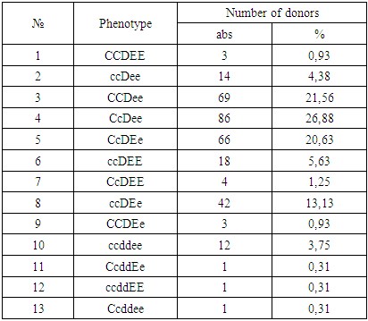
A donor test for the presence of incomplete immune antibodies using a 33% polyglucin and in an indirect Coombs test showed the following results: of 7878 antibody donors were detected in 19 serum samples. The titer of incomplete forms of immune antibodies ranged from 1: 2 to 1: 128.
Serum samples that yielded positive results in the determination of antibodies can contain both “pure” anti-D antibodies and simultaneously antibodies to several antigens: anti-C + D + E and others.
To determine the specificity of antibodies, a special study of serum with a set of standard erythrocytes, including the rare ccDee, Ccddee, ccddEe, ccddee groups, including both containing and not containing Kell (K) and Duffy (Fy) antigens, is necessary.
To determine the specificity of the antibody was carried out to develop a panel of standard erythrocytes. A total of 21 samples of erythrocyte O (I) blood group donors were selected and immunotyped (of which 20 are Rh-positive, 1 is Rh-negative, 1 is Kell-positive, 1 is CW-positive). Compiled electronic file of typed donors in alphabetical order. The results of the distribution of phenotypes of the "Rh-Hr" system among the donors of the city of Tashkent are given in the following table 1.
Table 1. Frequencies of distribution of phenotypes of the Rh-Hr system among donors in Tashkent
     
The results of the analysis showed that the most common among donors in Tashkent is the CcDee phenotype. The frequency of occurrence of the Rhesus antigens detected by us was as follows: C-71.56%, s-76.25%, E-43.13%, e-91.56%. In general, the prevalence rates of the above antigens were close to those of the Europeans, who according to Umnova M. (1989) data were: C-70%, E-30%, c-85%, e-97%. The combination of group antigens is individual for each person, and the frequency of occurrence of blood groups among representatives of different races and ethnic groups is not the same, which is a consequence of the genogeographic adaptation to one or another ecosystem during the evolution process.
The rhesus system is one of the most polymorphic antigenic systems of human erythrocytes, includes about 50 serological distinguishable antigens, not counting weak, transitional and partial forms. Being structural proteins of the erythrocyte membrane, the antigens of the "Rh-Hr" system are involved in the metabolic processes of the cell and the gas transport function of the body, and their absence may be accompanied by an irregular form of erythrocytes and an increased tendency of the body to hemolysis and anemia.
The formation of Rh antigens is encoded by three pairs of allelic genes D-d, E-e, C-c, while it should be noted that the existence of antigen d, the antithetic partner of antigen D, is supposed to be empirically.
Rhesus erythrocyte affiliation is determined by the presence in the phenotype of the Rho (D) antigen: people whose erythrocytes do not contain the antigen D are considered Rh-negative.
A different approach is used in assessing the Rhesus accessories of donors: the donor is considered Rh negative only if there are no D, E, C antigens on its erythrocytes; if the donor contains one of the three listed antigens, it is counted as a Rh-positive donor.
This separation of donors eliminates the possibility of sensitization of the recipient during transfusion of blood components and thereby reduces the risk of post-transfusion complications. Antigens C and E are rarely present separately.
Persons having only antigen C are found in 2.14%; antigen E - 0.27%; the combination of C and E in 0.08% of cases. In more than 95% of cases, these antigens are presented in combination with antigen D.
In addition to well-defined antigens, the "rhesus" system includes options under which the antigens are expressed weakly, or not at all are produced.
Du antigen, which is a variant of antigen D (the differences between antigens D and Du are quantitative, not qualitative), has the greatest clinical significance and is found in 1.5% of cases among positive individuals. Du is immunogenic for Rh-negative individuals, so donors who have Du identified are classified as Rh-positive, and recipients as Rh-negative.
If clinically significant Rhesus antigens are taken into account when transfusing erythrocyte-containing media, the risk of post-transfusion complications can be reduced by 98%. Current safety requirements for blood transfusions include the use of components identical or compatible with the main antigens.
The obtained information will help the attending physicians to orient in the selection of compatible donor-recipient pairs, reduce post-transfusion complications, and blood service specialists to regulate the production of the necessary blood-transfusion components and improve the blood immunohematological safety.
At present, in the republic, the group identity of a person is determined by the “ABO” system, denoting 0 (I), A (II), B (III), AB (IV) and Rh-Rh + (Rh-positive) and Rh- (rhesus negative). At the same time, the modern transfusiological doctrine declares identical blood transfusions for ABO, Rh-Hr and K. This means that erythrocytes transfused to the recipient should not contain ABO-system factors and Rhesus system factors (D, C, E, C, e) absent from the recipient [25].
At the same time, the erythrocytes of the donor should not contain antigen K.
The international standard for blood transfusion provides for a registry of typed donors in the blood service institutions, and a registry of typed patients in hospitals, but unfortunately, it has not yet been introduced into the practice of hospitals in the country.
An analysis of post-transfusion complications in the country recently confirms this statistics, and indicates the need for in-depth iso-serological studies on transfusion-dangerous erythrocyte minor antigens [20,23,24,26,27]. The solution of this technical problem will ensure the prevention of possible post-transfusion complications and disability associated with the transfusion of incompatible blood components and the early diagnosis and prevention of hemolytic disease of the newborn.

3. Conclusions

Among the donors in Tashkent, the distribution of the clinically important antigens of the Rh-Hr system (except for antigen D) corresponds to similar indicators in Europeans.
To prevent the development of post-transfusion disease, it is necessary to immunophenotype donors and recipients not only for antigens A, B and D, but also for antigens C, C, E, E.
The obtained data will help the attending physicians to orient in the selection of compatible donor-recipient pairs, reduce the risk of post-transfusion disease (PTD), and blood service specialists to regulate the production of the necessary blood-transfusion components and improve the blood immunohematological safety.

ACKNOWLEDGEMENTS

The author thanks the leadership of the Republican Center for Blood Transfusion of the MH RUz and personally to the Director Alonur Bakhtinurovich Saidov for assisting in the organization of the selection of the studied group of donors.
The author also thanks the head of the Clinical Diagnostic Department of the Research Institute of Hematology and Blood Transfusion of the MH RUz Nigora Akhrarova for their assistance in organizing the implementation of laboratory tests.

References

[1]  Bahramov S.M., Sabirov D.M., Donskov S.I. Transfusion medicine. - Tashkent, 2013. - 512s.
[2]  Gadylshina R.S., Belskaya E.E. Cases of alloimmunization with Rh-antigen D in patients with an uncomplicated transfusion history // Kazan Medical Journal, 2012. Volume 93, No.2, P.347-348.
[3]  Gerasimova N. D., Karavayev A. V. Risk of post-transfusion complications // Herald of the Blood Service of Russia. - 2010. - № 2. - p. 6-12.
[4]  Donskov S. I., Afonin N. I. Appeal to the immuno-serologists of the Blood Service of Russia. Bulletin of the Blood Service of Russia, 2006, No. 3, pp. 3-4.
[5]  Donskov S.I., Morokov V.A. Immuno-serologists of the Blood Service of Russia, conducting population studies // Herald of the Blood Service of Russia, 2008, No. 1, p. 9-11.
[6]  Donskov S.I., Morokov V.A., Dubinkin I.V. Group of human blood: a guide to immuno-serology. - M., 2011. - 1016 p.
[7]  Donskov S.I. On the origin of anti-erythrocyte antibodies and their ability to prevent the sensitization of puerperas // Herald of the blood service of Russia. 2010. - № 3. - P. 21–26.
[8]  Krobinets I., Chechetkin A., Slotina N. Features of allosensitization to erythrocyte antigens from donors in the Arctic // Transfusiology - 2011. T 12, - P. 204-205.
[9]  Lipatov I.S. Alloimmunization by group antigens of erythrocytes (individual and population features) // Abstract of dissertation candidate of medical sciences, Moscow, 2009. 23 p.
[10]  Mineeva N.V. Human blood groups (basics of immunohematology). - SPb., 2004. - p. 94-99.
[11]  Ryzhkova TV, Sofonova I.S., Kasyanov A.D., Chechetkin A.V. Estimation of the frequency of alloimmunization in blood component donors // Med. Immunology. - 2007. –T.9, No. 2-3. - p. 34.
[12]  Suvorov A.V. Nikitin E.N., Grakhova V.N., Udaltsova E.V., Penkina G.G. Distribution of homozygous phenotypes of erythrocyte antigens in the population // Transfusiology. - 2007. - V. 8, № 1-2. - p. 52.
[13]  Feofanova A.V., Volkova O.Ya. Study of the frequency of allosensitization to erythrocyte antigens of donors and cancer patients // Bulletin of the Blood Service of Russia. - 2010. - № 2. - P. 35–41.
[14]  Bhatti FA, Salamat N, Nadeem A, Shabbir N. Red cell alloimmunization in beta thalassemia major. J Coll Physicians Surg Pak. 2004. – Vol. 14, - P. 657–660.
[15]  Chao YH, Wu KH, Lu JJ, Shih MC, Peng CT, Chang CW. Red blood cell alloimmunisation among Chinese patients with β-thalassaemia major in Taiwan. Blood Transfus. 2013. – Vol. 11, - P. 71–74.
[16]  Chaudhary R, Agarwal N. Safety of type and screen method compared to conventional antiglobulin crossmatch procedures for compatibility testing in Indian setting. Asian J Transfus Sci. 2011. – Vol. 5, - P. 157–159.
[17]  Danieles G.L., Fletcher A., Garratty G., Henry S. et al. Blood group terminology 2004: from the International Society of Blood Transfusion committee on terminology for red cellsurface antigens // Vox Sang. – 2004. – Vol.87, № 4. – Р. 304-316.
[18]  Hoeltge GA, Domen RE, Rybicki LA, Schaffer PA. Multiple red cell transfusions and alloimmunization. Experience with 6996 antibodies detected in a total of 159,262 patients from 1985 to 1993. Arch Pathol Lab Med. 1995. – Vol. 119, - P. 42–45.
[19]  Ko KH, Yoo BH, Kim KM, Lee WY, Yon JH, Hong KH, et al. Frequency of unexpected antibody and consideration during transfusion. Korean J Anesthesiol. 2012. – Vol. 62, - P. 412–417.
[20]  Luzo ACM, Pereira FB, de Oliveira RC, Azevedo PR, Cunha Rd, Leonardi MI, et al. Red blood cell antigen alloimmunization in liver transplant recipients. Transplant P. 2010. – Vol. 42, - P. 494–495.
[21]  Makroo RN, Bhatia A, Gupta R, Phillip J. Prevalence of Rh, Duffy, Kell, Kidd and MNSs blood group antigens in the Indian blood donor population. Indian J Med Res. 2013. – Vol. 137, - P. 521–526.
[22]  Murao M, Viana MB. Risk factors for alloimmunization by patients with sickle cell disease. Braz J Med Bio Res. 2005. – Vol. 38, - P. 675–682.
[23]  Olujohungbe A, Hambleton I, Stephens L, Serjeant B, Serjeant G. Red cell antibodies in patients with homozygous sickle cell disease: a comparison of patients in Jamaica and the United Kingdom. Brit J Haematol. 2001. – Vol. 113, - P. 661–665.
[24]  Pahuja S, Pujani M, Gupta SK, Chandra J, Jain M. Alloimmunization and red cell autoimmunization in multitransfused thalassemics of Indian origin. Hematology. 2010. – Vol. 15, - P.174–177.
[25]  Poole J, Daniels G. Blood group antibodies and their significance in transfusion medicine. Transfus Med Rev. 2007. – Vol. 21, - P. 58–71.
[26]  Singer ST, Wu V, Mignacca R, Kuypers FA, Morel P, Vichinsky EP. Alloimmunization and erythrocyte autoimmunization in transfusion-dependent thalassemia patients of predominantly Asian descent. Blood. 2000. – Vol. 96, - P. 3369–3373.
[27]  Thakral B, Saluja K, Sharma RR, Marwaha N. Red cell alloimmunization in a transfused patient population: a study from a tertiary care hospital in north India. Hematology. 2008. – Vol. 13, - P. 313–318.
[28]  Tormey CA, Fisk J, Stack G. Red blood cell alloantibody frequency, specificity, and properties in a population of male military veterans. Transfusion. 2008/ - Vol. 48, - P. 2069–2076.