-
Paper Information
- Next Paper
- Previous Paper
- Paper Submission
-
Journal Information
- About This Journal
- Editorial Board
- Current Issue
- Archive
- Author Guidelines
- Contact Us
American Journal of Medicine and Medical Sciences
p-ISSN: 2165-901X e-ISSN: 2165-9036
2019; 9(8): 298-301
doi:10.5923/j.ajmms.20190908.05

Morphological Estimation of LED Impact to the Oral Mucosa at the Treatment of Prosthetic Stomatitis
 Abstract
 Reference
 Full-Text PDF
 Full-text HTML
Baibekov I. M.1, 2, Irkhanov M. M.1
1Republican Specialized Scientific and Practical Medical Center of Surgery named after Academician V.Vakhidov, Tashkent, Uzbekistan
2Tashkent State Institute of Stomatology, Tashkent, Uzbekistan
Copyright © 2019 The Author(s). Published by Scientific & Academic Publishing.
This work is licensed under the Creative Commons Attribution International License (CC BY).
http://creativecommons.org/licenses/by/4.0/

The effect of LED radiation of the oral mucosa at its use in the complex treatment of prosthetic stomatitis was investigated with the use of morphology methods. It has been shown that there are pronounced changes in the mucous membrane of the oral cavity in prosthetic stomatitis which expressed in inflammatory infiltration, predominantly by neutrophils and lymphocytes of the connective tissue base, by the appearance of edema zones and extravascular erythrocytes. It was revealed that the light emission of the Barva-Flex SIK device (λ-470 nm and λ-940) when used, reduces the inflammatory changes of the oral mucosa, reduces the relative volume of extravascular erythrocytes and zones of edema and has an evident stimulating effect on the healing of the oral mucosa erosions. LED radiation is promising for the use in the complex treatment of stomatitis.
Keywords: Prosthetic stomatitis, LED, Morphology
Cite this paper: Baibekov I. M., Irkhanov M. M., Morphological Estimation of LED Impact to the Oral Mucosa at the Treatment of Prosthetic Stomatitis, American Journal of Medicine and Medical Sciences, Vol. 9 No. 8, 2019, pp. 298-301. doi: 10.5923/j.ajmms.20190908.05.
1. Introduction
- LED - Light Emitting Diode (LED) is increasingly used in all fields of medicine. The first studies conducted under the auspices of NASA in the early 2000s showed a high efficiency of LED. Further researches and comparisons of LED with laser therapy have shown that LED, in terms of efficiency, is comparable with the action of low-intensity laser radiation [5-7,10]. However, there are few experimental studies on using LED in dentistry [1-3,5,7,10-12]. Although prosthetic stomatitis characterized by significant inflammatory changes of the oral mucosa with the appearance of ulcerations is quite common [2,4,8-9]. The efficiency of the use of LED in the treatment of prosthetic stomatitis has not been studied. Any inflammatory lesion of the oral mucosa (OM) causes the disorder of physiological and reparative regeneration processes, up to the appearance of ulcerative changes [4,8-9].LED, as well as laser radiation, is a powerful anti-inflammatory factor and stimulator of regenerative processes [1-3,5,7,10-12]. However, the effect of LED on the OM in these aspects has not been investigated, the possibilities of their use in the treatment of prosthetic stomatitis have not been studied.
2. Aim
- To study the effect of the LED matrix of Barva-Flex / SIC on OM inflammatory processes at prosthetic stomatitis.
3. Materials and Methods
- We studied the effect of LED - Barva - Flex / SIC (manufacturer “Center for Laser and Microwave Technologies” Joint-stock company of closed type, Kharkov, Ukraine) of blue and infrared range on OM in prosthetic stomatitis (PS) in 26 patients using light microscopy and morphometry. In 14 cases OM biopsy specimens outside of the ulcerative lesions and in 12 cases – from the ulcerative lesion zone were examined. The age of patients ranged from 45 to 70 years. Studies were conducted by taking oral or written consent of the patients. 10 volunteers with no OM and other parts of the digestive tract pathologies served as the control group. OM of patients with PS was irradiated daily for 3 min. during 7 days using the LED matrix Barva - Flex / SIC in the mode of maximum approaching to the OM surface. A thin polyethylene case was put on the matrix to prevent infection of the surface. The radiation power of each LED is 5 mW. OM biopsy specimens were examined 1 day after the last session. Tissue samples were fixed in a 10% phosphate buffer formalin solution for light microscopy. Paraffin sections were stained with hematoxylin and eosin. Stereomorphometric studies were performed according to G. G. Avtandilov’s method. This method has been modified and adapted for morphometry of objects from a computer monitor screen and for this purpose the transparent films were marked: for the study of discrete structures - points in the amount of 100 per each cell. Measurements were carried out when projecting a morphological object onto a screen [1].Statistical data processing was performed on a Pentium-IV computer using the BS-Statistica program, as well as the Microsoft Excel-Office and Microsoft Windows XP-Professional applied programs. All photomicrographs were processed and stored on a computer with the help of Microsoft- “Windows XP-Professional” applied programs.
4. Results and Discussion
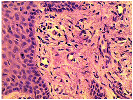
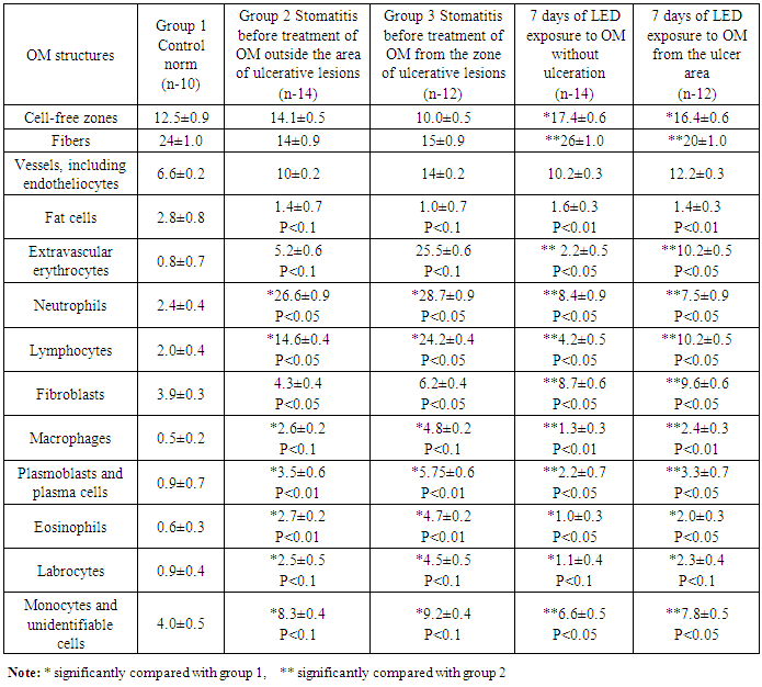
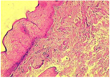
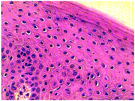
- There is an epithelial part and its own connective tissue layer in the OM separated by a basement membrane (Fig. 1).There is one row of basal cells on the basement membrane separating the epithelium from its own connective tissue layer. There are often mitosis figures here. A layer of prickly cells composed of 4-7 or more layers is the most developed. As a rule, the corneal layer is absent in most part of the OM (Fig.2).The basis of its own connective-tissued layer is loose connective tissue with a large number of fiber components, fibroblasts and other cells of the connective tissue, quite a lot of microvessels. (Fig. 1, Tab.1).
 | Figure 1. Oral mucosa, the control. HE staining 10x40 |
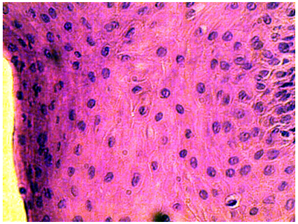 | Figure 2. Oral mucosa. Epithelial lining, control. HE staining 10x40 |
|
 | Figure 3. OM outside the area of the ulcerative defect. HE staining 10x40 |
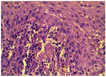
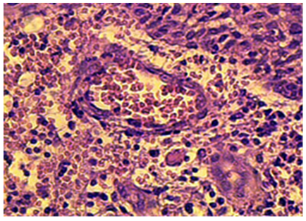 | Figure 4. OM at the ulcer. Pathological forms of erythrocytes in the lumen of vessels and outside of them. HE staining 10x40 |
 | Figure 5. OM after the course of LED exposure. HE staining 10x10 |
 | Figure 6. OM epithelium after a course of LED exposure. HE staining 10x10 |
5. Conclusions
- Prosthetic stomatitis is one of the most common lesions of the oral cavity. This pathology is more common in persons of mature age due to the use of prostheses in this age group. Prosthetic stomatitis is an inflammatory change in various parts of OM. Studying the effect of LEDs on wound healing and the course of various pathological processes has shown their evident properties to exert an anti-inflammatory effect and stimulate recovery processes [1-2,4,10,12,14-16]. This allows us to recommend LED as a means of stimulating OM reparative processes and normalizing the microbiocenosis of the oral cavity during prosthetic stomatitis.