American Journal of Medicine and Medical Sciences

p-ISSN: 2165-901X    e-ISSN: 2165-9036

2019;  9(8): 293-297

doi:10.5923/j.ajmms.20190908.04


Prevalence of Acromegaly in the Republic of Uzbekistan

Zamira Y. Khalimova, Adliya O. Kholikova, Umida A. Mirsaidova, Dildora H. Abidova

Department of Neuroendocrinology with Pituitary Neurosurgery, Center for the Scientific and Clinical Study of Endocrinology, Tashkent, Uzbekistan

Correspondence to: Adliya O. Kholikova, Department of Neuroendocrinology with Pituitary Neurosurgery, Center for the Scientific and Clinical Study of Endocrinology, Tashkent, Uzbekistan.

Email:

Copyright © 2019 The Author(s). Published by Scientific & Academic Publishing.

This work is licensed under the Creative Commons Attribution International License (CC BY).
http://creativecommons.org/licenses/by/4.0/

Abstract

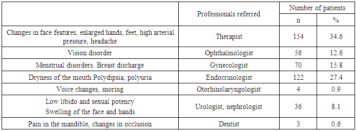
Acromegaly is a rare disease requiring extensive examination of population to get reliable epidemiological data which are of high value for adverse outcomes of the disease to be reduced, as well as for improvement of early diagnosis and results of treatment. The assessment of acromegaly prevalence in the Republic of Uzbekistan as per the National Registry has been performed. The data on 442 patients with the pituitary somatotrophic adenomas entered the Registry database from January 1, 2007 to January 2018. All patients were examined at the local level for the Registry Card to be filled in. The patients’ age varied from 18 to 75 years, mean age was 41 ± 21 years. The findings from the study demonstrated that acromegaly prevalence in the Republic of Uzbekistan is 1.4 cases per 100 000 of population with uneven distribution in the regions of the Republic. High prevalence of the pituitary somatotrophic adenomas was registered in Tashkent-city (2.5) and Namangan region (2.2) with the predominance among women. Late diagnosis of the disease delayed by 6-10 years with the peak at 40-49 years of age for men and at 50-59 years of age for women. Analysis of provoking factors demonstrated that manifestation of the disease was preceded by neuroviral infection (14.6%), psychological traumas and stresses (18.6%), pregnancy (10.1%) and craniocerebral injuries (15.4%). First health-seeking was found delayed corresponding to the height of the disease to be evidenced by the analysis of clinical signs of the disease’s manifestation. The vast majority of the patients were found to seek for help with specialists of other profiles, such as, neuropathologists, therapists and cardiologists; while 43.2% of patients visited endocrinologists, and this is thought to cause high prevalence of the disease and its early complications.

Keywords: Acromegaly, Epidemiology, Risk factors, Clinical features

Cite this paper: Zamira Y. Khalimova, Adliya O. Kholikova, Umida A. Mirsaidova, Dildora H. Abidova, Prevalence of Acromegaly in the Republic of Uzbekistan, American Journal of Medicine and Medical Sciences, Vol. 9 No. 8, 2019, pp. 293-297. doi: 10.5923/j.ajmms.20190908.04.

1. Introduction

Acromegaly is a rare disease characterized with growth hormone (GH) excess and increased insulin-like growth factor 1 (IGF-1) mostly associated with a pituitary adenoma. The disease results in early invalidization and higher mortality among the acromegalics which is 2-4 times higher than the one in the general population [1,2]. 60% and 25% of patients die due to cardiovascular and respiratory disorders, 15% because of malignancies [1,3,4,5]. GH levels < 5mU/l (< 2.5 μg/l) facilitate rapid improvement of acromegaly [4,6]. Maintaining GH and/or IGF-1 normal for the age was found to associate with the reduction in mortality rates regardless of concurrent complications [1,7,8].
In 1980-2001 acromegaly incidence was reported to range from 3.8 to 6.9 cases per 100 000 of population; the prevalence was from 0.28 to 0.4 cases per 100 000 of population [9-14]. Recently, in a number of reports from various geographical regions with various levels of public health care systems information on acromegaly incidence and prevalence (0.2-1.1 and 2.8-13.7 cases per 100 000 of population/year, respectively) was provided [15,16]. The epidemiological worldwide data vary. Thus, according to Daly et al., acromegaly prevalence in Belgium is 12.5 cases per 100 000 of population [17]; Tjörnstrand et al. report on 3.3 cases per 100 000 population in Sweden [18]. There are 3.4 cases per 100 000 population in Spain [19], 13.7 in Iceland [20], 12.4 in Malta [21], 7.8 in the USA [9] and 2.8 in the South Korea [10].
In epidemiology of acromegaly, it is necessary to elucidate possible geographic variance and effects of environmental factors, ethnicity, sex, level of public health care system and accessibility of medical service, as well as to get information on early onset of the disease, familial susceptibility and mixed GH-PRL secreting adenomas [22].
Delay in the diagnosis is still considerable; presence of the disease is usually confirmed in the 5th decade affecting condition of persons of working age, in its turn to result in the decreased working capacity, social and financial consequences, as well as in prolonged burden for the public health care system confirming necessity of higher awareness of medical professionals [23,24].
Acromegaly is a rare disease requiring extensive studies aiming at acquiring valid and reliable epidemiological data extremely necessary for reduction of adverse outcomes of the disease, improvement of early diagnosis and patient outcomes, for description of the disease patterns and hypotheses relevant to cause factors, for evaluation of the role the disease plays in the lives of patients, their families and society, as well as for assessment of acromegaly burden for the public health care system [25].
For ascertainment of early acromegaly forms and formulation of a strategy for management of the patients with somatotrophic pituitary adenomas the acromegaly registries making it possible to keep tracking of cases, to assess the factors determining manifestation, course and outcomes of the disease, are of great significance. Our work was initiated to analyze data of the National Uzbekistan Acromegaly Registry, which gives the opportunity to impartially assess a situation with diagnosis and efficiency of therapeutic interventions, and to develop the scientifically grounded strategy for prevention of complications and lethal outcomes, and for improvement of the acromegalics’ late-term quality of life.

2. Materials and Methods

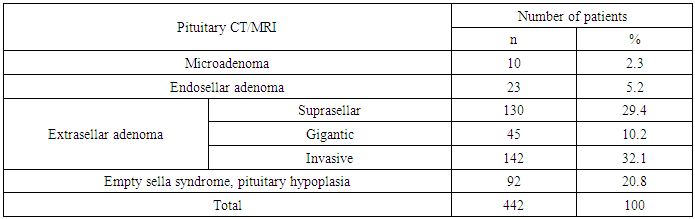
442 patients with the somatotrophic pituitary adenomas whose data were entered into Uzbekistan Acromegaly Registry database from January 1, 2007 to January 1, 2018, were the object of the study. All the patients were examined at the local level for the personal registry card to be filled up. The patients’ age varied from 18 to 75 years, mean age was 41±21 years.
All patients underwent
- the clinical examination,
- RIA of hormones, such as LH, FSH, TSH, GH, IGF-1, IGFBP-3, cortisol, testosterone, estradiol, free T4, prolactin/
- X-ray study including the sella spot filming, CT and/or MRI of the hypothalamo-pituitary region,
- US study of inner organs and the thyroid,
- Neuro-ophthalmological evaluation of vision acuity, fundus of the eye and fields of vision.
To characterize complications we categorized them as cardio-vascular, endocrine-metabolic, respiratory, osteoarticular and neuromuscular, and neoplasms, such as tumors of large intestine, breast, thyroid or prostate) [1,26].

3. Results and Discussion

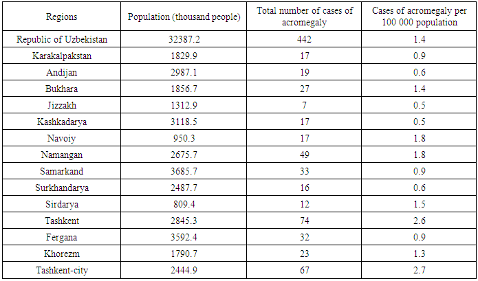
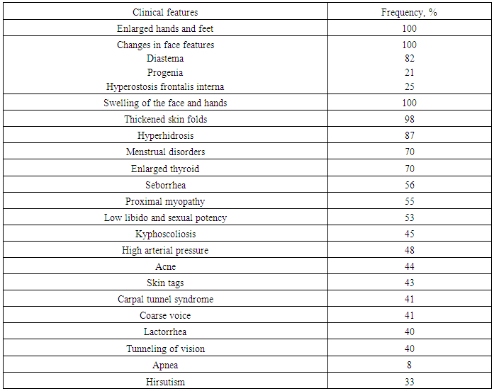
Of 442 acromegalics, there were 277 (62/6%) women and 165 (37.4%) men. The highest prevalence of the disease was found in patients aged from 30 to 44 years (43.4%), the lowest one could be seen in persons of age group of 75 years and older (2.1%); as a whole the trend persisted for both sexes. Mean prevalence of acromegaly in the Republic of Uzbekistan is 1.4 cases per 100 000 population (Table 1), varying from 0.5 cases in Djizak and Kashkadarya regions to 2.7 per 100 000 population in Tashkent-city. Due to varying acromegaly prevalence, we arbitrarily categorized the regions into those with low, intermediate and high frequency.
Table 1. Cases of acromegaly per 100 000 population in the Republic of Uzbekistan as per the Acromegaly Registry
     
Thus, Tashkent-city and Tashkent region turned out to be the regions of high prevalence (2.7 and 2.2 cases per 100 000 population, respectively). Bukhara (1.4), Namangan (1.8), Khorezm (1.3) and Navoyi (1.0) regions were among those with intermediate prevalence. Regions of low prevalence included Andijan (0.6), Djizak (0.5), Kashkadarya (0.5), Syrdarya (0.6), Samarkand (0.9) regions and Republic of Karakalpastan (0.9 cases of acromegaly per 100 000 population).
Growth hormone secretion is known to have sex and age peculiarities [27], so we studied the frequency of acromegaly cases by the factors (Fig.1).
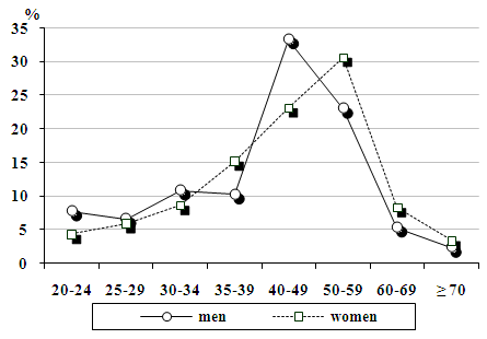
Figure 1. Frequency of acromegaly cases by sex and age
Our findings demonstrated that the acromegaly prevalence is higher in females, but the incidence rate turned out to be sex-specific. Thus, incidence in females starts increasing from the 4th decade of life (23.1%), peaking at 50-59 years of age (30.7%) and lowering to the value found in the age group of 30-34 years (8.6%). In men the disease peaks at 40-49 years of age.
As a whole, the data seems to be true, but according to the literature data, due to slow progression of acromegaly, its diagnosis lags behind its onset, and 5-15 years can pass between the symptoms onset and diagnosis of the disease [28]. In our study this period varied from 2 to 26 years; the diagnosis delayed in 19.5% by 3 years, in 24.7% by 3-5 years, in 45.6% by 6-10 years and in 10.2% by 10 years. Thus, in one fifth of patients the disease was diagnosed within a short time, while the diagnosis was found to delay in 80% of the acromegalics predisposing to complications.
Pathogenesis of pituitary adenomas is known to be multi-step one with chromosomal mutations within a cell [29,30]. But there are factors likely to accelerate the process; some authors report that nearly 15.1% of patients associate onset of the disease with a craniocerebral injury, ~ 6% of women attribute it to abortions or deliveries, while 20% of patients link their disease with chronic sinusites and otitides [26].
Most patients (n=164) in our cohort have associated onset of their disease with no event in particular; no data in medical history indicated either causes or factors facilitating adenoma growth. This indicates that formation of a tumor in the pituitary gland is genetically determined; the process starts subtly. Prior psychoinjuries and stresses were pointed out as the possible causes by 18.6% (n=82) (Fig.2).
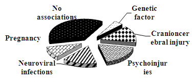
Figure 2. Risk factors in medical histories of patients in the period prior to acromegaly manifestation
Acute respiratory viral infection and inflammatory conditions in the brain preceded manifestation of acromegaly in 14.6% (n=70). 10.1% (n=28) female patients associated the disease onset with pregnancy.
Moreover, craniocerebral injuries (n=68, 15.4%) and hereditary burden (n=19, 4.3%) turned out to be significant factors.
As to the symptoms that caused patients to seek medical advice, headache was the most frequent complaint (72%), followed by changes in appearance, such as enlargement of the nose, earflaps, hands and feet (71%), hyperhidrosis (61%), swelling of the face and extremities (66%). In addition, 70% of women complained of menstrual disorders (Table 2).
Table 2. Acromegaly symptoms as per The National Acromegaly Registry (n=442)
     
Next, we analyzed frequency of clinical features typical of acromegaly to establish that at the moment of visiting a physician most patients had the dramatic ones (Table 3). Thus, changes in face features, enlarged hands and feet, swelling of the face and hands could be seen in all patients. 98% of patients had thickened skin folds, 87% complained of hyperhidrosis and 70% of women of menstrual disorders, such as oligomenorrhea or amenorrhea; lactorrhea was found in 40%. 53% of male patients had low libido and sexual potency. High arterial pressure could be seen in 30%.
Table 3. Clinical features of acromegaly as per cards of the Registry
     
Our findings demonstrated that most acromegalics sought medical advice of professionals from other than endocrinology area of expertise; only 27.4% visited an endocrinologist (Table 4).
Table 4. First symptoms of acromegaly (according to the patients) and professionals referred
     
Due to slow progression of the disease, paucisymptomatic clinical picture misled both the patients and physicians preconditioning delayed diagnosis and complicated course of the disease.
Among the complications, in Uzbekistan endocrine (96.1%), cardiovascular (54.2%) and osteoarticular and neuromuscular (65.6%) were the most frequent (Fig.3).
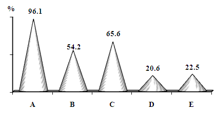
Figure 3. Complications in the acromegalics. A – endocrine, B – cardiovascular, C – osteoarticular and neuromuscular, D – respiratory, E - neoplasms
Next, computed tomography (CT) and magnetic resonance imaging MRI) were used to study the sizes of pituitary tumors (Table 5). Microadenomas were registered in 9 patients (3%), 68 patients (22.7%) had the adenoma with endosellar growth. Macroadenomas with extrasellar growth were found in 192 patients (64%), and those with suprasellar growth were registered in 102 patients (34%). 25 patients (8.3%) had gigantic pituitary adenomas. According to CT/MRI findings 32 patients (10.3%) had partially empty sella with the pituitary hypoplasia (Table 5).
Table 5. CT/MRI findings
     
According to our findings, large sizes and extrasellar growth are typical of somatotrophic pituitary adenomas (71.7%). In addition, in 32.1% of patients the tumor’s invasive growth was found. Merely by the character of an adenoma’s growth it is evident that more than 80% of patients should have got operated, 32.1% needed radiotherapy.
The evaluation of GH secretion, that is, single measurement of basal GH, which varied in our study from 2.3 to 115.4 mU/l (mean value 47.4±21.6 mU/l), was principal in the laboratory diagnosis. Herewith, “harmless” GH level < 5 mU/l was registered in 54 (12.2%) patients only. In other words, though most patients (87.8%) needed an intensive therapy they received some drug treatment or another. Thus, of 442 patients 44.5% received dopamine agonists, 28.4% underwent radiotherapy and only 27.1% got operated.

4. Conclusions

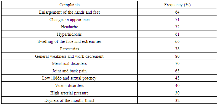
To sum up, acromegaly prevalence in the Republic of Uzbekistan is 1.4 cases per 100 000 population with non-uniform regional distribution. The highest frequency of the somatotrophic pituitary adenomas was found in Tashkent-city (2.5) and Namangan region (2.2). The female predominance could be clearly seen. The delayed diagnosis by 6-10 years with peaking at 40-49 years among men and at 50-59 years among women was found. As to potential triggering factors, neuroviral infections were found to precede the disease onset in 14.6% of the patients, psychoinjuries and stresses in 18.6%; 101% of women associated the disease onset with pregnancy, 15.4% of patients linked it with the craniocerebral injuries.
As the results from analysis of clinical presentation period demonstrated, the patients first sought medical attention late and at the height of the disease. Herewith, most acromegalics sought medical advice of professionals from other than endocrinology area of expertise, that is, of a neuropathologist, a therapist or a cardiologist, while only 27.4% of acromegalics visited an endocrinologist; of all others, this determined high frequency and early onset of complications. The point to be emphasized is that high frequency of complications with affection of endocrine, cardiovascular, and osteoarticular and neuromuscular systems caused by the delayed diagnosis and nonspecific manifestations of the disease which could be confused with arterial hypertension, cephalgia, menstrual disorders and infertility, hypothyroidism and diabetes mellitus.

References

[1]  Annamaria Colao, Diego Ferone, Paolo Marzullo, Gaetano Lombardi. Complications of Acromegaly: Epidemiology, Pathogenesis, and Management. // Endocrine Reviews 25.– 2004.- V. 1.- P. 102-152.
[2]  Holdawey I.M., Rajasoorya C. Epidimiology of acromegaly // Pituitary.-1999.-V. 2(1).- P.29-41.
[3]  Holdaway M., Raja C., Rajassoorya G. Factors influencing mortality in acromegaly. // J. of Clin. Endocrin. and metabab. - 2004.- Vol.89, N.2.- P.677-674.
[4]  Melmed S. Acromegaly and cancer: not a problem.// J. Clin. Endocrinol. Metab. – 2001. – V.86. – P.2929–2934.
[5]  Giustina A., Casanueva F.F., Cavagnini F., et all. Diagnosis and treatment of acromegaly complications.// J Endocrinol Invest. 2003; 26: 1242–7.
[6]  A. Giustina, A. Barkan, F.F. Casanueva et all. Criteria for cure of acromegaly: a consensus statement // J. Clin. Endocrinol. Metab. -2000.- V.85.- P.526–529.
[7]  Harris A. Acromegaly and its managemeht. // Philadelphia, USA: Lippincott Raven Publishers, 1996.- P.175.
[8]  Orme S.M., McNally R.Y., Cartwright R.A. Mortality and cancer incindence in acromegaly: a retrospective cohort stude.// J. Clin. Endocr. and Metab.-1998. - V.83. – P. 2730-2734.
[9]  Burton T, Le Nestour E, Neary M, Ludlam WH (2016) Incidence and prevalence of acromegaly in a large US health plan database. // Pituitary 19(3):262–267.
[10]  Kwon O, Song YD, Kim SY, Lee EJ, Rare Disease Study Group, Science and Research Committee, Korean Endocrine Society (2013) Nationwide survey of acromegaly in South Korea.// Clin. Endocrinol (Oxf) 78(4):577–585.
[11]  Bengtsson B, Eden S, Ernest I, Oden A, Sjogren B (1988) Epidemiology and long-term survival in acromegaly: a study of 166 cases diagnosed between 1955 and 1984. // Acta Med Scand 223:327–335.
[12]  Ritchie CM, Atkinson AB, Kennedy AL, Lyons AR, Gordon DS, Fannin T, Hadden DR (1990) Acertainment and natural history of treated acromegaly in Northern Ireland. // Ulster Med J 59:55–62.
[13]  Etxabe J, Gaztambide S, Latorre P, Vazquez JA (1993) Acromegaly: an epidemiological study. // J Endocrinol Invest 16:181–187.
[14]  Davis JR, Farrell WE, Clayton RN (2001) Pituitary tumours. // Reproduction 121:363–371.
[15]  Ntali G, Karavitaki N (2015) Recent advances in the management of acromegaly.// F1000Res. doi: 10.12688/f1000research.7043.1
[16]  Katznelson L, Laws ER Jr, Melmed S, Molitch ME, Murad MH, Utz A, Wass JA, Endocrine Society (2014) Acromegaly: an endocrine society clinical practice guideline.// J Clin Endocrinol Metab 99(11):3933–3951.
[17]  Daly AF, Rixhon M, Adam C, Dempegioti A, Tichomirowa MA, Beckers A (2006) High prevalence of pituitary adenomas: a cross- sectional study in the province of Liege, Belgium.// J ClinEndocrinolMetab 91(12):4769–4775.
[18]  Tjörnstrand A, Gunnarsson K, Evert M, Holmberg E, Ragnarsson O, Rosén T, Filipsson Nyström H (2014) The incidence rate of pituitary adenomas in western Sweden for the period 2001–2011.// Eur J Endocrinol 171(4):519–526.
[19]  Mestron A, Webb SM, Astorga R, Benito P, Catala M, et all (2004) Epidemiology, clinical characteristics, outcome, morbidity and mortality in acromegaly based on the Spanish Acromegaly Registry (RegistroEspanol de Acromegalia, REA). //Eur J Endocrinol 151(4):439–446.
[20]  Agustsson TT, Baldvinsdottir T, Jonasson JG, Olafsdottir E, Steinthorsdottir V, Sigurdsson G, Thorsson AV, Carroll PV, Korbonits M, Benediktsson R (2015) The epidemiology of pituitary adenomas in Iceland, 1955–2012: a nationwide population-based study. //Eur J Endocrinol 173(5):655–664.
[21]  Gruppetta M, Mercieca C, Vassallo J (2013) Prevalence and incidence of pituitary adenomas: a population based study in Malta. // Pituitary 16(4):545–553.
[22]  A. Lavrentaki, A. Paluzzi, J.A. H. Wass, N. Karavitaki et all. Epidemiology of acromegaly: review of population studies // Pituitary February 2017, Volume 20, pp 4–9.
[23]  Wass J.A.H. Handbook of Acromegaly. // Bristol, 2004. - P.19-27.
[24]  Lugo G, Pena L, Cordido F. Clinical manifestations and diagnosis of acromegaly. // J Endocrinol 2012.
[25]  A. Ben-Shlomo, M. C. Sheppard, J. M. tephens, S. Pulgar, and S. Melmed. Clinical, quality of life, and economic value of acromegaly disease control// Pituitary. 2011 September; 14(3): 284–294.
[26]  Марова Е.И. Заболевания гипоталамо-гипофизарной системы - Москва, 2001.- С. 4-9.
[27]  Colao A., Amato G., Pedroncelli A.M. et all. Gender- and age-related differences in the endocrine parameters of acromegaly. // J. Endocrinol. Invest.- 2002.- V. 25.- P.532–538.
[28]  Barcan A.L. Acromegaly: diagnosis and therapy. // Endocrin. and Metab.Clin. of North America. – 1990.- V.18. - P.277-310.
[29]  Shimon I., Melmed S. Pituitary tumor pathogenesis.// J. of Clin. Endocr. andmetab. - 1997. - Vol.82, N.6. - P.1675-1681.
[30]  Melmed S. Acromegaly pathogenesis and treatment.// J Clin Invest. 2009; 119:3189-3202.