American Journal of Medicine and Medical Sciences

p-ISSN: 2165-901X    e-ISSN: 2165-9036

2019;  9(7): 262-269

doi:10.5923/j.ajmms.20190907.08


Metabolic Effectiveness of Laparoscopic Sleeve Gastrectomy in Morbid Obesity

F. G. Nazirov, Sh. Kh. Khashimov, U. М. Makhmudov, Z. R. Khaybullina, O. D. Tuychiev

Department of Endovisual Surgery, State Institution “Republican Specialized Scientific-Practical Medical Centre of Surgery named after Academician V.Vakhidov”, Tashkent, Uzbekistan

Correspondence to: U. М. Makhmudov, Department of Endovisual Surgery, State Institution “Republican Specialized Scientific-Practical Medical Centre of Surgery named after Academician V.Vakhidov”, Tashkent, Uzbekistan.

Email:

Copyright © 2019 The Author(s). Published by Scientific & Academic Publishing.

This work is licensed under the Creative Commons Attribution International License (CC BY).
http://creativecommons.org/licenses/by/4.0/

Abstract

Metabolic effectiveness of laparoscopic sleeve gastrectomy (LSG) have been evaluated in 3 month after surgery according plasma lipidomic profile, fasting glucose, levels of proinflammatory cytokines (interleukin-6 (IL-6), tumor necrosis factor alpha (TNF-alpha), C-reactive protein (CRP), as well as oxidative stress marker malondialdehyde (MDA), vasculo endothelial growth factor (VEGF) at patients with morbid obesity (MO). It was established, that LSG is not accompanied by activation of systemic inflammation in the early postoperative period and leads to decreasing in the initially elevated levels of pro-inflammatory factors in serum at 3 months after the intervention. The results indicate a positive effect of LSG on the elimination of inflammation and metabolic disorders in obese patients. LSG is efficient in reducing the amount of visceral fat by reducing body mass index and waist circumference (WC) at 3 months after the intervention. LSG reduces cardiometabolic risk due to normalization of the lipid profile and indicators of oxidative stress (OS) in blood, helps to optimize the paracrine and endocrine functions of adipose tissue by reducing inflammation in it.

Keywords: Laparoscopic sleeve gastrectomy, Inflammation, Metabolic effectiveness

Cite this paper: F. G. Nazirov, Sh. Kh. Khashimov, U. М. Makhmudov, Z. R. Khaybullina, O. D. Tuychiev, Metabolic Effectiveness of Laparoscopic Sleeve Gastrectomy in Morbid Obesity, American Journal of Medicine and Medical Sciences, Vol. 9 No. 7, 2019, pp. 262-269. doi: 10.5923/j.ajmms.20190907.08.

1. Introduction

Obesity is the most important component of metabolic syndrome, an independent risk factor for a number of socially significant diseases - arterial hypertension, ischemic heart disease (IHD), type 2 diabetes, and a risk factor for premature death [8,29]. It is certain, that with a body mass index (BMI) of more than 35 kg/m2 in combination with a waist circumference (WC) of more than 90 cm and a triglyceride (TG) level of more than 2,3 mmol/l, the risk of cardiovascular diseases (CVD) increases 20 times [24]. At the same time, “metabolically healthy individuals” are found among obese people, and a BMI without consideration WC cannot be criteria for the CVD risk [36]. In a study of 15547 patients with IHD, the highest risk of death was observed in patients with the normal BMI, and increased WC [7]. The magnitude of WC and all-cause mortality are linearly dependent [15]. WC reflects the amount of visceral fat. According to M.R. Salazar et al. (2014), an increase in WC, more than 90 cm, corresponds to visceral adipose tissue, having an area of more than 130 cm2, and the excess of this value is combined with metabolic disorders [35].
However, it is not precisely established that exactly obesity causes comorbidity and increases cardiometabolic risks - high visceral fat, adipocyte dysfunction, elevated levels of TG and free fatty acids, impaired adipo-cytokines secretion, low-intensity inflammation, oxidative stress (OS) or other factors [21].
Possibly, cardiometabolic risk in obesity is associated with the condition of the functional activity of visceral adipose tissue and the intensity of inflammation in it [34]. Visceral adipose tissue has a high metabolic activity and is considered as a part of endocrine system, that produces leptin, apelin, adiponectin, angiotensinogen, insulin-like growth factor, insulin-binding protein, monobutyrin, TNF-alpha, IL-6, plasma activator of plasminogen-1 inhibitor (PAI-1), resistin, lipoprotein lipase, acetylation stimulating protein, cholesterol ester transfer protein, retinol-binding protein, estrogens [5]. When TG are accumulated in adipocytes, macrophage cells start to infiltrate the adipose tissue, meanwhile activated macrophages, as well as adipocytes, are considered as producers of pro-inflammatory cytokines — TNF-alpha and IL-6 [4], triggering and maintaining low-intensity inflammation [21]. Macrophages of adipose tissue are represented by two phenotypes: M1 and M2. M1 phenotype is “activated” macrophages that blocks adipocyte differentiation, as a result, occurs their hypertrophy, adipokines secretion is deteriorated and ectopic leptin cumulates in the liver, muscles and other tissues. M2 phenotype is an anti-inflammatory phenotype of macrophages, it is formed from M1 under the action of a PPAR gamma agonist (peroxisomal proliferative factor receptor agonist) [12]. Adipocytes and macrophages of adipose tissue (AT) produce inflammatory cytokines, including TNF-alpha, IL-1, PAI-1, monocytic chemoattractant protein-1 [5]. Adipokines, primarily leptin and adiponectin, are also involved in the regulation of the inflammatory process [10,25,36]. Adipose tissue dysfunction and adipocytokine dysregulation, associated with inflammation, consequently makes for insulin resistance and the formation of metabolic disorders [38].
In this regard, the study of the intensity of inflammation in obesity and weight loss in dynamics after surgical bariatric intervention is of considerable interest.
Of all the methods of treating obesity, surgical is the most effective [26]. A meta-analysis of surgical treatment of obesity, in which studied effectiveness of weight loss (89 studies), incidence (134 studies) and complications (128 studies) of surgical treatment of obesity, showed that weight loss after surgery is 20-30 kg in patients with an initial BMI of more than 40 kg/m2 and is resistant (up to 10 years). Weight loss allows controlling comorbid conditions in obesity [27]. Data from a randomized trial of the effectiveness of laparoscopic gastric bypass showed that weight loss by 13.6% in 5 years after surgery was accompanied by a decrease in cardiometabolic risk markers: high-density lipoprotein (HDL), cholesterol (CH), TG, glucose [33]. After bariatric surgery, manifestations of type 2 diabetes are stopped in 86.6% of cases [19]. The most effective weight loss is achieved by using biliopancreatic and duodenal bypass, however, after these operations, physiological digestion is disturbed, vitamins deficiency and malabsorption were developed [33,41], and the intensity of systemic inflammation syndrome does not decrease [23].

2. Aim of the Study

The purpose of this study was to evaluate metabolic outcomes of the LSG, as well as the intensity of systemic inflammation and oxidative stress at patients with morbid obesity (MO) after bariatric surgery – LSG.

3. Materials and Methods

The study involved 37 patients (29 female and 8 male) with MO, operated in State Institution “RSSPMC of Surgery named after acad. V.Vakhidov” in the period from 2016 to 2019. All individuals were non-smoking, the average age was 36,5±2.4 years, and BMI = 51,2±2,3 kg/m2. The control group consisted of 10 female volunteers aged 38.4 ± 1.9 years, without obesity (BMI = 23.4 ± 0.3 kg/m2), having an WC = 76.1 ± 1.0 cm. Minimally invasive surgery – LSG – was performed to 37 patients. Interventions were performed by the OR-1 endoscopic surgical complex and the toolset of Karl Storz GMBH & CO.KG (Germany). During surgical endovisual intervention were used: the Force Triad energy platform with LigaSure technology from Covidien (USA), the Harmonic G11 ultrasonic scalpel (Johnson & Johnson, USA), and the endoscopic stapling-cutting device from Ethicon Endo Surgery (Johnson & Johnson, USA). This endovisual intervention is considered as a restrictive bariatric surgical procedure. The technique of laparoscopic surgery was to remove most of the stomach, located along gastric curvature major, with preservation of the cardiac sphincter and pylorus and the formation of a narrow gastric tube with a volume of 60-150 ml, located along the gastric curvature minor.

3.1. Operational Technique

LSG was performed under general endotracheal anesthesia. The operation is usually involved three surgeons. The patient was placed in the position of Trendelenburg (with an elevated head end), while the legs were separated so that the operating surgeon can be placed between them.
As a rule, used 5-port access. A 30° optics was inserted through a trocar placed on 10–12 cm of supraumblical area. The remaining trocars were inserted on approximately the same horizontal line. On the left midclavicular line, a 12-mm trocar was installed for the instrument in the surgeon’s right hand, on the contralateral same area was placed a 12-mm trocar for the surgeon’s left hand and the subsequent stapling device, on the left anterior axillary line - the 5-mm trocar for the assistant’s instrument. The next trocar was inserted for use of the hepatic retractor, it was placed subxiphoidal along the middle or right anterior axillary line.
Using the harmonic scalpel Harmonic G11 (Johnson & Johnson, USA) and LigaSure Atlas of ForceTriad energy platform (Covidien, USA), a full mobilization of the stomach along the curvature major was performed: from the pyloric part to the angle of the Hiss, dissected the gastrocolic ligament, were cut short gastric vessels, completely mobilized the fundus of the stomach on the back wall (Figure-1).
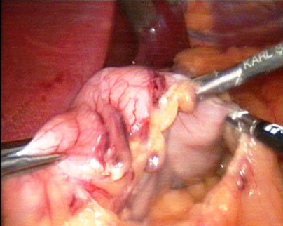
Figure 1. Intraoperative photo. Stomach mobilization along greater curvature (gastric curvature major)
The anesthesiologist inserted into the stomach a calibration bougie with a diameter of 36-40 Fr, which served as a kind of carcass for the formation of the sleeve in order to avoid stenosis. Focusing on the bougie located along the lesser curvature, the stomach was gradually stapled with 3-row staples and simultaneously transected with the help of Echelon (Ethicon - Endo Surgery) or Covidien devices starting at the antrum, 2-4 cm from the pylorus (Figures - 2-3).
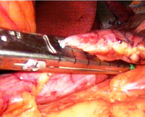
Figure 2. Intraoperative photo. Stapling device during firing
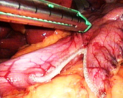
Figure 3. Intraoperative photo. The stage of forming sleeve with firing and excising device
We did not adhere to any particular distance from the pylorus; the main criteria was the creation of a uniformly narrow gastric pouch with preservation of a small part of the antrum.
As a rule, for the first two firings of the staple, green cartridge with 60 mm length and 4.1 mm staples height was used, the subsequent firing was performed with blue cartridges with a staple height of 3.5 mm. When stapling and crossing, the exposure to compress was held before and after firing for 15-20 seconds. For every operation, we used 5-6 cartridges.
After transsection most of the stomach, the initial bougie was replaced with a smaller one (32 Fr) in diameter, along the lesser curvature, over which the staple line was peritonized for all extent from the angle of His to the antrum. For this aim, a continuous serous-muscular suture was applied with a special absorbable “barbed” V-Loc 180 thread (Covidien, USA) (Figure-4). Thus, a uniformly narrow sleeve was formed, i.e. gastric tube with a volume of 60 to 120 ml depending on the length of the sleeve (Figure -5).
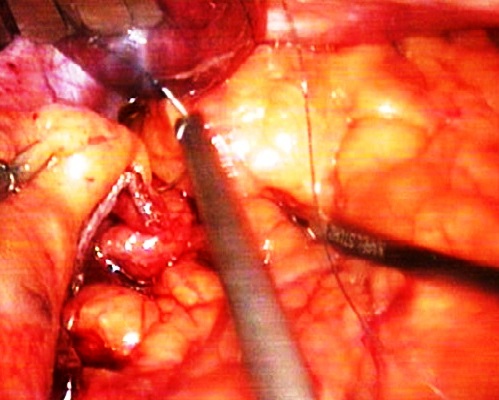
Figure 4. Intraoperative photo. Maintaining the staple line
Figure 5. Intraoperative photo. Final appearance of gastric sleeve
Placed one drainage along the staple line of the stomach pouch through a trocar place in the left hypochondrium. In cases of gallstone disease, according to the preoperative ultrasound data, simultaneous cholecystectomy was performed.
The patients activated in 4-6 hours after the surgery. In the first 2 days, they were allowed to drink up to 500 ml per day fractionally, with further increase in the liquid food volume up to 1.5 liters/day by the end of the 1st week. In postoperative diet, mashed food permitted from the 4th-5th days after the surgery, solid food, with thoroughly chewing, was allowed from the 3rd week after the surgery. Patients were discharged on the 5-7th day, followed by outpatient follow-up and removal of sutures on the 5-7th days.
Patients were provided with special recommendation blanks on nutrition and behavior. In early period after surgery (1-5 days) antibiotic therapy was prescribed; low-molecular-weight heparins were prescribed for 1 month, as well as anti-ulcer drugs for 2 months. Taking into account international clinical recommendations, patients undergoing LSG were prescribed vitamin and mineral support (multivitamins, calcium and vitamin D supplements daily, vitamin B12 injections monthly). The algorithm of postoperative surveillance corresponds to the commonly accepted rules: every 3 months during the first year, every six months during the second year, then annually. Laboratory control (complete blood count and urine analysis, a comprehensive biochemical blood test) was an integral part of the postoperative surveillance and are tested at the same time.

3.2. Laboratory Assessment

Data of plasma lipid profile: total CH, TG, HDL, very low-density lipoproteins (VLDL), as well as C-reactive protein (CRP), fasting glucose, uric acid (UA), total protein (TP), and albumin was obtained from an automatic biochemical analyzer "VITROS-350" (Ortho Clinical Diagnostics, USA). Low-density lipoprotein (LDL) was calculated by Frivald’s: LDL = CH-(HDL-TG/2.2), and atherogenic index (AI) - according to Klimov manner: AI = (CH - HDL)/HDL. Total blood count includes the amount of WBC, monocytes (M), lymphocytes, that performed an automated hematology analyzer BC 5800, Mindray (China). Serum cytokines: IL-6, TNF-alpha, as well as vasculoendothelial growth factor (VEGF), were determined by ILISA kits of "Vector-BEST" (Russia) on the ST-360 IFA analyzer (China). Malon dialdehide (MDA) was determined by Ohkawa in the modification of Al-Gayyar (2007). Catalase activity was investigated according to M.A. Korolyuk. (1988).

3.3. Statistical Methodology

Statistical processing of amount data was performed using ANNOVA test, pare t-Test by the Microsoft Excel software package. The data are presented as M±m, the differences were considered significant at a level of p <0,05.

4. Results and Discussion

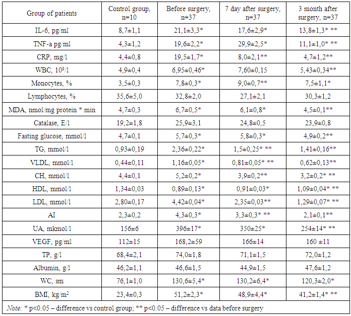
At patients with MO metabolic status before surgery includes increasing TG level 2,7 times vs control group, increasing CH level (p<0,05), decrease HDL level (p<0,05), increasing UA level in 2,5 times vs to control group. The level of CRP, IL-6 and TNF-alpha in the blood was increased by 3,5, 2,7 and 5,3 times vs to control group, respectively. WBC number was within the reference interval (4-9*109/l), however, it was significantly higher (p<0,05) vs the control group. Also in patients with obesity, there was an increase in monocytes by 2.0 times relative to the control group (Table 1).
Table 1. Metabolic profile and proinflammatory factors after LSG
     
The data, given above, suggests the presence of systemic inflammatory response in patients with MO, enrolled to our research.
Discussing the findings, we noted that monocytes, being precursors of tissue macrophages, are actively involved in inflammation, infiltrating tissues and secreting pro-inflammatory cytokines that activate the immune response and maintain chronic inflammation of low intensity in obese people [16,17,18,39]. Synthesis of pro-inflammatory mediators can intersect with the metabolism of the insulin receptor and glucose transporter GLUT-4 (phosphorylation of serine of the insulin receptor blocks tyrosine phosphorylation there in response to insulin, which makes the receptor insulin-insensitive) [32,42]. The presence of inflammation and the accumulation of pro-inflammatory cytokines disrupt adipocyte production of its own hormones: an increase in resistin production while reducing adiponectin and leptin causes the development of insulin resistance (IR), and not only in adipocytes, but also in hepatocytes and muscle cells [10,30].
Approximately 30% of total plasma IL-6 has been originated from adipose tissue (Mohammed Ali et all, 2007), and many researchers consider a TNF-alpha increase as a sign of insulin resistance in obese patients [12,42]. The consequence of an increase in TNF-alpha is a deterioration of insulin signaling in peripheral tissues, beta-cell dysfunction, and activation of inflammasomas [9].
Various factors may act as initiators of inflammation in AT: an increase in the mass of fat cells and their hypertrophy; local hypoxia of hypertrophied adipocytes with hyperproduction of ROS, stimulating infiltration with T-lymphocytes and macrophages; impaired signaling of adipocytes [2], some viruses and microorganisms, including CMV, Ch. pneumoniae and H. pillory [31,37].
In adipose cells, congenital immunity receptors have been found that recognize the molecular components of bacteria, viruses, fungi and other pathogens, and activate pro-inflammatory signaling pathways in response to microbial pathogens. These are Toll-like receptors (TLR), primarily TLR4 [37]. The specific ligand for TLR4 is lipopolysaccharide (LPS) from the wall of gram-negative bacteria, and saturated fatty acids [43]. Activation of TLR4 stimulates intracellular kinases, which enables translocation of nuclear factor NF-κB in core of the cell with the subsequent stimulation of transcription of many pro-inflammatory genes coding inflammatory regulatory biomolecules, including cytokines, chemokine, adipokines, IL-6, TNF-alpha, resistin [3]. Activation of TLR-2 occurs under the action of lipid degradation products, formed as a result of oxidation under the action of ROS, and activation of macrophages, which is a consequence of TLR-2 activation, maintains the connection between OS and inflammation [13,40]. All these data indicate a close relationship of cell receptor metabolism, lipolysis reactions with the activation of pro-inflammatory cascades in cells [22]. Some authors consider this as “metabolic inflammation” [11], and, inflammation in adipose tissue is a self-sustaining process: once initiated, it progresses without additional support.
The marker of lipoperoxidation and ROS overproduction - MDA in individuals with MO was significantly higher than in the control group (p<0,05), while the catalase activity did not differ from the control. Perhaps, this is due to the high reserve capacity of the enzyme and the weak severity of the OS (increase in MDA 1,3 times, p<0,05) at our patients.
One of the indicators characterizing endothelial dysfunction and neoangiogenesis – vasculo-endothelial growth factor - VEGF in obese patients varied in statistically insignificant ranges, indicating the absence of disorders (p>0,05).
Thus, as our results showed, MO is characterized by changes in serum lipid spectrum, hyperuricemia, and inflammation with increasing of plasma IL-6, TNF-alpha, CRP levels. At the same time, MO has a property of weak expression of OS and normal endothelial function.
As our studies have shown, performing LSG contributed to effective weight loss, decreasing WC, normalizing not only the parameters of lipid and carbohydrate metabolism, but also reducing the intensity of systemic inflammation, probably due to a decrease in the amount of visceral fat and a reduction of local inflammation in the tissue.
In this case, the normalization of TG, CH, VLDL, and LDL occurred on day 7 after surgery, long before clinically significant weight loss. At the same period, the concentration of IL-6 and CRP decreased significantly relative to the preoperative rate. After 3 months of surgery, WC were decreased from 130,6±5,4 to 120,3±2,0; BMI – from 51,2±2,3 to 41,2±1,4.
The patients, however, did not experience discomfort, felt subjectively better, and the level of TP, albumin and lymphocytes in the blood corresponded to those before surgery. This indicates the absence of malabsorption, protein-energy deficiency and immunodeficiency after LSG. 3 months after surgery, the level of fasting glucose, TG, VLDL was not significantly different from the control group (p>0,05), and the total CH content was significantly lower than in the control. It should be noticed, the decrease in UA concentration by 32% from baseline, the normalization of MDA level, a steady downward trend in CRP, IL-6 and TNF-alpha. 3 months after LSG, the level of CRP was not significantly different from the control, and the content of IL-6 and TNF-alpha decreased 1,6 and 2,2 times from the baseline, although they were significantly higher than in the control.
Discussing the obtained data, we noticed that the decrease in the intensity of inflammation and the level of pro-inflammatory cytokines IL-6 and TNF-alpha, as a result of surgical treatment, is a favorable prognostic sign. Besides, such decrease is one of the factors contributing to the normalization of metabolic processes of carbohydrate and lipid metabolism, manifested in decline of TG, proatherogenic lipoprotein fractions, glucose and UA. The reduction of glucose after LSG can be explained by the restoration of insulin sensitivity in peripheral tissues due to the lowering of inflammation in AT, as well as an increase in incretin hormones and glucagon like peptides [25]. Because of the fact that as a result of the operation, the volume of the stomach decreases and the patient intakes food in small portions and frequent intervals, which stimulates the secretion of incretins in the gastrointestinal tract. Incretins, in turn, have receptors on beta-cells and cause enhanced early insulin production in response to food ingestion in the gastrointestinal tract and postprandial hyperglycemia [8]. Incretins, in particular, glucagon-like peptide-1 (GLP-1) reduces gluconeogenesis and glycogenolysis in the liver, increases the consumption and utilization of glucose in muscle tissue, thereby contributing to an increase in insulin sensitivity; the hypothalamus, pituitary gland, and bottom of the fourth ventricle are rich in GLP-1 receptors, stimulation of which decreases the appetite [1].
In addition, dietary features and reduced animal fat intake, coupled with weight loss and reduced visceral fat, may optimize the paracrine and endocrine function of adipose tissue, restoring adipocytokines levels and reducing the production of pro-inflammatory factors IL-6 and TNF-alpha, which are consistent with literature data [15,42]. The normalization of the plasma lipid profile, found after surgery, may be determined by the optimization of adipocytokines synthesis.
Another advantage of LSG is its minimally invasive nature; diminish the inflammatory processes in the surgical wound, as well as its physiology due to the lack of a radical reconstruction of the gastrointestinal tract, typical of shunting bariatric operations [6,28].
The obtained positive results of using LSG are consistent with the reference data on the effectiveness of bariatric surgery. It is estimated, that bariatric surgery changes the secretion of adipocytokines, and is most pronounced when the BMI is reached 26-30 kg/m2, i.e. with a significant reduction in the initial weight. As a result, weight loss after biliopancreatic shunting operations decreases the level of leptin in the blood, which contributes to the normalization of the insulin signaling and the recovery of insulin sensitivity after surgery, causing a decrease in hyperglycemia [8]. Bariatric surgery contributes to an increase in insulin-dependent glucose uptake by 33–36% relative to the primary level, a decrease in fatty infiltration of liver by 76%, a decline in liver gluconeogenesis by 19–26%, which the authors consider as evidence of insulin sensitivity refreshment in the postoperative period [14,29]. Over the next 10 years after bariatric surgery, the overall morbidity decreases by 40% (myocardial infarction is reduced by 56%, diabetes is reduced by 92%, cancer is reduced by 60%) [6,20]. However, the use of shunting bariatric operations is not accompanied by a decrease in the intensity of inflammation, as shown by our previous experience [23]. When assessing the effect of the treatment method on the intensity of systemic inflammation in MO, it can be argued, that restrictive bariatric surgery - LSG has advantages over bypass bariatric surgery.

5. Conclusions

Morbid obesity is characterized by changes in the serum lipid spectrum, hyperuricemia, and inflammation with increasing serum levels of IL-6, TNF-alpha, CRP. At the same time, a weak expression of oxidative stress and normal endothelial function are typical in morbid obesity.
Restrictive bariatric surgery - laparoscopic sleeve gastrectomy helps to reduce the intensity of systemic inflammation via reduction the plasma CRP level to the reference range, decreased IL-6 and TNF-alpha in 1.6 and 2.2 times from data before surgery.
Laparoscopic sleeve gastrectomy contributes to effective weight loss, decreasing WC, normalizing lipid and carbohydrate metabolism parameters, reducing asymptomatic hyperuricemia and oxidative stress.

References

[1]  Baggino L., Drucker D. biology of incretins: GLP-1 and GIP // Gastroenterology.- 2007.-№ 132: 2131-57.
[2]  Bouloumié A., Curat C.A.., Sengenès C. et al. Role of macrophage tissue infiltration in metabolic diseases.// Curr Opin Clin Nutr Metab Care, 2005, no.l; 8(4), pp.347–354.
[3]  Bouwman J.J., Visseren F.L., Bouter K.P. et al. Infection-induced inflammatory response of adipocytes in vitro.// Int J Obes (Lond), 2008, no.32(6), pp. 892–901. doi: 10.1038/ijo.2008.36.
[4]  Calabro P., Yeh E.T. Intra-abdominal adiposity, inflammation, and cardiovascular risk: new insight into global cardiometabolic risk. //Curr Hypertens Rep., 2008, no. 10(1), pp.32–38.
[5]  Calabro P., Yeh E.T. Obesity, inflammation, and vascular disease: the role of the adipose tissue as an endocrine organ. // Subcell Biochem. 2007, no. 42, pp. 63–91.
[6]  Chevallier JM. From bariatric to metabolic surgery: 15 years experience in a French university hospital.[Article in French] // Bull Acad Natl Med. 2010 Jan;194(1):25-36; discussion 36-8.
[7]  Countinbo T., Goel K., Correa de SA D. Combining body mass with measurment of central obesity in the assesment of mortality with coronary disease – role of normal weight central obesity // J Cardiol 2013.-61(3).- 553-560.
[8]  Dedov I.I., Yashkov Yu.I., Ershova E.V. Incretins and their effect on the course of type 2 diabetes in patients with morbid obesity after bariatric operations // Obesity and Metabolism. 2012.-№2.- P. 3-10.
[9]  Esser N., Legrand-Poels S., Piette J. et al. Inflammation as a link between obesity, metabolic syndrome and type 2 diabetes. //Diabetes Res Clin Pract. 2014, no. 105(2), pp.141–150. doi: 10.1016/j.diabres.2014.04.006.
[10]  Fernández-Bergés D, Consuegra-Sánchez L, Peñafiel J, Cabrera de León A, Vila J, Félix-Redondo FJ6, Segura-Fragoso A7, Lapetra J8, Guembe MJ9, Vega T10, Fitó M11, Elosua R3, Díaz O12, Marrugat J3.Metabolic and inflammatory profiles of biomarkers in obesity, metabolic syndrome, and diabetes in a Mediterranean population. DARIOS Inflammatory study.// Rev Esp Cardiol (Engl Ed). 2014 Aug;67(8):624-31. doi: 10.1016/j.rec.2013.10.019. Epub 2014 Apr 3.
[11]  Furuhashi M., Saitoh S., Shimamoto K. et al. Fatty Acid-Binding Protein 4 (FABP4): Pathophysiological Insights and Potent Clinical Biomarker of Metabolic and Cardiovascular Diseases. // Clin Med Insights Cardiol. 2015, no. 2;8 (Suppl 3), pp. 23–33. doi: 10.4137/CMC.S17067.
[12]  Heilbronn L.K., Campbell L.V. Adipose tissue macrophages, low grade inflammation and insulin resistance in human obesity.// Curr Pharm Des., 2008, no. 14(12), pp.1225–1230.
[13]  Holvoet P.Relations between metabolic syndrome, oxidative stress and inflammation and cardiovascular disease.// Verh K Acad Geneeskd Belg. 2008;70(3):193-219.
[14]  Immonen H, Hannukainen JC, Iozzo P, Soinio M, Salminen P, Saunavaara V, Borra R, Parkkola R, Mari A, Lehtimäki T, Pham T, Laine J, Kärjä V, Pihlajamäki J, Nelimarkka L, Nuutila P. Effect of bariatric surgery on liver glucose metabolism in morbidly obese diabetic and non-diabetic patients.// J Hepatol. 2014 Feb;60(2):377-83. doi: 10.1016/j.jhep.2013.09.012. Epub 2013 Sep 20.
[15]  Jacobs E.J. Newton C.C., Wang Y Waist circumference increasing cause mortality in large US cohort // Arch Internal Med 2010.-17.-123-130.
[16]  Janket S.J., Ackerson LK What is passing through toll gate 4: Lipids or infection?// Arch Oral Biol. 2015 Jan 20. pii: S0003-9969(15)00018-7. doi:10.1016/j.archoralbio.2015.01.007.
[17]  Jarvis M.A., Nelson JA. Human cytomegalovirus persistence and latency in endothelial cells and macrophages.// Curr Opin Microbiol. 2002 Aug;5(4):403-7.
[18]  Ji YN, An L, Zhan P, Chen XH.Cytomegalovirus infection and coronary heart disease risk: a meta-analysis.// Mol Biol Rep. 2012Jun;39(6):6537-46.doi: 0.1007/s11033-012-1482-6. Epub 2012 Feb 4.
[19]  Kasalický M. Contemporary overview of the bariatric-metabolic surgery // Rozhl Chir.- 2012 Jan;91(1):5-11.
[20]  Kelishadi R, Mansourian M, Heidari-Beni M. Association of fructose consumption and components of metabolic syndrome in human studies: a systematic review and meta-analysis//Nutrition. 2014 May;30(5):503-10. doi: 10.1016/j.nut.2013.08.014.
[21]  Khaybullina Z., Zyamutdinova Z., Suleymanova G., Kosnikova I. Biomarkers of the system inflammation and blood lipids spectrum changes at the patients with obesity of various degree // Universum - Medicine et Pharmacology - 2015.-№12(23).
[22]  Khaybullina Z.R. Inflammation and oxidative stress – critical role for metabolic syndrome// J Vasc Med Surg 2017; 5: 302. doi: 10.4172/2329-6925.1000302/.
[23]  Kosnikova I.V., Bazhenov L.G., Nizamkhodzhayev Z.M., Ligay R.E. The metabolic effects of surgical treatment of patients with morbid obesity // Bulletin of the Association of Doctors of Uzbekistan, 2012, № 2, p. 84-86.
[24]  Lemieux I, Poirier P, Bergeron J, Alméras N, Lamarche B, Cantin B, Dagenais GR, Després JP. Hypertriglyceridemic waist: a useful screening phenotype in preventive cardiology?// Can J Cardiol. 2007 Oct;23 Suppl B:23B-31B.
[25]  Litvinova L.S., Vasilenko M.A., Zatolokin P.A. et al. Adipocytokine production in patients with morbid obesity with various treatment methods // Diabetes. - 2014. - № 3. - P. 51–59. doi: 10.14341 / DM2014351-59.
[26]  Lima KV, Costa MJ, Gonçalves Mda C, Sousa BS. Micronutrient deficiencies in the pre-bariatric surgery //Arq Bras Cir Dig. 2013;26 Suppl 1:63-6.
[27]  Maggard M.A. MD, MSHS; Lisa R. Shugarman, PhD; Marika Suttorp, MS; Margaret Maglione, MPP; Harvey J. Sugerman, MD; Edward H. Livingston, MD; Ninh T. Nguyen, MD; Zhaoping Li, MD, PhD; Walter A. Mojica, MD, MPH; Lara Hilton, BA; Shannon Rhodes, MFA; Sally C. Morton, PhD; and Paul G. Shekelle, MD, PhD.- Meta-Analysis: Surgical Treanment of Obesity (Guidelines) // Ann Int Med.-2003.-V.142.-№ 7.-Р.547-559.
[28]  Makhmudov U M, Khashimov Sh Kh Khaybullina Z R, Sharapov N U, Sadikov N S, Kabulov T M and Tashkenbaev F R. Effect of intra-gastric balloon installation and laparoscopic sleeve gastrectomy to comorbidity reduction and cardiometabolic disease staging in 6 month after procedure // J Obes Weight Loss Ther 2018, Volume 8.-P.43. DOI: 10.4172/2165-7904-C6-073.
[29]  Mancini MC. Bariatric surgery - An update for the endocrinologist. // Arq Bras Endocrinol Metabol. 2014 Dec;58(9):875-88. doi: 10.1590/0004-2730000003413.
[30]  Martínez-Martínez E, Jurado-López R, Cervantes-Escalera P, Cachofeiro V, Miana M. Leptin, a mediator of cardiac damage associated with obesity//Horm Mol Biol Clin Investig. 2014 Apr;18(1):3-14. doi: 10.1515/hmbci-2013-0060.
[31]  Morre S.A., Stooker W., Lagrand W.K., van den Brule AJC, Niessen HWM. Microorganisms in the aetiology of atherosclerosis // J Clin Pathol.-2000.-53: 647-654.
[32]  Oliver E., McGillicuddy F., Phillips C. et al. The role of inflammation and macrophage accumulation in the development of obesity-induced type 2 diabetes mellitus and the possible therapeutic effects of long-chain n-3 PUFA.// Proc Nutr Soc. 2010, no. 69(2), pp.232–243. doi: 10.1017/S0029665110000042.
[33]  Risstad H, Søvik TT, Engström M, Aasheim ET, Fagerland MW, Olsén MF, Kristinsson JA, le Roux CW, Bøhmer T, Birkeland KI, Mala T, Olbers T. Five-Year Outcomes After Laparoscopic Gastric Bypass and Laparoscopic Duodenal Switch in Patients With Body Mass Index of 50 to 60: A Randomized Clinical Trial // JAMA Surg. 2015 Feb 4. doi: 10.1001/jamasurg.2014.3579.
[34]  Ruf W1, Samad FTissue factor pathways linking obesity and inflammation//Hamostaseologie. 2015 Jan 27;35(2).
[35]  Salazar M.R., Carbajal H.A., Espeche W.G. et al. Identification of cardiometabolic risk: visceral adiposity index versus triglyceride/HDL cholesterol ratio. // Am J Med. 2014, no. 127(2), pp.152–157. doi: 10.1016/j.amjmed.2013.10.012.
[36]  Samorodskaya I.V., Bolotova E.V., Boytsov S.A. "The obesity paradox" and cardiovascular mortality // Cardiology.-2015.-№55.-P.31-36.
[37]  Schwarz V.Y. Inflammation of adipose tissue (part 4). Is obesity a new infectious disease? (literature review) // Problems of endocrinology.-2011.-№5.-С.63-71.
[38]  Spritzer PM1, Lecke SB2, Satler F3, Morsch DMAdipose tissue dysfunction, adipokines and low-grade chronic inflammation in PCOS. // Reproduction. 2015 Jan 27. pii: REP-14-0435.
[39]  Tsiotra P.C., Boutati E., Dimitriadis G. et al. High insulin and leptin increase resistin and inflammatory cytokine production from human mononuclear cells. // Biomed Res Int. 2013, no. 20, P. 487081. doi: 10.1155/2013/487081.
[40]  Vahdat K, Azizi F, Zandi K, Assadi M, Nabipour I. Chronic inflammation is correlated with percentage of body fat independent of the burden of infection.// Inflammation. 2012 Aug;35(4):1322-9. doi: 10.1007/s10753-012-9445-6.
[41]  Valente DS1, Padoin AV. Post-bariatric surgery body contouring treatment in the public health system: cost study and perception by patients // Plast Reconstr Surg. 2015 Feb 9.
[42]  Wieser V, Moschen AR, Tilg H. Inflammation, cytokines and insulin resistance: a clinical perspective. //Arch Immunol Ther Exp (Warsz). 2013 Apr;61(2):119-25. doi: 10.1007/s00005-012-0210-1. Epub 2013 Jan 10.
[43]  Xiao S, Zhao L. Gut microbiota-based translational biomarkers to prevent metabolic syndrome via nutritional modulation.// FEMS Microbiol Ecol. 2014 Feb;87(2):303-14. doi: 10.1111/1574-6941.12250. Epub 2013 Dec 12.