-
Paper Information
- Previous Paper
- Paper Submission
-
Journal Information
- About This Journal
- Editorial Board
- Current Issue
- Archive
- Author Guidelines
- Contact Us
American Journal of Medicine and Medical Sciences
p-ISSN: 2165-901X e-ISSN: 2165-9036
2019; 9(4): 148-150
doi:10.5923/j.ajmms.20190904.04

The State of Perivascular and Pericellular Spaces in the Cerebral Hemispheres of Individuals with Death from Hemorrhagic Shock
 Abstract
 Reference
 Full-Text PDF
 Full-text HTML
Jumanov Ziyadulla Eshmamatovich1, Indiaminov Sayit Indiaminovich2, Blinova Sofya Anatolyevna3
1Senior Lecturer at the Department of Pathological Anatomy, Samarkand State Medical Institute, Uzbekistan
2Head of the Department of Forensic Medicine, Doctor of Medical Sciences, Professor, Samarkand State Medical Institute, Uzbekistan
3Professor of the Department of Histology, Doctor of Medical Sciences, Samarkand State Medical Institute, Uzbekistan
Correspondence to: Jumanov Ziyadulla Eshmamatovich, Senior Lecturer at the Department of Pathological Anatomy, Samarkand State Medical Institute, Uzbekistan.
| Email: |  |
Copyright © 2019 The Author(s). Published by Scientific & Academic Publishing.
This work is licensed under the Creative Commons Attribution International License (CC BY).
http://creativecommons.org/licenses/by/4.0/

Relevance of the research problem: The study of various aspects of hemorrhagic shock remains an urgent problem of medicine to date. Brain morphological study at the General Staff have established of tanatogenetic significant changes in its structure. Objective: To study the state of pericellular and perivascular spaces in the IV-ventricle of the brain in persons who died from hemorrhagic shock. Materials and methods: Investigated field 6 from the region of the cerebral cortex and the white matter adjacent to it from 12 corpses of persons who died in the hospital with a clinically established diagnosis of hemorrhagic shock caused by knife wounds of internal organs and blood vessels. In the blood and urine of these individuals, ethyl alcohol was not detected. The duration of hospital stay of such victims was 9±4.2 hours. Results of the study: In the cortex of the hemispheres and the white matter of the hemorrhagic shock of people adjacent to it, people who died from hemorrhagic shock, there is a moderate spasm of intracerebral arteries of large and medium caliber. Conclusions: In the case of a single lesion of peripheral vessels in the thanatogenesis in hemorrhagic shock, neurons and intracerebral vessels are equally involved, and in case of multiple damage, the role of damage to intracerebral vessels increases.
Keywords: Brain, Perivascular space, Pericellular space, Hemorrhagic shock
Cite this paper: Jumanov Ziyadulla Eshmamatovich, Indiaminov Sayit Indiaminovich, Blinova Sofya Anatolyevna, The State of Perivascular and Pericellular Spaces in the Cerebral Hemispheres of Individuals with Death from Hemorrhagic Shock, American Journal of Medicine and Medical Sciences, Vol. 9 No. 4, 2019, pp. 148-150. doi: 10.5923/j.ajmms.20190904.04.
Article Outline
1. Relevance of the Research Problem
- The hemorrhagic shock is an extreme degree of the above hemodynamic and metabolic disorders resulting from severe blood loss. It is a type of hypovolemic shock and requires a number of conditions for development: the interval of time for the development of the body's responses; inadequate tissue perfusion; disorders of cell metabolism; the potentially lethal nature of the injury [3]. The study of various aspects of hemorrhagic shock (HSh) remains an urgent problem of medicine to date [1, 3]. Morphological studies of the brain in hemorrhagic shock allowed us to establish significant changes in its structures [2]. However, a forensic assessment of changes in the nervous and vascular structures of the brain according to the state of the perivascular and pericellular spaces in HSh has not yet been carried out. The expansion of these spaces in pathological conditions is considered as perivascular and pericellular edema [4].
2. Objective
- To study the state of pericellular and perivascular spaces in the IV-ventricle of the brain in persons who died from hemorrhagic shock.
3. Materials and Methods
- Investigated field 6 (Brodman) from the region of the cerebral cortex and the white matter adjacent to it from 12 corpses of persons who died in the hospital with a clinically established diagnosis of HSh caused by knife wounds of internal organs and blood vessels. In the blood and urine of these individuals, ethyl alcohol was not detected. The duration of hospital stay of such victims was 9 ± 4.2 hours. All victims were transfusedtherapy in the amount of from 1000 to 6150 ml. In 5 cases, HSh was caused by damage to the heart and main vessels (aorta and pulmonary trunk), in the remaining 7 cases, blood loss occurred due to damage to peripheral vessels and internal organs (except the heart). By the number of injuries: single injuries - 4 cases, multiple injuries - 8 cases. In all cases, the injury was accompanied by internal and external blood loss. Brain pieces are fixed in 10% neutral formalin, passed through an alcohol battery, embedded in paraffin and painted: with hematoxylin and eosin, according to Van-Gieson and Mallory. Histological preparations of the brain were first investigated qualitatively, then quantitatively. The state of perivascular and perivascular of spaces (edema) was investigated by the point method according to G.G. Avtandilov. For mathematical data processing, the Student’s – Fisher method was used with the determination of the arithmetic mean M, the mean error of the relative values, and the coefficient of confidence of the difference t.
4. Results of the Study
- In the cortex of the hemispheres (field 6) and the white matter of the brain of people adjacent to it, people who died from hemorrhagic shock, there is a moderate spasm of intracerebral arteries of large and medium caliber. They determine the content consisting of either loosely arranged erythrocytes with a large number of leukocytes and plasma admixture, or of fresh non-deformed erythrocytes, separated by narrow gaps from the vessel wall. At the same time, the arteries of small calibers resemble tissue cords with slit-like gaps, they show little signs of erythrocyte aggregation and plasma coagulation. Expanded perivascular space and a rarefaction of the brain tissue are noted around them.For an objective characterization of the state of the structural components of the cerebral hemispheres (field 6), morphometric parameters were used for HSh. A study of pericellular (PCE), perivascular (PVE) edema and white matter edema was performed.A study of the severity of edema in the cerebral hemispheres of brain showed that with HSh caused by injury of the heart and great vessels the edema is more pronounced in the white matter than in the cortex (table 1).
|
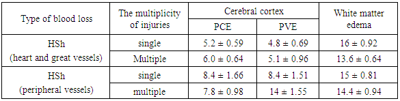
5. Conclusions
- We have found that by the volume of the PCE and PVE in the cortex of the cerebral hemispheres it is possible to determine the role of the nervous tissue and the vascular component in thanatogenesis in HSh. It differs depending on the type of damaged vessels, as well as the frequency of injuries. When death from HSh caused by a single and especially multiple damage to the heart and great vessels, the defeat of neurons prevails in thanatogenesis. In the case of a single lesion of peripheral vessels in the thanatogenesis in HSh, neurons and intracerebral vessels are equally involved, and in case of multiple damage, the role of damage to intracerebral vessels increases.