American Journal of Medicine and Medical Sciences

p-ISSN: 2165-901X    e-ISSN: 2165-9036

2018;  8(2): 355-359

doi:10.5923/j.ajmms.20180812.01


Analysis of the Significance of IIE 105Val Polymorphism of the GSTP1 Gene in the Development of Allergodermatosis in Uzbekistan

Shahnoza Zakirovna Mavlyanova1, Kodirjon Tukhtabaevich Boboev2, Sevara Muradovna Alimova3

1Department of Dermatology, Republican Specialized Scientific and Practical Medical Center of Dermatology and Venereology of the Ministry of Health of the Republic of Uzbekistan, Tashkent, Uzbekistan

2Department of Molecular Medicine and Cell Technologies, Scientific Research Institute of Hematology and Blood Transfusion of the Ministry of Health of the Republic of Uzbekistan, Tashkent, Uzbekistan

3Department of Dermacosmetology, Republican Specialized Scientific and Practical Medical Center of Dermatology and Venereology of the Ministry of Health of the Republic of Uzbekistan, Tashkent, Uzbekistan

Correspondence to: Sevara Muradovna Alimova, Department of Dermacosmetology, Republican Specialized Scientific and Practical Medical Center of Dermatology and Venereology of the Ministry of Health of the Republic of Uzbekistan, Tashkent, Uzbekistan.

Email:

Copyright © 2018 The Author(s). Published by Scientific & Academic Publishing.

This work is licensed under the Creative Commons Attribution International License (CC BY).
http://creativecommons.org/licenses/by/4.0/

Abstract

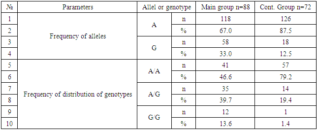
The purpose of research was to establish the role of polymorph variant IIe 105Val gene GSTP1 enzyme biotransformation of xenobiotic in the mechanism of formation and development of allergic skin diseases to substantiate genetic and biochemical criteria for assessing the risk of dermatosis. Thus, allele G and hetero/homozygous genotypes of polymorphism IIe 105 Val of GSTP1 gene are significant markers of increased risk of allergic skin diseases in Uzbekistan (P <0.05). Allele A and the functionally beneficial genotype A/A are reliable protective markers for the development of pathology (χ2=16.5; P<0.05; OR=0.2; 95%CI 0.1186-0.4868).

Keywords: Allergodermatosis, Enzyme biotransformation of xenobiotic, Polymorph variant IIe 105Val gene GSTP1

Cite this paper: Shahnoza Zakirovna Mavlyanova, Kodirjon Tukhtabaevich Boboev, Sevara Muradovna Alimova, Analysis of the Significance of IIE 105Val Polymorphism of the GSTP1 Gene in the Development of Allergodermatosis in Uzbekistan, American Journal of Medicine and Medical Sciences, Vol. 8 No. 2, 2018, pp. 355-359. doi: 10.5923/j.ajmms.20180812.01.

1. Introduction

The ubiquitous high prevalence of allergic skin diseases in the structure of skin pathology, the tendency to continuing growth, makes it necessary to further study the pathogenetic features of the formation of allergic dermatoses to identify genetic and biochemical markers of predisposition. To date, special attention has been paid to genomic and proteomic studies on the study of genetically biochemical polymorphic systems and the relationship of individual allelic variants of genes with various pathological processes, as well as the intensity of biochemical reactions [A. I. Archakov, 2002, V. M. Govorun, 2003, V. A. Spitsyn, 2006, L. P. Kuzmina, 2010, K. O. Mironov, 2010].
It should be noted that in the pathogenesis of allergic diseases, along with the main trigeric factors, so-called modifier genes are involved, the effect of which is largely determined by environmental factors. Among these genes, the genes of glutathione-S-transferase-GST, which encode enzymes of the second phase of biotransformation of xenobiotics, are of particular interest. These enzymes are responsible for biotransformation of incoming chemical, biological agents, medicines. Particular attention is paid to the enzyme of the second phase of detoxification of glutathione-transferase – the product of the GSTP1 gene. The polymorphism of the GSTP1 gene is due to the replacement of nucleotides at positions 313 and 341, which leads to the appearance of three functionally different forms of the enzyme GSTP1 * a, * b, * c.
However, the results of studies on these genes are contradictory, which requires further study and refinement of their contribution to the predisposition to the formation and development of allergic dermatoses.

2. Main Body

2.1. The Purpose of Our Research

Was to establish the role of polymorph variant IIe 105Val gene GSTP1 enzyme biotransformation of xenobiotic in the mechanism of formation and development of allergic skin diseases to substantiate genetic and biochemical criteria for assessing the risk of dermatosis.

2.2. Material and Methods of Study

The subject and the subject of the study were patients with allergic dermatoses (AlD), DNA samples of patients and healthy donors, glutathione-transferase gene GSTP1 (IIe 105 Val).
The study included 88 patients with AlD Disease at the age of 5 to 67 years, observed on the basis of the clinic of dermatology сenter. Of these, 41 are women, 50 are men. The diagnosis in all patients is confirmed by the results of the clinical examination and laboratory tests. All patients were examined, observed and treated in the department of dermatology. Molecular-genetic examination of biomaterials (DNA) was carried out on the basis of the Department of Molecular Medicine and Cell Technologies of the Research Institute of Hematology and Blood Transfusion of the Ministry of Health of the Republic of Uzbekistan.
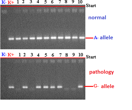
Figure 1. Electrophoregram of detection of polymorphism (A/G) of mutation-1 glutathione-S-transferase P1 (rs--): K - Negative control; K + Positive control; 1,3,8,9 - wild genotype A / A; 2,4,5,6,7,10 - heterozygous genotype A / G
In carrying out genetic studies, a population control was used as the comparison group, which was represented by DNA samples (n = 72) of conditionally healthy donors (without any signs of atopic diseases) from the DNA bank of this department. DNA samples were isolated from peripheral blood lymphocytes in accordance with a modified procedure. The concentration and purity of the isolated DNA were evaluated by measuring the optical density of DNA-containing solutions at a wavelength of 260 and 280 nm against TE on a NanoDrop 2000 spectrophotometer (USA). Genotyping of GSTP1 polymorphism was carried out by PCR on programmable thermal cycler CG-1-96 Corbett Research (Australia) and 2720 Applied Biosystems (USA), using the test systems of OOO Liteh (Russia), according to the manufacturer's instructions. Temperature regime: 94°C - 4 min; 94°C - 30 s, 60°C - 30 s, 72°C - 30 - 35 cycle; 72°C - 7 min.
Statistical analysis of the results was carried out using the statistical software package "OpenEpi 2009, Version 2.3".

2.3. Results of the Study

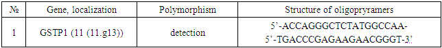
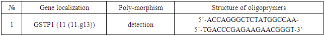
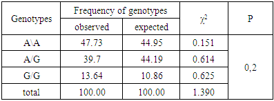
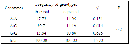
Among 88 patients with Alder Disease at the age of 14 years were 13, 15-20 years old - 12, 21-30 - 17, 31-40 years - 14, over 40 years - 32 patients. Allergic dermatitis was diagnosed in 88 patients with atopic dermatitis in 49 (55.7%) patients, 28 (31.8%) with urticaria and 11 (12.5%) with allergic dermatitis, respectively. Information on gene sequences and the structure of primers was obtained taking into account the original literature source [5] and GeneBank
The characteristics of the genetic marker and the sequence of synthesized oligopramers are given in Table 1.
Table 1. The sequence of oligonucleotide primers used for PCR
     
As can be seen from the table, a comparative analysis of the distribution frequencies of alleles and genotypes of polymorphism IIe 105 Val of the xenobiotics gene GSTP1 among 176 DNA samples in 88 patients with AlD revealed a normal allele A in 67.0% of cases and a G allele in 33% of cases, respectively. Whereas, in the control group, the frequency of occurrence of the mutant allele IIe 105 Val of the xenobiotic gene GSTP1 was 12.5%, which was 2.6 times lower in comparison with the main group (P<0.05).
We investigated the distribution of IIe 105 Val polymorphism of the gene of the xenobiotic enzyme GSTP1 in patients with AlD (Table 2; Table 3).
Table 2. Frequency of distribution of alleles and genotypes of polymorphism IIe 105 Val of the GSTP1 gene in patient groups and controls
     
Table 3. Differences in the incidence of alleles and genotypes of polymorphism IIe 105 Val of the GSTP1 gene in the main and control groups
     
The distribution of the frequencies of the genotypes of this polymorphism also revealed significant differences between the main and control comparison groups in the general sample (P <0.05). Increased associations of "functionally unfavorable" genotypes of A/G and G/G were revealed - 39.7% and 13.6%, respectively. According to the odds ratios, the risk of developing AlD. A in the main group in the presence of polymorphism is G/A (OR=2.6, 95%CI 1.264-5.382) and G/G (OR=11.2, 95%CI 1.421-88.43) in 1.9 and 12 times higher in comparison with the control healthy group. Such indicators in the groups studied had a statistically significant character (χ2=6.9; P<0.05; χ2=8.0; P<0.05).
As follows from Table 4, the frequency distribution of genotypes by RXV polymorphism IIe 105 Val of the GSTP1 gene in the main group of patients with Alder showed that the observed frequency of A/A genotypes was 47.7%, heterozygous A/G genotypes 39.7% and homozygous - G/G - 13.6%, respectively, whereas the expected frequency of genotypes of the A/A group and heterozygous - met 44.9 and 44.2%, respectively, and G/G - in 10.8% of cases.
Table 4. Expected and observed frequency of distribution of genotypes by RXV polymorphism IIe 105 Val of the GSTP1 gene in the main group of patients with allergodermatosis
     
Whereas in the control group, the observed and expected frequency of A/G heterozygote genotypes occurred in 19.4% and 19.7% of cases, and homozygous non-functional genotypes of G/G were 1.4 and 1.2%, respectively. (table 5).
Table 5. Expected and observed frequency of distribution of genotypes by RXV polymorphism IIe 105 Val of the GSTP1 gene in the main group of patients with allergodermatosis
     
As can be seen from Table 6, in patients with allergic dermatoses, the frequency of observed heterozygosity of polymorphism Iie 105 Val of the GSTP1 gene was 39.7%, which is 2.05 times higher than the control group, and the frequency of expected heterozygosity was 44.2%, which is 2,2 times higher than those of healthy individuals. (P<0.05).
Table 6. Difference between the expected and observed heterozygosity frequencies of polymorphism IIe 105 Val of the GSTP1 gene
     
An analysis of the results shows that the distribution of all the genotypes of polymorphism IIe 105 Val of the GSTP1 gene in the group of patients and control corresponds to RXV, indicating the absence of the influence of systematic or random factors that can change the genetic structure of populations. The study of the genetic structure of this marker revealed a relatively high level of expected heterozygosity in the main group of patients in relation to the control group (39.7% and 19.4%, respectively). In both groups the indicator D is to the left of 0, that is, it is negative (D<0). The revealed fact testifies to higher frequencies of expected heterozygotes, and not actually calculated heterozygotes.
When analyzing the frequency distribution of the occurrence of alleles and genotypes of a given polymorphism in the group of patients with allergic dermatoses, significant differences were found in comparison with the control group. The functionally unfavorable allele G of the GSTP1 gene 3.4 times statistically significantly prevailed in the studied chromosomes of patients with allergic dermatoses compared to the population sample (χ2 = 10.8; P <0.05; OR = 3.4; 95% CI 1.6- 7.4). The distribution of genotype frequencies of this polymorphism also revealed significant differences between the main group and the comparison group in the general sample (P <0.05). The associations of "functionally unfavorable" A/G genotypes were identified (χ2 = 6.9, P <0.05, OR=2.6, 95%CI 1.264-5.382) and G/G (χ2=8.0; P<0.05; OR=11.2; 95%CI 1.421- 88.43) with the development of allergic dermatoses.

3. Conclusions

Thus, allele G and hetero / homozygous genotypes of polymorphism IIe 105 Val of GSTP1 gene are significant markers of increased risk of allergic skin diseases in Uzbekistan (P <0.05). Allele A and the functionally beneficial genotype A/A are reliable protective markers for the development of pathology (χ2=16.5; P<0.05; OR=0.2; 95%CI 0.1186-0.4868).

References

[1]  AlievaV. Sh., Nazarov A. A, Arifov S. S., Karimov Kh. Ya., Boboev K. T. 2009, Analysis of the genes of anti-inflammatory cytokines associated with the risk of atopic allergic rhinitis. Medical Journal of Uzbekistan, – Tashkent, 3, 29-31.
[2]  Alimkhodzhaeva P. R., Karimov Kh. Ya., Muminova S. R. with co-workers. 2012, Development of systems for molecular testing and analysis of the relationship of the -590C> T polymorphism of the IL4 gene with atopic dermatitis. // Uzbekiston tibbyot reporter, 1, 19-21.
[3]  Sergeev Yu. V. 2002, Atopic dermatitis. A guide for doctors. edited by Medicine for all., 182 p.
[4]  Bochkov N. P. 2004, Clinical genetics. Textbook. 3rd ed. - Moscow: GEOTAR-MED, 480 p.
[5]  Bragina E. Yu. 2005, Comparative analysis of the structure of the hereditary component of susceptibility to bronchial asthma and tuberculosis according to the genes of enzymes of xenobiotics metabolism: Author's abstract. dis. ... cand. honey. sciences. Tomsk, 23 pp.
[6]  Gavalov S. M., Ryabova O. A., Vavilin V. A., Lyakhovich V. V., Makarova S. I. 2000, Association of gene polymorphisms of biotransformation and detoxification of xenobiotics with peculiarities of bronchial asthma in children, Allergology, 3, 14-21.
[7]  Karimov Kh. Ya., Saidov A. B., Boboev K. T., Assessorova Yu. Yu. et al. 2016, Fundamental and applied aspects of molecular genetics in medicine., Scientific publication. - Tashkent: IPTD "Uzbekistan", 352 p.
[8]  AlievaV. Sh., Nazarov A. A., Arifov S. S., Karimov Kh. Ya., Boboev K. T. 2009, Analysis of the genes of anti-inflammatory cytokines associated with the risk of atopic allergic rhinitis. Medical Journal of Uzbekistan, Tashkent, 3, 29-31.
[9]  Alimkhodzhaeva P. R., Karimov Kh. Ya., Muminova S. R. with co-workers. 2012, Development of systems for molecular testing and analysis of the relationship of the -590C> T polymorphism of the IL4 gene with atopic dermatitis. Uzbekistontibyotet reporter., 1, 19-21.
[10]  Davydova N. A., Dmitrieva A. I., Sevostyanova N. V. and others 2007, Analysis of polymorphic variants of glutathione-S-transferases T1, M1 and P1 genes in patients with prostate cancer. "XI Russian Cancer Congress": materials of the Congress. M., 224.
[11]  Sergeev Yu. V. 2002, Atopic dermatitis. A guide for doctors. Medicine for all. 182.
[12]  Bragina E. Yu. 2005, Comparative analysis of the structure of the hereditary component of susceptibility to bronchial asthma and tuberculosis according to the genes of enzymes of xenobiotics metabolism: Author's abstract. dis. ... cand. honey. sciences. Tomsk, 23 pp.
[13]  Vavilin V. A., Makarova S. I., Lyakhovich V. V., Gavalov S. M. 2002, Association of polymorphic genes of enzymes of biotransformation of xenobiotics with predisposition to bronchial asthma in children with hereditary complication and without it. Genetics. 38(4), 539-545.
[14]  Gavalov S. M., Ryabova O. A., Vavilin V. A., Lyakhovich V. V., Makarova S. I. 2000, Association of gene polymorphisms of biotransformation and detoxification of xenobiotics with peculiarities of bronchial asthma in children. Allergology, 3, 14-21.
[15]  Gorlanov I. A., Milyavskaya I. R. 1999, Features of the HLA system in atopic dermatitis and its relationship with clinical characteristics, endocrine and immune status in children. Allergology., 2, 17-20.
[16]  Gulyamova G. Sh., Mavlyanova Sh. Z., Boboev K. T. 2015, Role of the polymorphic version of the tumor necrosis factor gene-alpha in the development of atopic dermatitis in the population of Uzbekistan. Clinical dermatology and venereology. 14(4), 79-83.
[17]  Name E. N., Hanson K. P. 2003, The role of DNA diagnostics in modern oncology. Bulletin of the Russian Academy of Medical Sciences., 10, 3-5.
[18]  Karimov Kh. Ya., Saidov A. B., Boboev K. T., Assessorova Yu.Yu. et al. 2016, Fundamental and applied aspects of molecular genetics in medicine. Scientific publication. - Tashkent: IPTD "Uzbekistan", 352 p.
[19]  Lyakhovich V. V., Vavilin V. A., Makarova S. I. and others. 2000, The role of enzymes biotransformation of xenobiotics in predisposition to bronchial asthma and the formation of features of its clinical phenotype. Bulletin of the Russian Academy of Medical Sciences. 12, 36-41.
[20]  Mavlyanova Sh. Z. 2014, Atopic dermatitis. Monograph. Tashkent, 168s.
[21]  Popova S. N., Slominsky P. A., Galushkin S. N. 2002, Polymorphism of glutathione-8-transferases M1 and T1 in a number of Russian populations. Genetics. 3s(2), 2S1-2S4.
[22]  Shlyakhto E. V., Konradi A. O. 2002, The role of genetic factors in remodeling the cardiovascular system in GB., Arterial hypertension. 8(3), 107-112.
[23]  Abdel-Rahman S., El-Zein R. A., Anwar W. A., Au W. W. 1996, A multiplex PCR procedure for polymorphic analysis of GSTM1 and GSTT1 genes in population studies. Cancer. Lett. - 229-233.
[24]  Ali-Osman F., Akande O., Antoun G. et al. 1997, Molecular cloning, characterization, and expression in Escherichia coli of full-length cDNAs of three human glutathione S-transferase pi gene variants., J. Biol. Chem. 272. 10004-10012.
[25]  Altmuller J., Palmer L. J., Fischer G. et al. 2001, Genome wide scans of complex human diseases: true linkage is hard to find., Am. J. Hum. Genet., 69, 936-950.
[26]  Bartsch H., Nair U., Risch A. et al. 2000, Genetic polymorphism of CYP genes, alone or in combination, as a risk modifier of tobacco-related cancers., J. Cancer. Epidemiol. Prev., l, 3-28.
[27]  Ding X., Kaminsky L.S. 2003, Human extrahepatic cytochromes P450: Function in xenobiotic metabolism and tissue-selective chemical toxicity in the respiratory and gastrointestinal tracts., Annu. Rev. Pharmacol. Toxicol. 43. 149-173.
[28]  Gambaro G., Anglani F., D'Angel A. 2000, Association of genetic polymorphisms and complex disease., Lancet. 355, 308-311.
[29]  Hayes J.D., Pulford D.J. 1995, The glutatione S-transferase super-gene family: regulation of GST and the contribution of the isoenzymes to cancer chemoprevention and drug resistance // Crit. Rev. Biochem. Mol. Biol., 30. 445-600.
[30]  Hayes J. D., Flanagan J. U., Jowsey I. R. 2005, Glutathione transferases., Annu. Rev. Pharmacol. Toxicol., 45, 51-88.
[31]  Hayes J. D., McLellan L. I. 1999, Glutathione and glutathione-dependent enzymes represent a coordinately regulated defense against oxidative stress., Free Radic. Res., 31, 273-300.