-
Paper Information
- Next Paper
- Paper Submission
-
Journal Information
- About This Journal
- Editorial Board
- Current Issue
- Archive
- Author Guidelines
- Contact Us
American Journal of Medicine and Medical Sciences
p-ISSN: 2165-901X e-ISSN: 2165-9036
2018; 8(5): 91-95
doi:10.5923/j.ajmms.20180805.01

Liver Enzymes and Micro Hepatic Histoarchitecture in Adult Male Wistar Albino Rats treated with Methanol Leaf Extract of Securidaca longipedunculata Fres (Polygalaceae)
 Abstract
 Reference
 Full-Text PDF
 Full-text HTML
Anyebe Sunday Simon1, Emmanuel Nachamada Solomon2, Ogweje Abigail Egena2, Olubiyi Makinde2, Ejiogu Chioma Deborah2, Makena Wusa3, Yaduma Gaiuson Wandiahyel4, Chima Chinonso Nicodemus2, Samaila Saminu2, Mustafa Ibrahim Oladayo2, Ajibade Victoria2, Abubakar Sani Buba2
1Ahmadu Bello University Teaching Hospital Shika, Zaria, Nigeria
2Department of Human Physiology, Ahmadu Bello University, Zaria, Nigeria
3Department of Human Anatomy, Ahmadu Bello University, Zaria, Nigeria
4Department of Chemistry, School of Sciences, Adamawa State College of Education Hong, Nigeria
Correspondence to: Anyebe Sunday Simon, Ahmadu Bello University Teaching Hospital Shika, Zaria, Nigeria.
| Email: |  |
Copyright © 2018 The Author(s). Published by Scientific & Academic Publishing.
This work is licensed under the Creative Commons Attribution International License (CC BY).
http://creativecommons.org/licenses/by/4.0/

This study was designed to evaluate the hepatotoxic effect of Securidaca longipedunculata in adult wistar rats. Twenty four male (24) rats were randomized into four groups of six rats each (n=6) and treated as follows: Group one was administered normal saline (10 ml/kg), groups two, three and four were administered 60, 120 and 240 mg/kg doses of methanolic leaf extract of securidaca longipedunculata (MLESL) respectively. Administration was carried out by oral gavage for a period of twenty eight (28) days. At the end of experiment, animals were euthanized and blood samples collected, from which sera was obtained and used to assay for liver enzymes levels; Aspartate aminotransferase (AST), Alanine amino transferase (ALT) and Alkaline phosphatase (ALP). Liver tissues were harvested and used for histological study. Serum AST level was significantly elevated (P< 0.05) in all the extract treated groups; 60 mg/kg (49.50±1.23), 120 mg/kg (51.17 ±1.17) and 240 mg/kg (51.50±0.65) when compared to the control (44.17±1.74). A significant increase (P< 0.05) in serum ALT and ALP levels were observed in the 240 mg/kg extract treated group; (56.25±1.38) and (86.75±1.11) respectively when compared to the control (49.50±1.38). In conclusion, the extract has a good safety profile at low to moderate dose but has shown toxic potential at high dose over long use.
Keywords: Hepatic, Liver enzymes, Securidaca longipedunculata
Cite this paper: Anyebe Sunday Simon, Emmanuel Nachamada Solomon, Ogweje Abigail Egena, Olubiyi Makinde, Ejiogu Chioma Deborah, Makena Wusa, Yaduma Gaiuson Wandiahyel, Chima Chinonso Nicodemus, Samaila Saminu, Mustafa Ibrahim Oladayo, Ajibade Victoria, Abubakar Sani Buba, Liver Enzymes and Micro Hepatic Histoarchitecture in Adult Male Wistar Albino Rats treated with Methanol Leaf Extract of Securidaca longipedunculata Fres (Polygalaceae), American Journal of Medicine and Medical Sciences, Vol. 8 No. 5, 2018, pp. 91-95. doi: 10.5923/j.ajmms.20180805.01.
Article Outline
1. Introduction
- The liver is the primary site of drug metabolism; other tissues, such as kidney, brain, skin, blood, lung and gastrointestinal mucosa, also contribute [1]. The liver is a large, complex organ that is well designed for its central role in carbohydrate, protein and fat metabolism. It is the site where waste products of metabolism are detoxified through processes such as amino acid deamination, which produces urea. In conjunction with the spleen it is involved in the destruction of spent red blood cells and the reclamation of their constituents. It is responsible for synthesizing and secreting bile and synthesizing lipoproteins and plasma proteins, including clotting factors [2]. ALP is present in mucosal epithelia of small intestine, proximal convoluted tubule of kidney, bone, liver and placenta. It performs lipid transportation in the intestine and calcification in bone. The serum ALP activity is mainly from the liver with 50% contributed by bone [3]. ALT is found in kidney, heart, muscle and greater concentration in liver compared with other tissues of the body. ALT is purely cytoplasmic catalyzing the transamination reaction [3]. AST catalyze transamination reaction. AST exist two different isoenzyme forms which are genetically distinct, the mitochondrial and cytoplasmic form. AST is found in highest concentration in heart compared with other tissues of the body such as liver, skeletal muscle and kidney [3, 4]. Securidaca longepedunculata is a semi deciduous shrub or small tree that grows between six to twelve metres high. It has a pale grey, smooth bark with leaves that grow in clusters. The branches are small and have very fine hairs when young but they lose them as they mature. The leaves are variable in size and shape, alternate, often in cluster. The tree produces flowers that grow in the early part of summer and change from pink to purple. They are sweetly scented and grow in bunches on a peduncle. The fruit of this tree is round with a wing on one side that can grow up to forty millimetres long. It can be seen between April and August [5]. The plant’s geographical spread over Africa includes South Africa, Mozambique, Kenya, Tanzania, Zimbabwe, Nigeria, Sudan and Ethiopia. It is highly regarded medicinal and magical tree [6]. Therefore this study was designed to evaluate the hepatotoxic effect of Securidaca longipedunculata in adult wistar rats.
2. Materials and Methods
2.1. Animals
- A total of twenty four (24) Wistar rats of both sexes were obtained from the Animal House of the Department of Pharmacology and Therapeutics, Ahmadu Bello University, Zaria for the study. The animals were housed under standard laboratory conditions of temperatures (25±2°C), and light approximately 12/12 hours light/dark cycle and fed on standard diet and given water ad libitum. The animals were observed for one week prior to the study to acclimatize.
2.2. Chemicals and Drugs
- All drugs and chemicals that were used were purchased from a registered company and of analytical grade. Some of which include; Extraction solvent; absolute alcohol (Sigma-Aldrich, USA), Methanol (Sigma-Aldrich, USA), Piroxicam (Neimeth, Nigeria), Normal saline (0.9% Normal saline Isotonic Solution).
2.3. Preparation of Plant Extract
- The leaves were pruned while fresh, air dried at room temperature for two weeks, pulverized in a mortar, and sieved to fine powder. Five hundred grams (500 g) of the powdered material were extracted with methanol for 72 hours. The extract was concentrated using rotary evaporator and finally evaporated to dryness in a water bath at 35-40°C. This gave a yield of 126g (25.2% w/w). The product which was coffee brown in colour was regarded as the methanol leaf extract of Securidaca longepedunculata (MLESL) and was stored in desiccator for use.
2.4. Experimental Protocol
- A total of twenty four (24) adult Wistar rats of both sexes were used for this study were randomly grouped into four (4) groups of six animals each (n=6) treated as follows;

2.4.1. Effect of Methanolic Leaf Extract of Securidaca longepedunculata on Liver Enzymes
- Blood samples were collected in plain anti-coagulant free bottles and taken to the Department of Chemical Pathology, Ahmadu Bello University Teaching Hospital, Shika, Zaria, where they were centrifuged at 3000 rpm, using a centrifuge (Centurion model GP (CD295-30) for 10 minutes and a clear serum obtained. The levels of serum liver enzymes such as alanine aminotransferase, (ALT), alkaline phosphatase (ALP), aspartate aminotransferase (AST), were determined using Autoanalyser selectra XL (Vital scientific, Netherland) and results obtained were recorded.
2.4.2. Effect of Methanolic Leaf Extract of Securidaca longepedunculata on Liver Histology
- The histological examination was carried out in the Department of Human Anatomy, Ahmadu Bello University, Zaria. The tissues were dehydrated in ascending grades of ethanol (50-100%), cleared in xylene and portion of the tissues were embedded in paraffin wax. Histological sections of 5-6 µm in thickness were prepared according to standard micro techniques into glass slide and stained with haematoxylin and eosin. Photomicrographs under light microscope at high power magnifications (x 250) were examined for histopathological changes.
3. Statistical Analysis
- Results were expressed as Mean ± Standard Error of Mean. Data were analysed using one way analysis of variance (ANOVA) and repeated measure ANOVA test and were followed by Bonferroni post hoc test. Results were considered significant at P≤ 0.05. Statistical Package for Social Sciences (SPSS) version 20 was used.
4. Results
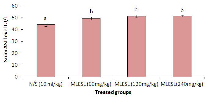 | Figure 1. Showing the result of serum AST. Different superscripts (a,b) indicate statistical significance compared to control (normal saline N/S) at P< 0.05, (One-way ANOVA followed by Tukey Post Hoc Test). MLESL = methanol leave extract of Securidaca longepedunculata |
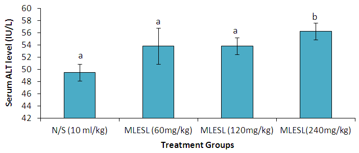 | Figure 2. Showing the result of serum ALT. Different superscripts (a,b) indicate statistical significance compared to control (normal saline N/S) at P< 0.05, (One-way ANOVA followed by Tukey Post Hoc Test). MLESL = methanol leave extract of Securidaca longepedunculata |
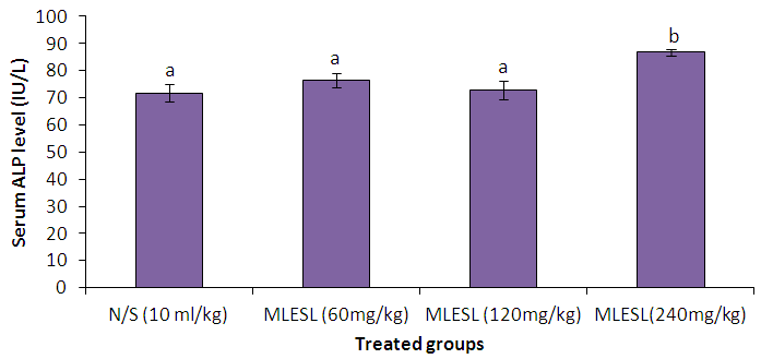 | Figure 3. Showing the result of serum ALP. Different superscripts (a,b) indicate statistical significance compared to control (normal saline N/S) at P< 0.05, (One-way ANOVA followed by Tukey Post Hoc Test). MLESL = methanol leave extract of Securidaca longepedunculata |
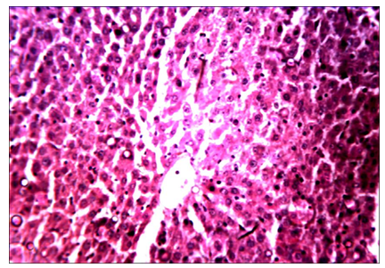 | Plate I. Liver section of rats administered normal saline for 28 days, showing normal liver histoarchitecture (H & E, x250) |
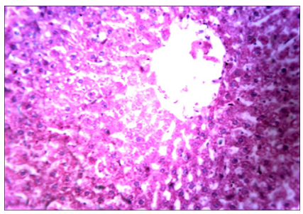 | Plate II. Liver section of rats treated with 60 mg/kg of MLESL for 28 days showing intense vacuolation of the hepatocytes with necrosis (H & E, x250) |
 | Plate III. Liver section of rats treated with 120 mg/kg of MLESL for 28 days showing mild perivascular necrosis (H & E, x250) |
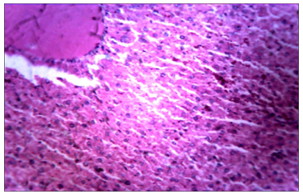 | Plate IV. Liver section of rats treated with 240 mg/kg of MLESL for 28 days showing venous and sinusoidal congestion, kupfer cell hyperplasia and moderate perivascular necrosis (H & E, x250) |
5. Discussion
- Elevated serum transaminase activities have been used in the diagnosis of pathological conditions the likes of myocardial infarction and hepatic necrosis [7]. Alanine Amino transaminase (ALT) and Aspartate amino transaminase (AST) are non-plasma specific enzymes involved in the deamination of aspartic acid and alanine respectively [8]. In this study, increase serum AST as shown in figure 1 above is a marker of aggression against hepatocytes [9]. This could have been from the activities of deleterious metabolites formed in the liver during the metabolism of the extract. This could also be the reason for the increased level of serum ALT and ALP as observed in this study. Serum ALT is known to increase when there is liver cell damage and it has been employed as a tool for measuring hepatic necrosis. AST however, is not a liver specific enzyme as high levels of the enzyme in serum can also be found in skeletal and cardiac muscle injury as well as red blood cell haemolysis. High level of alkaline phosphatase in the serum is usually indicative of cholestasis which may also result in progressive liver disease [10]. Repeated treatment with the extract in this study caused a statistically significant rise in ALT, AST and ALP. The finding of this study is in consonance with the findings [11]. The elevated liver enzymes are a pointer to hepatocellular toxicity of the extract. This is further substantiated by the histopathological findings in the liver.Histopathological studies have been concluded to help establish casual relationships between drug substances and various pathophysiological responses in the liver. It has proven to be a sensitive tool to detect effects of chemical compounds within target organs. The histopathological picture of the organs can corroborate with the biochemical changes accounting for the functional disruptions in the activity of the organs due to cellular damage [11]. From this study, the result of liver histology from the extract treated groups show intense vacuolation of the hepatocytes and mild perivascular necrosis. With increasing dose of the extract, there was observed, increased sinusoidal congestions, kupfer cells hyperplasia and moderate perivascular necrosis. This result suggests that at high doses, the extract could be hepatotoxic.
6. Conclusions
- This study was designed to evaluate the hepatotoxic effect of Securidaca longipedunculata in adult wistar rats using a total of 24 Wistar rats, and administered 60, 120 and 240 mg/kg dosages of the extract. Liver enzymes; ALT, AST and ALP were assayed alongside the histology of the liver tissue. Serum AST level was significantly elevated (P< 0.05) in all the extract treated groups; 60 mg/kg (49.50±1.23), 120 mg/kg (51.17 ±1.17) and 240 mg/kg (51.50±0.65) when compared to the control (44.17±1.74). In conclusion, the extract has a good safety profile at low to moderate dose but has shown toxic potential at high dose over long use.