-
Paper Information
- Paper Submission
-
Journal Information
- About This Journal
- Editorial Board
- Current Issue
- Archive
- Author Guidelines
- Contact Us
American Journal of Medicine and Medical Sciences
p-ISSN: 2165-901X e-ISSN: 2165-9036
2018; 8(3): 49-53
doi:10.5923/j.ajmms.20180803.03

The Relationship between Umbilical Obesity and Coronary Atherosclerosis Study in Monozygotic Goat Twins
 Abstract
 Reference
 Full-Text PDF
 Full-text HTML
Evren Burşuk1, Omer Mehmet Erzengin2, Esra Erzengin Ozdemir3, Faruk Erzengin4
1Program of Biomedical Technologies, University of Istanbul, Vocational School of Technical Sciences, Istanbul, Turkey
2Department of Gynecology, University of Istanbul, Veterinary Faculty, Istanbul, Turkey
3Department of Business (Statistical), University of Beykent, Faculty of Economics and Administrative Sciences, Istanbul, Turkey
4Department of Cardiology, Previous Dean, University of Istanbul, Istanbul Medical Faculty, Istanbul, Turkey
Correspondence to: Faruk Erzengin, Department of Cardiology, Previous Dean, University of Istanbul, Istanbul Medical Faculty, Istanbul, Turkey.
| Email: |  |
Copyright © 2018 Scientific & Academic Publishing. All Rights Reserved.
This work is licensed under the Creative Commons Attribution International License (CC BY).
http://creativecommons.org/licenses/by/4.0/

We have recently proved that; there is a close relationship between umbilical obesity and coronary atherosclerocalcifications. We have investigated 36 monozygotic male goat twins relationship between umbilical obesity and coronary atherosclerosis. Randomly selected 12 goats were left to graze in open pasture (B), and the remaining 24 were kept in stables and pens (A), and fed the identical feed. The A group goats were observed having all umbilical obesity and also after slaughter process, more prominent epicardial fat tissue were seen macroscopically. The remaining unslaughtered 12 goats of Group A were treated for atherosclerosis for 6 months, using a combination of drug. All of the Group A goats had indicators of obesity in their body and visible epicardial fat tissue was observed, atherosclerocalcific plaques were found in the coronary artery, and some narrowing was observed in artery lumens. Making a comparison between Group A and Group B was found statistically significant (p=00001) our results. The remaining 12 goats were treated for atherosclerosis for 6 months, using a combination of drugs. Following the treatments, very little epicardial fat tissue was observed, and 8 of the goats had perfectly normal coronary arteries with the remaining four having minimal atherosclerosis plaques. The Group A goats had significant obesity unlike Group B as well as epicardial fat tissue and significant coronary atherosclerocalcifications. Moreover, the unslaughtered ones of Group A were treated with ideal drugs, epicardial fat tissue and coronary atherosclerosis declined significantly compared to their co-twins that did not receive this treatment.
Keywords: Umbilical obesity, Coronary atherosclerosis, Pericardial fat
Cite this paper: Evren Burşuk, Omer Mehmet Erzengin, Esra Erzengin Ozdemir, Faruk Erzengin, The Relationship between Umbilical Obesity and Coronary Atherosclerosis Study in Monozygotic Goat Twins, American Journal of Medicine and Medical Sciences, Vol. 8 No. 3, 2018, pp. 49-53. doi: 10.5923/j.ajmms.20180803.03.
1. Introduction
- Despite the steady progress in the treatment of atherosclerotic and atherothrombotic cardiovascular diseases, coronary artery diseases and myocardial infarctions continue to be significant causes of morbidity and mortality, especially in industrialized countries. The term atherosclerosis refers to the thickened and hardened lesions, which have lipids and at the end calcifications in the intimae and media of elastic and muscular arteries. To date, the primary cause of arterial atherosclerotic calcifications has not yet been elucidated. It is generally accepted that atherosclerotic and calcified lesions first appear and develop within the innermost layer of the arteries (at the intimae) (Figure 1).
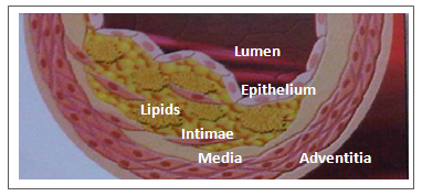 | Figure 1. Classical pathway and accumulation of the lipids core in the intimae have been shown in this figure |
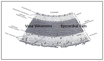 | Figure 2. This figure shows epicardial fats tissue, vasa vasorums and tunicas of an artery (endothelium, intimae, tunica elastica interna, media, tunica elastica externa and adventitia) |
2. Subjects and Methods
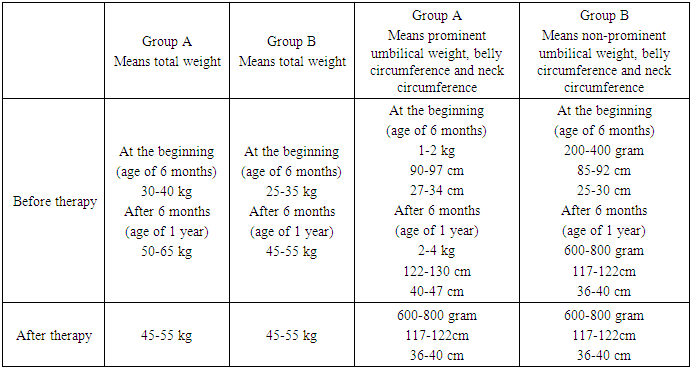
- This study was conducted with 36 monozygotic male goat twins older than 6 months, and lasted two years. At the beginning of the study (at age of 6 months), weights of the goats were between 25-40 kilograms and after 6 months (at the age of 1 year) between 45-65 kg. At the end of the study (after therapy) their means weights became between 45 and 55 kilograms same as non-prominent umbilical group (Table 1). Ethical committee approval was received from Istanbul University, Medical Faculty of the Advanced Research Laboratory.
|
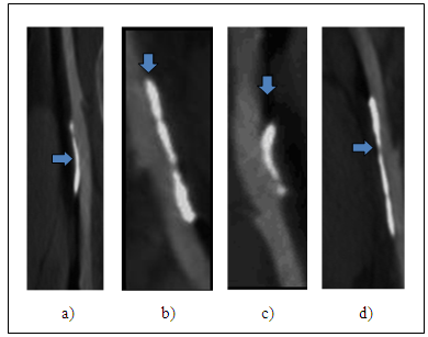 | Figure 3. Adventitial atherosclerocalcifications (atheromatous components) of on the femoral arteries of a) Guinea pig, b) Rabbit, c) Goat, d) Mouse. The grey areas are soft plaque (lipids core), the white areas are calcified plaques |
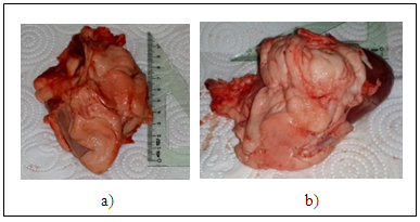 | Figure 4a-b. These figures show more prominent epicardial fat tissue were seen goats having umbilical obesity |
|
|
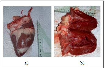 | Figure 5a-b. This figures show no epicardial fat tissue were seen goats having less umbilical obesity treated with medically |
3. Results
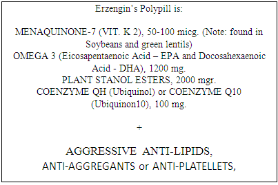
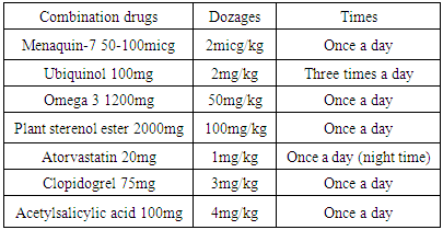
- All of the grazing goats (Group B) and 12 of the stable goats (Group A) were slaughtered at the end of six months (when they turned one); none of the 12 goats in the grazing group (Group B) had any indicators of obesity in their waist, neck or abdominal regions, and no trace of epicardial fat was found in their hearts. In addition, during properly conducted coronary artery cross sectional analysis, macroscopic and microscopic inspection of the layers (Adventitia, Media, Intima) of the coronary artery showed that there were no atherosclerotic or calcific plaques in any of the layers, and the coronary arteries were found to be perfectly normal (Group B, Figure 5a-b).All of the goats kept in stables and pens (Group A) had indicators of obesity in their waist, neck and abdominal regions, and in heart examinations following the slaughter of randomly selected 12 of these goats that turned one, visible epicardial fat tissue was observed, atherosclerocalcific plaques at various stages of development were found in the adventitia and Intima layers of the coronary artery, and significant (medium-advanced) narrowing was observed in artery lumens (Group A). The comparison between Group A and Group B showed that differences in epicardial fat tissue and coronary artery plaques were statistically very significant (p=00001).The remaining 12 goats were treated for atherosclerosis for 6 months, using a combination of (4+3=7) drugs (Menaquinone-7 50-100micg, Ubiquinol 100mg, Omega-3 1200mg, Plant Stanol Ester 2000mg, Atorvastatin 20mg, Clopidogrel 75mg, Acetylsalicylic Acid 100mg) (Table 2a-b). Following the slaughter at the end of six months of treatment, very little epicardial fat tissue was observed, and properly conducted coronary artery cross sectional analysis showed that 8 of the goats had perfectly normal coronary arteries both in macroscopic and microscopic terms, with the remaining four having minimal atherosclerosis plaques. Significant narrowing was not observed in any of the goats in this group. The differences between the results from this group and the pasture grazing group were found to be statistically insignificant.
4. Discussion
- Thus, our surprising findings were that the formation of the atherosclerotic and calcified plaques begin and develop not only in the intimae or media (Mönckeberg’s Sclerosis), but also on the adventitia (Erzengin’s Arterial Atherosclerotic Calcifications) of the coronary arteries and/or all of the medium and large arteries [11-12]. We suggest that the formation of the Adventitial Atherosclerotic calcifications is caused by vasa-vasorums and/or diffusion from the epicardial adipose tissues around the pericardium.
5. Conclusions
- The main findings of this study, which was conducted over a two-year period with 36 (18 pairs) identical monozygotic goats, are as follows:1) Twin subjects raised in stable and pen, had significant obesity in their bodies, unlike those raised in mobile conditions in open pasture, as well as epicardial fat tissue and significant coronary adventitial atherosclerosis.2) It was found that when twins raised in stable and pen, were treated with ideal drugs, epicardial fat tissue and coronary atherosclerosis declined significantly compared to their co-twins that did not receive this treatment. 3) This study is the first in the literature which is conducted with monozygotic twins.4) In this study, we couldn’t assessed the other criteria of the Metabolic syndrome (such as blood pressure, blood cholesterol and sugar levels etc) (for that time being), but we can also assume that this study suggests the connection between Metabolic syndrome and atherosclerosis.