-
Paper Information
- Next Paper
- Previous Paper
- Paper Submission
-
Journal Information
- About This Journal
- Editorial Board
- Current Issue
- Archive
- Author Guidelines
- Contact Us
American Journal of Medicine and Medical Sciences
p-ISSN: 2165-901X e-ISSN: 2165-9036
2017; 7(6): 242-247
doi:10.5923/j.ajmms.20170706.03

The Secret of the Silence of the Silent Maxillary Sinus Syndrome
Abdullah M. Nasrat1, Randa M. Nasrat2, Mohammad M. Nasrat2
1Department of Surgery, Zytona Center, Medina, KSA
2Department of Internal Medicine, Helwan General Hospital, Helwan, Egypt
Correspondence to: Abdullah M. Nasrat, Department of Surgery, Zytona Center, Medina, KSA.
| Email: |  |
Copyright © 2017 Scientific & Academic Publishing. All Rights Reserved.
This work is licensed under the Creative Commons Attribution International License (CC BY).
http://creativecommons.org/licenses/by/4.0/

The study aimed todemonstrate a hidden influence of the bacterium Helicobacter pylori in the pathogenesis of the silent maxillary sinus syndrome.Sinusitis is a disease with significant discomfort affecting health and quality of the patient’s life, it is one of the most common chronic diseases involving different age groups.Diagnosis of sinusitis is clinical and the standard of choice for the detection of micro-organisms that cause sinusitis is culture of sinus discharge drainage. The etiology of chronic sinusitis is not completely known and due to the fact that there is no standard treatment for the disease, routine cultures are often made to assist empirical antibiotic prescribing therapy. As the etiology of chronic sinusitis is not clearly understood, the frequency of all causative agents of the disease must be adequately determined. H. pylori could migrate or get forced to migrate to the maxillary sinus under the influence of antibiotic violence leading to local tissue inflammatory reaction. H. pylori was detected in the nasal and maxillary sinus tissue specimens of some patients with chronic sinusitis associated with gastric existence of H. pylori. The study of H. pylori DNA extracted from patients with gastric reflux disease and chronic rhino-sinusitis emphasized prevalence of H. pylori in the oral and nasal cavities and similarity of the same H. pylori strain genotype among the same family but whether the strain genotype of gastric H. pylori is mostly identical with that of the oro-nasal strains and whether H. pylori is leading to chronic rhino-sinusitis or its existence in the maxillary sinus is a result of rino-sinusitis remained rather indefinite for some investigators.Sixteen middle aged patients with resistant symptoms of recurrent maxillary sinusitis in spite of adequate medications and sinus drainage were included in the study. H. pylori DNA extraction was done for the drained sinus discharge. Patients who proved positive for existence of H. pylori in the maxillary sinus discharge were encouraged for following the traditional habit of using the chewing stick even after every small bite of food in order to interfere with the nutrition of H. pylori from remnants of food particles in the mouth. Inhalation of the smell of white vinegar once or twice per day and daily mouth wash with diluted white vinegar was requested from them in order to disappoint H. pylori from the atmosphere of its new secondary habitat in the oro-nasal cavity. H. pylori was detected in the maxillary sinus drainage in fourteen patients. All patients showed improvement of symptoms within three days while twelve of them demonstrated disappearance of all symptoms in one week with clearance of the maxillary sinus in X-ray. Interestingly, the two patients where H. pylori was not detected in the sinus preferred also to follow the same traditional therapy and they quit after reasonable improvement of their symptoms. On conclusion, H. pylori could migrate and exist in the maxillary sinus as a secondary habitat particularly in those patients with resistant symptoms of sinusitis; H. pylori in this situation most probably accounts for the symptoms of sinusitis. Existence of H. pylori in the maxillary sinus could be the hidden reason behind creep up of a maxillary sinus syndrome in silence among some people. Hence, regular mouth hygiene could be an integral measure to protect from developing silent collapse of the maxillary sinus in association with existence of abnormal-behavior H. pylori strains in the stomach.
Keywords: Arak, Chewing stick, Helicobacter pylori, Maxillary sinus syndrome, Miswak, Vinegar
Cite this paper: Abdullah M. Nasrat, Randa M. Nasrat, Mohammad M. Nasrat, The Secret of the Silence of the Silent Maxillary Sinus Syndrome, American Journal of Medicine and Medical Sciences, Vol. 7 No. 6, 2017, pp. 242-247. doi: 10.5923/j.ajmms.20170706.03.
Article Outline
1. Introduction
- Helicobacter pylori colonized the stomach since an immemorial time as if both the stomach and the bacterium used to live together in peace harmless to each other. [1, 2] As prevalence of the migrating abnormal behavior H. pylori strains due to the antibiotic violence and their existence in unusual secondary reservoirs other than the common gastric habitat is associated with local tissue pathology and symptomatic disorders; therefore, it should be of vital significance to identify the secondary reservoirs of this bacterium. The maxillary sinus is one of the common and most critical sites among these secondary reservoirs of H. pylori as anti-H. pylori antibiotics are seldom effective against extra-gastric H. pylori strains and it is difficult to force or attract H. pylori to return back to the stomach again. [1-3] Maxillary sinusitis is a common finding and an important issue in dentistry and maxillofacial surgery. [4] The silent maxillary sinus syndrome or silent maxillary atelectasis is rare but serious clinical event; it is due to accumulation of profuse amounts of mucus leading to obliteration of the sinus cavity. Chronic maxillary collapse is characterized by progressive enophthalmos secondary to maxillary sinus hypoventilation. [5, 6] The silent maxillary sinus syndrome presents with unilateral enophthalmos without any particular frank sinus symptoms. The reason the sinus component of the disease remains mostly asymptomatic and is discovered only after thorough evaluation of enophthalmos is unclear. [6-8] The most effective treatment of the silent maxillary sinus syndrome is endoscopic maxillary antrostomy and reconstruction of the orbital floor. [5, 7] The chewing stick or the Arak stick (miswak) which is an organic natural green toothbrush that requires no toothpaste is obtained from the Salvadora persica (the Arak) tree. Various studies have demonstrated strong antibacterial activity of the chewing stick with immediate effect against oral and cariogenic bacteria. Further reports have emphasized significant in vitro influence of the crude chewing stick extract on the activity and growth of oral pathogens. The world health organization has suggested and encouraged the use of the chewing stick as an effective remedy for oral hygiene. The traditional reputation guidance recommends the chewing stick for purification of the mouth. [9-12]Dietary vinegar (acetic acid 5%) has been recently shown to be an effective and decisive measure for the clinical eradication of the abnormal-behavior H. pylori strains with an immediate dramatic relief of patient’s symptoms. [2, 13-15] The complex nutritional requirements of H. pylori are achieved mainly via utilization of pyruvate. As acetate is demonstrated as an end product among the metabolic pathway of H. pylori and the activity of the pyruvate dehydrogenase complex is controlled by the rules of product inhibition and feedback regulation; hence, the addition of acetic acid to the atmosphere around H. pylori could compromise the energy metabolism of H. pylori or interfere with the bacterium respiratory chain metabolism. [16-19] As long the matter includes interference with the energy metabolism and the respiratory chain metabolism of H. pylori; an immediate lethal effect on the bacterium could be considered.
2. Aim
- Demonstration of a hidden influence of the bacterium H. pylori in the pathogenesis of the silent maxillary sinus syndrome.
3. Design & Settings
- Prospective study done in Jeddah, Saudi Arabia between October 2014 and May 2015.
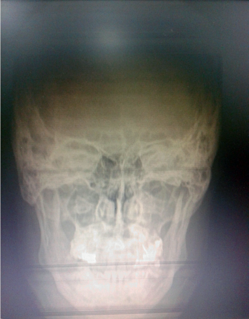
 | Figure 1. Shows veiling of the maxillary antrum, more on the right side, of one patient before starting therapy |
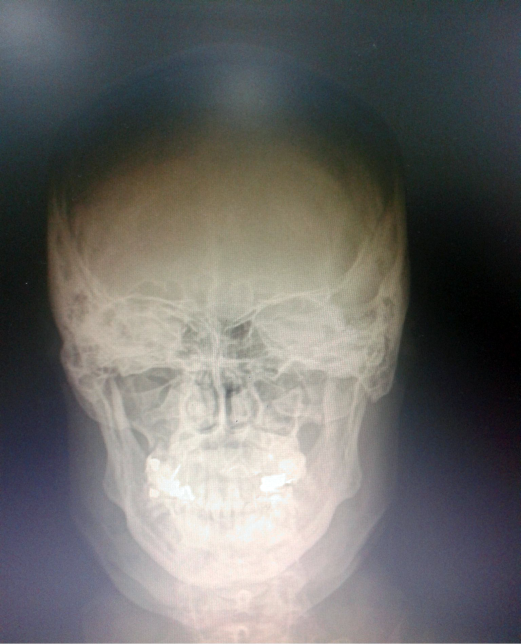
 | Figure 2. Demonstrates persistence of opacity of the maxillary sinus of the same patient with faint improvement in spite of two weeks of adequate therapeutic medication including potent antibiotics |
4. Patient & Method
- Sixteen middle aged patients with resistant symptoms of recurrent maxillary sinusitis in spite of adequate medications and sinus drainage were included in the study. H. pylori DNA extraction was done for the drained sinus discharge. Patients who proved positive for existence of H. pylori in the maxillary sinus discharge were encouraged for following the traditional habit of using the chewing stick even after every small bite of food in order to interfere with nutrition of H. pylori from remnants of food particles in the mouth. Inhalation of the smell of white vinegar once or twice per day and daily mouth wash with diluted white vinegar was requested from them in order to disappoint H. pylori from the atmosphere of its new secondary habitat in the oro-nasal cavity.
5. Results
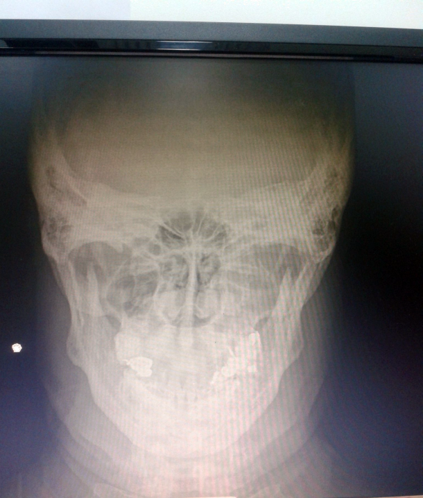
- H. pylori was detected in the maxillary sinus drainage in fourteen patients. All patients showed improvement of symptoms within three days while twelve of them demonstrated disappearance of all symptoms in one week with clearance of the maxillary sinus in X-ray. Interestingly, the two patients with negative H. pylori existence in the sinus preferred to follow the same traditional therapy and they quit the study after fair improvement of their symptoms in few days.
 | Figure 3. Shows clearance of the maxillary sinus shadow after completing one week therapy with the natural remedy |
6. Discussion
- H. pylori colonized the stomach as its main habitat since an immemorial time. H. pylori could migrate or get forced to migrate to the maxillary sinus under the influence of antibiotics. [1, 2] H. pylori escapes from the antibiotic aggression and hides in the maxillary sinus and other secondary sites in the body as antibiotics are seldom effective against extra-gastric H. pylori strains. [20] The reason that the investigators missed to detect H. pylori in the sinus discharge in cases of chronic sinusitis might be due to the conventional practice of performing routine bacterial cultures without much attention towards the specific tests for detecting H. pylori like urease (CLO) test or DNA extraction. [7, 8] H. pylori survives in the stomach since unrecognized time leading the attitude of natural bacteria as if both the stomach wall and the bacterium used to live together in peace harmless to each other. The attitude of H. pylori in the stomach simulates the behavior of natural bacteria as supported by the observational facts of its existence since an immemorial time, having majorly harmless long history inside the stomach before the anti-H. pylori antibiotics, its huge biological talents of survival among the hell fire of the strong gastric acid and its unavoidable gastric recurrence, [1, 2, 15] that is in addition to its protective influence against low acidity-related carcinoma of the cardia of stomach and the possibility of its defensive role towards esophageal reflux disease. [2, 21-24] Hence H. pylori can survive in the colon; what forces a weak bacterium that suffocates due brief exposure to a weak acid to select by its own a shelter inside the hell fire of the gastric acid unless it is a natural bacterium and is obliged for a natural biological function in the stomach!! So long H. pylori could survive in the mouth, if the matter is up to its own; why it does not choose the mouth and enjoy fun and company with millions of bacteria there!! Maxillary sinusitis includes the cardinal signs of pathologic inflammatory sequels such as pain, fever and odor. [4] Silence of the sinus syndrome which is related to H. pylori existence should mean absence of these cardinal signs with consequent creep of the sinus syndrome in silence towards atelectasis without any apparent symptoms like fever, pain or bad smell. Actually, the matter is not totally silent as there should be negligible recurrent unilateral nasal obstruction, nasal discharge and recurrent odorless faintly-brownish post-nasal discharge; it is apparently odorless and even leaves a slightly sweetish sensation in the throat due to non-change of its muco-polysaccharide component by any infective element. These negligible manifestations (recurrent nasal obstruction/discharge and recurrent post-nasal secretions) could be easily missed or ignored by the patients even the attention of their physicians could miss that these symptoms may overlie a serious sequel. The reason that H. pylori can reside in the maxillary sinus in silence without causing remarkable pathologic signs is the observational finding that H. pylori is not essentially pathologic by its own; a normal-behavior H. pylori is only recognized by the stomach wall tissues, it is mostly peaceful and useful to the stomach protecting it via elaboration of ammonia from its gastric acid if it goes in excess. Migration of H. pylori to extra-gastric sites will render the bacterium a foreign structure to the tissues leading to initiation of local tissue response or inflammatory reaction. [1, 2, 15] H. pylori in the sinus would remain surrounded with ammonia at its immediate vicinity, the local irritation caused by this ammonia stimulates mucus secretion; therefore, existence of H. pylori in the maxillary sinus will cause continued mucus production either directly or via the effect of shear stress until obliteration or collapse of the sinus cavity happens in silence. [25-27] For the same reason that H. pylori is essentially a natural bacterium and non-pathogenic in nature, the mucus produced in the sinus is not infected, hence; it is odorless and is not associated with fever or other constitutional symptoms of maxillary sinusitis such as pain, a matter that explains the silent consequences of the sinus syndrome. Usually obstruction favors infection, hence; what causes absence of infection in an obstructed maxillary sinus unless the causative reason is not essentially an infective pathogen. H. pylori could also migrate to the middle ear; in a study of chronic otitis media with effusion associated with gastro-esophageal reflux disease, although H. pylori was readily detected in the middle ear discharge, the role of the bacterium in chronicity of the ear effusion was missed and reported as not clear while chronicity of the middle ear pathology was attributed by the investigators to the associated reflux disease. [28] This might indicate that as much as miscorrelation in etiology of symptoms may happen in some conditions such as H. pylori-related otitis media, misdiagnosis could further happen in other pathologic situations like the silent maxillary sinus syndrome.Concerning criteria of inclusion of patients, the patients were selected in this study so that their symptoms were limited to recurrent negligible unilateral nasal obstruction which lasts for short periods or few time of the day and a post-nasal discharge which did never smell bad. It was demonstrated also among patients of the study the association of un-explained occasional recurrent bronchial irritation with smooth or sticky bronchial secretions and transient chocking sensation which usually lasts for few time. The recurrent sensation of light-headedness or as expressed by some patents ‘heaviness of the head’ and the increased desire to sleep or desire to go back to sleep after short time from waking up were considered as part of the constitutional symptoms of the sinus obstruction. In addition, easy fatigue was found constant in those patients; it was attributed to loss of the physiologic nature and function of the sinus or to the original H. pylori dyspeptic disease. These symptoms were constant in all patients in spite of adequate medical therapy and all of them had got used to these symptoms, even some of their physicians considered these symptoms negligible and they re-assured their patients that they can survive with these negligible residual symptoms. The patients were selected in this study in purpose according to this particular clinical history as those patients are the candidates who are possibly expected to proceed and progress into silent collapse of the maxillary sinus. A smart questionnaire had been raised by an elegant research study as concerns the undesired sequels of existence of oral H. pylori strains, a question that is still remaining without answer by the researcher; “oral H. pylori, is it possible to stomach it again!!” [29] That was exactly the actual purpose and idea of methodology of this study; H. pylori as all living structures needs nutrition, in the stomach it feeds on remnants of food after travel of the meal from gastric lumen, when H. pylori resides in the maxillary sinus it similarly picks up remnants of food particles from the mouth and then escapes back to the sinus, [1, 2] the roaming of the bacterium in the mouth seeking its food could account for the recurrent bronchial secretions and the transient chocking sensation due to spreading of the irritant ammonia related to the immediate vicinity of the bacterium. Amazingly, H. pylori in the sinus is leading the same natural behavior as in the stomach where it does not exist in the oral cavity during presence of food but it picks up its nutrition after end of the meal; [1, 2, 15] hence, symptoms of bronchial irritation were noticed after finishing the food. Therefore, this study intended to interfere with nourishment of H. pylori via continuous purification of the mouth by using the strong antiseptic chewing stick after any food. It was found in this study that the chewing stick extract has got a direct lethal effect on H. pylori culture media. In addition, H. pylori was also repeatedly rendered disappointed towards the atmosphere of the mouth by inhaling the smell of vinegar and by the mouth wash with diluted vinegar. The expected outcome is that the maxillary sinus would become no longer a favorite shelter for H. pylori anymore; which is consistent with the results of this study. [9-12] Hence; the main purpose of employing the chewing stick (miswak) is to ensure purification of the mouth after intake of food in order to disappoint the bacterium from the oral atmosphere without the need of elimination of all remnants of food particles from the mouth. Brushing the teeth with the chewing stick at bed times even the tooth paste and brush are being used should be therefore integral. Accordingly, the habit of purification of the mouth by using the chewing stick, the smell of vinegar and the diluted vinegar mouth wash could be the answer for that question “how to render oral H. pylori strains return back to the stomach!!”. The principle of employing vinegar in this study was supported by the results of previous literature which reported that a brief exposure to high dilutions of acetic acid (0.03%) is sufficient to suffocate H. pylori. It has been also demonstrated that 20 times dilution of acetic acid 6% has got an immediate lethal influence on H. pylori culture media; [14, 30-33] That is an intense fast effect that explains why the mere smell of vinegar could be disappointing and terrifying to H. pylori. The smell of vinegar is strong and was found in this study instantly related to relief of nasal obstruction; it is not yet definite whether the relief of nasal symptoms related to the smell of vinegar was due to a local physical effect or a metabolic influence on H. pylori, the effect of the smell of vinegar was definite in this study but the exact mechanism of its effect still needs further adequate assessment. It should be also considered that vinegar according to the traditional reputation is a food that includes health but not a medicine to dilute in water and drink otherwise it is worse than antibiotics on gut flora. [2, 15] Those disadvantaged patients who develop H. pylori-related maxillary sinusitis should learn that they are liable for recurrences whenever they neglect their colonic condition or mouth hygiene; therefore, they should return to colon care/colon clear and keep strictly careful about oral hygiene with the chewing stick and white vinegar whenever needed. [14, 15, 34] Interestingly, all patients of this study were demonstrated to have one or more spots of bleeding gums that could ensure a source of organic urea and pyruvate for H. pylori nutrition. [2, 15] Therefore; those patients should treat any bleeding spots of the gum in order to deprive oral H. pylori strains from a constant source for its nutrition in the mouth. Persistent sinusitis with recurrent unilateral nasal obstruction and discharge, morning brownish odorless post-nasal discharge or at wake-up times, recurrent un-explained irritant cough upon having few bites of food that stops with smelling the vinegar or washing the mouth with diluted white vinegar and the recurrent development of sticky or soft bronchial secretions without apparent reason could be sufficient symptomatic predictors in a person for the possibility of progress and developing silent collapse of the maxillary sinus. H. pylori DNA detection in the post-nasal discharge for those patients could be a new health care predictor for the sinus syndrome.
7. Conclusions
- Migration and escape of H. pylori inside the maxillary sinus hiding from the antibiotic violence towards it could be the possible reason for maxillary sinusitis to creep in silence into obliteration and collapse of the sinus. The world misconception, medical attitudes and aggressive antibiotic behaviors towards H. pylori might be in need of serious revision and accurate re-determination. Regular mouth hygiene should be the integral shield to protect from developing silent collapse of the maxillary sinus in association with existing abnormal-behavior oral H. pylori strains. Improvement of the symptoms of chronic sinusitis upon inhaling the smell of vinegar could be an indication for a new health care predictor to screen patients with persistent nasal discharge and recurrent bronchial secretions for H. pylori DNA detection. The natural measures suggested in this study seem safe, effective and protective from developing silent collapse of the maxillary sinus.
ACKNOWLEDGEMENTS
- The study appreciates the continuous support offered by Abdul-Aziz Al-Sorayai Investment Company (ASIC) in Jeddah/Saudi Arabia and the true brotherhood friendly encouragement of Mr. Abdul-Aziz Al-Sorayai is extremely valued and appreciated.
 Abstract
 Reference
 Full-Text PDF
 Full-text HTML