-
Paper Information
- Previous Paper
- Paper Submission
-
Journal Information
- About This Journal
- Editorial Board
- Current Issue
- Archive
- Author Guidelines
- Contact Us
American Journal of Medicine and Medical Sciences
p-ISSN: 2165-901X e-ISSN: 2165-9036
2015; 5(2): 105-111
doi:10.5923/j.ajmms.20150502.08
Comparative Analysis of a Retrospective Vs Prospective Study Ascertaining Spectrum of Renal Lesions: Experience of a Tertiary Care Teaching Centre in North India
 Abstract
 Reference
 Full-Text PDF
 Full-text HTML
Rahul Mannan1, Manisha Sharma1, Manish Chandey2, Pramela A. Singh3, Vatsala Misra3, Mamta Singh3, Ravi Mehrotra4, Mridu Manjari1
1Department of Pathology, Sri Guru Ram Das Institute of Medical Sciences and Research, Amritsar (Punjab), India
2Department of Medicine, Sri Guru Ram Das Institute of Medical Sciences and Research, Amritsar (Punjab), India
3Department of Pathology, MLN Medical College, Allahabad (Uttar Pradesh), India
4Institute of Cytology and Preventive Oncology, NOIDA (Uttar Pradesh), India
Correspondence to: Rahul Mannan, Department of Pathology, Sri Guru Ram Das Institute of Medical Sciences and Research, Amritsar (Punjab), India.
| Email: |  |
Copyright © 2015 Scientific & Academic Publishing. All Rights Reserved.
The present study was undertaken over a period of thirteen years in two halves: 10 years retrospective and 3 years prospective at a single tertiary care centre in the northern part of India, so as to take make a comparison in the same area over a time period and also to over all take a glimpse into the pattern of the disease. Ten year retrospective data in this study showed that glomerulonephritis (57.55%) were the most common of the 172 renal lesions followed by chronic pyelonephritis on nephrectomy specimen (20.93%). Membranous glomerulonephritis (MG) was commonest glomerulopathy, accounting for 14.14% of all glomerulopathies. In the prospective study glomerulonephritis (78.39%) was the most common lesion. Similar to retrospective study, CPN on nephrectomy followed the glomerulopathies as the next most common renal lesion (10.55%). End stage renal disease (ESRD) was however the most common glomerulonephritis in the prospective study. The study thus concludes that ESRD along with CPN (both leading to dialysis dependant renal failure) continues be the major challenge to patients suffering from renal disorders. The incidence of dialysis dependant kidney has increased over the years.
Keywords: Comparative, Epidemiology, Prospective, Retrospective, Renal diseases, Spectrum
Cite this paper: Rahul Mannan, Manisha Sharma, Manish Chandey, Pramela A. Singh, Vatsala Misra, Mamta Singh, Ravi Mehrotra, Mridu Manjari, Comparative Analysis of a Retrospective Vs Prospective Study Ascertaining Spectrum of Renal Lesions: Experience of a Tertiary Care Teaching Centre in North India, American Journal of Medicine and Medical Sciences, Vol. 5 No. 2, 2015, pp. 105-111. doi: 10.5923/j.ajmms.20150502.08.
Article Outline
1. Introduction
- Epidemiologic studies are used to assess the burden of a particular disease and also to ascertain the trend/pattern of that entity, in order to monitor the population at risk. These studies are not only helpful in imparting important information for the treatment and diagnostic purposes but also in planning and implementing various health related programs. Apart from various biochemical, serological and urine investigations; renal biopsy (histopathological examination) which in the field of nephrology remains the gold standard for diagnosing various renal disorders [1]. The present study was undertaken over a period of thirteen years in two halves: 10 years retrospective and 3 years prospective at a single tertiary care centre in the northern part of India catering to urban as well as rural population in an area of poor human developmental indices, so as to make a comparison in the same area over a time period and also to take an overall glimpse into the pattern of the disease. The study also compares the findings with studies done in rest of India and the neighbouring countries of Asia and the world.
2. Material and Methods
- The present study comprised of both retrospective and prospective spectra of renal lesions from in and around the city of Allahabad, Uttar Pradesh (India), having a population of 5,959,798 people (According to 2011 national Census). The retrospective study comprised of 172 patients presenting with various renal disorders from the record section of histopathology unit, Department of Pathology, M.L.N Medical College, Allahabad. 164 patients with suspected lesions of renal system, attending the outpatient department (OPD) of Medicine, Surgery and Paediatrics; S.R.N and its allied hospitals, Allahabad were included in the prospective study during the period of January 2006 to December 2008. Most of the patients who were included in the study were of the adults, with very few paediatric cases. Therefore, no subanalysis for the adult versus the paediatric age group was attempted in the present study, owing to the lesser number of paediatric samples. For renal biopsies 4 percutaneous core (specimen) biopsies were retrieved after the ultra-sonographic localization of the kidneys in each individual case. Post procedure complaints were noted. Both renal biopsies and nephrectomy specimen were subjected to light microscopic studies (with Haematoxylin and Eosin, Periodic Schiff, Masson’ Trichrome and Periodic Methenamine Silver). The renal biopsy samples were considered as satisfactory for making a diagnosis if they contained five or more glomeruli (were put under a category of ‘inadequate for diagnosis’ if the glomeruli were less than 5).
3. Results
3.1. Retrospective Analysis
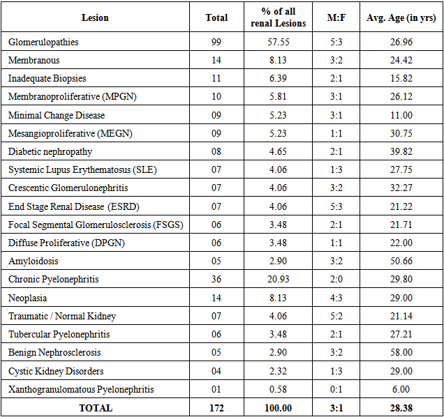
- OverallTen year retrospective data in this study (Table-1) showed that glomerulopathies (57.55%) were the most common of the 172 renal lesions followed by chronic pyelonephritis (20.93%). The age of the patients ranged from 6 years to 58 years. Apart from some genetic disorders such as cystic kidney and few non neoplastic reactive processes like xanthograulomatous pyelonephritis, statistics revealed a clear male preponderance in renal disorders; male: female (M: F) ratio being 3:1.
|
|
|
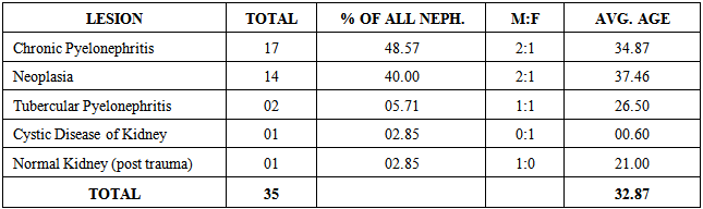
3.2. Prospective Analysis
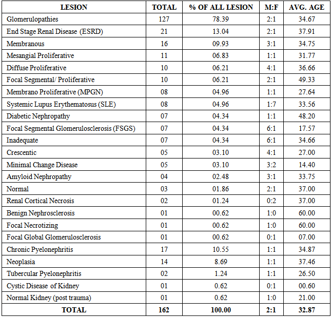
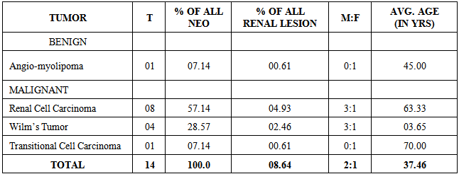
- OverallIn the prospective study glomerulonephritis (78.39%) was the most common lesion as shown in Table-4. Chronic pyelonephritis followed the glomerulopathies as the next most common renal lesion (10.55%). Cystic disease of kidney and post traumatic kidney completed the pool of these cases by having one case (0.62%) each. Of the 162 cases in the prospective study males accounted for 107 (66.04%) and females for 53 (32.71%) patients. In all the subcategories of renal lesions there was an overall male preponderance with exception of cystic disease of kidney where the only single recorded case was of a female child. The average age recorded in the present study was 32.87 years.Subcategorizing as done in Table-4 of all the renal lesions revealed that ESRD followed by CPN were the most common renal lesions in the prospective study pointing towards the dominance of the chronic and dialysis dependant nature of these lesions. Both the chronic lesions together accounted for 23.45% of all the renal lesions in the present study. The male predominance trend was seen in virtually every lesion (overall, 109 males for 53 females), however; more female cases were recorded in SLE, renal cortical necrosis, tubercular pyelonephritis, focal global glomerular sclerosis and renal cystic disease accounting for 8, 2, 1, 1 and 1 case each respectively. Overall, the age groups ranged from 0.6 years in case of renal cystic disease to 60.0 years in cases of benign nephrosclerosis and focal necrotizing glomerulonephritis. The average age seen in all the cases of renal lesions in the prospective study was 32.89 years.Glomerulopathies on Renal Core Biopsies Distribution of various types of glomerulopathies is shown in Table-4. In the prospective study end stage renal disease (ESRD) accounted for majority of glomerulopathies (16.53%), followed by membranous glomerulopathy (12.59%). Males outnumbered females by a ratio of 2:1. Apart from SLE and renal cortical necrosis (both post puerperal) which were common in females, all the other glomerulopathies showed a male predominance. The average age in glomerulopathies was 34.90 years, with age ranging from 7.0 years to 60.0 years. Focal global glomerulosclerosis, focal necrotizing glomerulonephritis and benign nephrosclerosis were the least common of all the glomerulopathies accounting for one case (0.62%) each.
|
|
|
4. Discussion
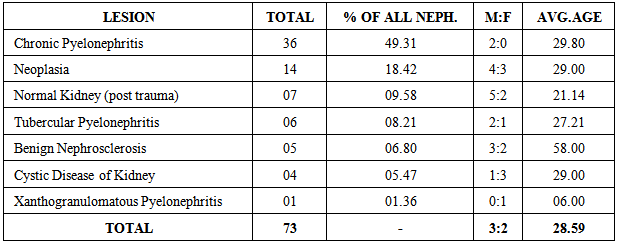
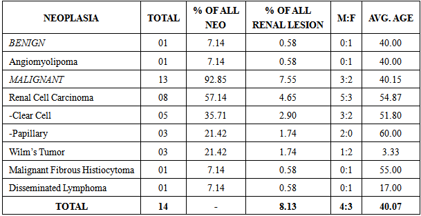
- OverallTen year data generated in this study showed that glomerulopathies diagnosed on core biopsies (57.55%) were the most common of the 172 renal lesions followed by chronic pyelonephritis (20.93%) and renal neoplasms (8.13%) on nephrectomy specimen. This correlated well with the prospective study of 162 renal lesions in which glomerulonephritis (78.39%) were by far the most common lesion followed by chronic pyelonephritis (CPN), (10.55%) and renal neoplasms (8.69%) as next most common renal lesions. An exhaustive study conducted by Deshpande et al at Department of Pathology, Armed Forces Medical College, Pune (India) in year 2000 based on only light microscopic findings were in accordance with the present study who found that the glomerulopathies were the commonest entity and comprised of 61.5 percent of all the nephropathies, the next most common renal lesions comprised of 22.2 percent cases of tubulointerstitial diseases (CPN) and 5.5 percent cases of malignant kidney tumours [2].The average age of presentation in the 10 year retrospective study was 28.38 years and in prospective study 32.87 years. In all the subcategories of renal lesions in both the studies, there was an overall male preponderance with few exceptions. This is corroborated with studes done elsewhere [2-4]. Glomerulopathies on Renal Core BiopsiesRetrospectively MGN was the commonest cause of glomerular disease followed by MPGN and MeGN (14.14 %, 11.11% and 10.10 % respectively). In prospective studies it was ESRD which was the commonest cause of glomerular diseases followed by MGN and MeGN (13.04 %, 9.13 % and 6.83 % respectively). Overall, among the 226 cases of various glomerular diseases in combined retrospective and prospective study, MGN was the commonest pattern of injury which was noted (13.2%). This was found to be on a higher side in comparison to the findings of other published Indian studies conducted in the southern and western parts of India (7.41% and 7.0% respectively) [2, 3]. MGN was shown to be a male prevalent lesion, which was in accordance with the findings of other studies. MGN remains the main cause of the nephrotic syndrome in European adults, China and Indonesia. It is the second commonest lesion in Italy, Japan, Hong Kong, Singapore and Taiwan after IgA glomerulonephritis [5-8].In India, however; MEGN is the commonest pattern of glomerular injury which is seen in south India [9] , primary IgA glomerulonephritis is the commonest in western India [10] and Minimal Change Disease (MCD) is the commonest lesion in northern India [11, 12]. The significant point which was noted in the present study was that the combined second commonest lesion overall and in prospective study which was seen on the core biopsy specimen was that of ESRD (12.83% and 13.04% respectively). It is worth while to note if the sub-categories of diabetic nephropathy and amyloidosis kidney were added in primary ESRD, then the combined ESRD would become the leading pattern of the glomerular injury. The patients with primary ESRD presented clinically with hypertension and elevated creatinine levels. Some presented with proteinuria and even nephrotic syndrome. The mean age of these patients was 37 years, with a male preponderance. In contrast to the findings of our study; ESRD is not prevalent to such a degree in other studies which were done in various geographical regions of India, as has been discussed above [9-12]. Overall Lesions on Nephrectomy SpecimenIn the present study, overall; CPN was seen in 20.93% cases retrospectively and 10.49% cases prospectively with a M: F ratio of 2: 0 and 2: 1, respectively. CPN was thus, the commonest lesion in the retrospective study whereas it was the second most common renal lesion in the prospective study in patients who underwent nephrectomy. Clinically patients presented with fever and backache similar to that observed by other researchers [13]. Most of the patients (60.8%) had history of renal stones. In the present study, CPN accounted for the most common cause of nephrectomy in both the ten year and prospective studies (49.37 % and 48.57% respectively). Renal Neoplasia on Nephrectomy SpecimenRenal Cell Carcinoma (RCC)RCC was the commonest neoplasm recorded in the nephrectomy specimens in both ten years and prospective study (57.14% in both). In the present study, mean age of patients was 54.87 years in the ten year study and 63.3 years in prospective study for RCC. Motzer et al, (1996) observed that average age range of diagnosis of RCC was 55 to 66 years in the present study. Male: Female (M: F) ratio was 5: 3 retrospectively and 2: 1 prospectively, almost similar to that reported by Rosai, (2004) [14, 15].Wilm’s Tumour (WT)WT comprised of the second most common neoplasia in both retrospective and prospective groups (21.42% and 28.57% respectively). In the present study the tumour was seen exclusively in the paediatric age group with average age in the retrospective study being 3.33 years and 3.67 years in the prospective study. According to Crist, (1991); 50% of cases occur before the age of 3 years and 90% before the age of 6 years [16]. M: F ratio in ten year study was 1: 2 whereas in the prospective study it was 3: 1. Investigators have stressed upon a female predominance in cases of WT [17]. Angiomyolipoma (AML)In the benign renal neoplasia category only lesion encountered was AML. In both ten year and prospective study only one case each was diagnosed (0.58% and 0.61%) of all renal lesions. Both the cases were seen in females with average reported age of 40 years and 45 years respectively in ten year and prospective study. This correlates with the findings of Steiner et al, 1993 [18].
5. Conclusions
- A high prevalence of ESRD in this study in both retrospective and prospective study along with CPN (as the commonest cause of nephrectomy) which was conducted at a tertiary care centre in the north Indian gangetic plain, speaks volumes about the delayed presentation and the patient ignorance being a great challenge to nephrologists in the developing countries of Asia and Africa. This is also explained by the fact that glomerular diseases continue to be the key factor, resulting in a large number of individuals suffering from renal disability/failure, resulting in considerable mortality and morbidity. This is more so compounded in the third world countries, where despite the advances in health care, additional factors such as environmental ones (both infectious and non -infectious), the standard of living and the access to health care and health education ( awareness on the renal diseases) have a role to play.The study thus, concludes that ESRD along with CPN (both leading to dialysis dependant renal failure) continues be the major challenge to patients suffering from renal disorders. The incidence of dialysis dependant kidney has increased over the years. The finding of ESRD as the most common glomerulopahy in the region under investigation should prompt both the practising physicians and the pathologist in this region to be ever vigilant against a possibility of glomerulopathy in patients attending outpatients so that early action can be initiated to preserve kidney function and to avoid renal replacement therapies which add on to the morbidity and economic burden to the patients.