American Journal of Medicine and Medical Sciences

p-ISSN: 2165-901X    e-ISSN: 2165-9036

2015;  5(1): 20-25

doi:10.5923/j.ajmms.20150501.05

The Association among Serum Leptin, Insulin and Insulin Resistance in Iraqi Patients with Cholelithiasis

Bushra F. Hasan1, Samer A. Muhi2, Noor Alhuda Kh. Ibrahim1

1College of Science for Women, University of Baghdad, Iraq

2AL-YarmoukTeaching Hospital, Baghdad, Iraq

Correspondence to: Bushra F. Hasan, College of Science for Women, University of Baghdad, Iraq.

Email:

Copyright © 2015 Scientific & Academic Publishing. All Rights Reserved.

Abstract

Gallstone disease (GD): is a common condition all over the world as well as in Iraq .It is a chronic recurrent hepatobiliary disease, the basis for which is the impaired metabolism of cholesterol, bilirubin and bile acids. The aim of this study is to investigate the association between serum leptin and insulin, insulin resistance and cholelithiasisand serum electrolyte and cholelithiasis.Fifty patients with GD, aged between 16 to 83 years attending in AL-Yarmouk teaching hospital in Baghdad, Iraq (between December 2013 and May 2014) were included in the present study, also forty normal persons, aged (19-77 years) who serve as control group. The result shows a significant increasing in the mean of fasting serum glucose, Homeostatic Model Assessment [HOMA], VLDL, AST, ALP and iron, and a significant decreasing in the means of HDL, sodium, chloride of patients compared to controls. From the present study, the following can be concluded: hyperinsulinemia, hyperglycaemia, insulin resistance, increase leptin level, abnormal changes of lipoprotein, abnormal changes in liver functions, mineral and increase in BMI. All of these parameters are associated with cholelithiasis.

Keywords: Gallstone, Leptin, Insulin resistance

Cite this paper: Bushra F. Hasan, Samer A. Muhi, Noor Alhuda Kh. Ibrahim, The Association among Serum Leptin, Insulin and Insulin Resistance in Iraqi Patients with Cholelithiasis, American Journal of Medicine and Medical Sciences, Vol. 5 No. 1, 2015, pp. 20-25. doi: 10.5923/j.ajmms.20150501.05.

1. Introduction

Cholelithiasis is a common condition all over the world as well as in Iraq. It's a real problem in Baghdad, because of increasing number of females attending the hospital due to gallstone complications; the disease seems to be more prevalent than expected (Taher, 2013).
It is a chronic recurrent hepatobiliary disease, the basis for which is the impaired metabolism of cholesterol, bilirubin, gallbladder function and disorder in entero-hepatic circulation of bile acid, which is characterized by the formation of gallstones as crystalline. Its can occur due to the super saturation of bile and cholesterol precipitation in the hepatic bile duct, common bile duct, or gallbladder. There are various types of stones; mixed stones are cholesterol predominate, black and blackish brown as pigment calculi and brown pigment stones are formed as a result of infections which convert soluble bilirubin into insoluble state leading to formation of soft brown stones (Belousov, 2006) Among the risk factor of GD; age, gender, obesity, diet and dietary cholesterol, drugs, pills and contraceptive serum factors (Al-Obaidi et al., 2006).
Leptin is a peptide hormone comprising 167 amino acids sequences containing one disulphide bond with a 16 KDa. It is one of the most important hormones produced by adipose tissue.
Leptin action in the central nervous system (CNS) promotes weight loss by decreasing food intake and increasing energyexpenditure.
The Ob (Lep) gene (Ob for obese, Lep for leptin) is located on chromosome 7 in humans. It's one of the main genes that linked to the obesity phenotype in humans (Oswaland Yeo, 2010).
Obesity is associated with increased secretion of cholesterol into the bile. The excess cholesterol accumulates in the gallbladder, which can lead to the formation of gallstones (Ruhl and Everhart, 2001 and Mendhez –Sanchez et al., 2005).
The effect of increased leptin is not clear on biliary secretion in human. It is suggested that hyperinsulinemia and insulin resistance increases leptin level and there are an important association with HOMA index (has been regarded as the mathematical expiration of insulin sensitivitys) (Mattews et al., 1985; Reaven et al., 2005).

2. Material and Methods

Blood samples (5ml) were obtained from the antecubital vein and collected in evacuated plastic tubes after overnight fasting. The samples were subsequently analyzed at a certifiedlaboratory. This study was carried out in AL-Yarmouk teaching hospital in Baghdad-Iraq, between December 2013 and May 2014. Ninety subject were enrolled in the study. Fifty patients with gallstone disease (44 female and 6 male) with age range between of 16 and 83 years and forty (20 female and 20 male) healthy subjects as control group with age range between of 19 and 77years.
All patients with gallstone disease were diagnosed clinically by ultrasound examination and some laboratory investigation before surgery.
Patients with hepatic, renal, endocrine disease, uncontrolled hypertension, acute blood loss, alcohol intake, on medications for lowering lipid, and smokers were excluded from the study.
The routine biochemical parameters fasting serum glucose, liver enzymes (AST, ALT, and ALP), bilirubin, total cholesterol, high-densitylipoprotein (HDL) cholesterol, and triglyceride (TG) levels were measured with an enzymatic colorimetric method using a (ARCHITECT c4000, ABBOTT,USA), also calcium, iron, and electrolytes were determined immediately by the same instrument.
Low-density lipoprotein (LDL), very low-density lipoprotein (VLDL) were calculated by the Friedewald formula (Friedewald et al., 1972).
Insulin resistance was calculated by means of the homeostasis model assessment (HOMA-IR).
HOMA-IR = [fasting insulin μU/mL) fasting glucose (mg/dl)]/405(Matthews et al., 1985) high index of insulin resistance a value >2.5. Serum fasting insulin and leptin hormones level were measured by ELISA (sandwich) using a kit supplied by (DRG- Germany) and (CUSA Bio – China) kits.
Statistical analysis
The data processing and statistical analysis were done by the SPSS (Statistical Package for Social Science – version20) and Microsoft Excel 2010.Data was presented in simple statistical measures of percentage, mean and standard deviation. The following statistical procedures were done: student’s T – test for comparing the significance of difference for the quantitative data, and simple linear correlation was used for determination of correlation between two quantitative parameters for the different groups. P-value was used to determine statistical significance and taking (p≤0.05) as the lowest limit of significance.

3. Results and Discussion

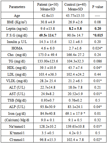
The mean of patient's age was 42.6±2 years which matched to that of healthy control (43.8±13.6) years. Regarding the sex distribution of patients there was a higher incidence in female (88%) than male (12%).
Table (1) show the of age factor in GD patients and control group, our result agree with Sharma et al., (2012)who found that the age is a risk factor of GD and the incidence of gallstone was increased along with advancing age. This result could be attributed to the negative correlation between age and the amount of synthesized bile acids and a positive correlation between cholesterol levels and age. Furthermore, hemoperfusion of the gallbladder wall is noted to be reduced with age due to the presence of sclerotic changes. This contributes to the dysfunction of the gallbladder, its infection and inflammation with exudation into the lumen of the organ (Reshetnyak, 2012).
Table 1. Comp ration of anthropometric measurements and biochemical parameters between cholelithiasis and control group
     
In the current study, gallstone disease was predominantly seen in females (88%) as compared to male (12%), this agree with the findings of previous studies (Marschalland Einarsson, 2007; Sun et al., 2009) who reported that female was also a major risk factor for gallstone disease. The commonly perceived opinion that women are at greater risk of developing gallstone disease than men may largely be due to extraneous risk factors, such as length of fertility period, abortions, number of pregnancies.
Sex hormones are most likely to be responsible for the increased risk, active role of progesterone and estrogens increases biliary cholesterol secretion causing cholesterol super saturation of bile, also hormone replacement therapy in postmenopausal women has been described to be associated with an increased risk for gallstone. (Tassaduqe et al., 2004; Marschall and Einarsson, 2007; Sharma et al., 2013)
The difference between mean of BMI in patients (30.8 ± 4.9 Kg/m2) and the mean of control group (26.9±2.6 Kg/m2) was not significant (p=0.08) (Table 2). This result disagreed with some studies (Tirziui et al., 2008; Wittenburg, 2010) which confirmed that the obese patients were more at risk to develop GD as compared with non-obese, as obesity playing an important role in the pathogenesis of increasing hepatic secretion of cholesterol.
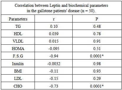
Table 2. Correlation between Leptin and other parameters in the gallstone disease patients
     
Table (1) show that the difference in serum leptin hormone between patients (9.6±13.8 ng/ml) and control group (5.8± 8.2 ng/ml)was not significant (p=0.20) and this result was in agreement with result obtained by Chenet al., (2012) who found that leptin level was an important risk factor for GD.
Also serum insulin hormone level in patients (14.3±15.6 µ U/L) was not differed significantly as compared with control group (12.1 ±6.5 µ U/L) (p=0.20) (Table 1). This result was in agreement with result obtained by Atamer et al., (2013). On the other hand, the results showed a significant increase (p=0.015) in the mean of serum glucose levels for patients (114.7 ±49.5mg/dl) in comparison with healthy control group (90.0± 14.7 mg/dl). Similar results were reported by Atamer et al., (2013) and Chen et al., (2012).
In the current study, there was a significant increase (p=0.024) in the insulin resistance levels (HOMA) for patients (4.6±6.0) in comparison with that of healthy control group (2.7±1.6), and this result agreed with the previous studies (Chen et al., 2012; Atamer et al., 2013).
Hyperinsulinemia, hyperglycemia and insulin resistance are common factors linking cholesterol gallstone. Insulin may increase gallstone formation through a mechanism in which insulin increases the activity of hydroxyl-3- methyglutaryl-coenzyme a reductase and stimulates bile acid-independent flow of bile into perfused liver, which deposited with cholesterol molecules to be gallstones formation (Kim, 2011).
Regarding the lipid profile, results revealed that there was a significant increase (p=0.01) in the mean of VLDL levels for patients (28.2 ± 25.4mg/dl) in comparison with that of healthy control group (21.2 ± 6.5 mg/dl), while there was a significant decrease (P=0.04) in the mean of HDL levels for patients (39.5±10.9 mg/dl) in comparison with that of healthy control group (43.7±7.4mg/dl), as shown in table (1). These results were consistent with result reported by Atamer et al., (2013).
Results showed that there was a non-significant increase (p=0.44), (p=0.086) (p=0.24), in the mean of LDL, TG and cholesterol levels for patients (103.4±36. 3 mg/dl), (133.0±123.6 mg/dl), (173.0±49.4 mg/dl) respectively, in comparison with that of control group (102.4±24.6 mg/dl), (104.25±32.3mg/dl), (166.4±27.2 mg/dl) respectively (Table 1). These results confirmed the previous results obtained by Misciagna et al., (2000) and Channa et al., (2010). Some investigators found a positive association between gallstone disease and increased levels of serum triacylglycerol, LDL cholesterol and decreased HDL cholesterol. The actual mechanism behind it is still unknown, this may be due to two mechanisms, one gallstone patients have abnormal secretary mechanism for bile acids and phospholipids and some of gallstone patients present with metabolic syndrome which is associated with altered lipid levels (Channa et al., 2010; Atamer et al., 2013).
In the liver function analysis there was a significant increase (p=0.01), (p=0.04) in the means of AST and ALP levels for patients (24.9±8.2 U/L), (93.8±30.9U/L) in comparison with healthy control group (20.52±5.9 U/L), (83.1±24.1U/L) respectively, as shown in table (1). Similar results were reported by Channa et al., (2005) and Al-Allaf et al., (2013). While there was a non-significant differences (p=0.21), (p=0.5) in the mean of ALT and TSB levels between patients and healthy control group.
Regarding of the mineral study there was a significant increase (p=0.01) in the mean of iron levels for patients (84.9±40.8 µg/dl) in comparison with that of healthy control group (69.1 ± 17.9 µg/dl), while there was a non-significant difference (p=0.32) in the mean of calcium levels between patients and healthy control group. This result was in contrast with those found by Kumari and Krishna, (2010). These differences could be attributed to difference in the criteria between two studies as well as methodology.
However the electrolytes study showed a significant decrease (P=0.05), (P=0.02) in the means of sodium and chloride levels for patients (128.2±10.2mmol/l), (96.8 ± 15. 3mmol/l) in comparison with that of healthy control group (139.65±10.8 mmol/l), (102.4 ± 7.8mmol/l) respectively.
Results showed a non-significant difference (p = 0.5) in the mean of potassium levels for patients (3.5 ± 0.5 mmol/l) in comparison with that of healthy control group (4.2 ± 0.5mmol/l). This result disagreed with the result obtained by Kumari and Krishna (2010), who reported a significant increase in serum sodium level for patients compared to control group.
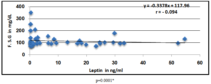
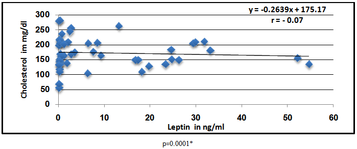
In the current study, the correlation coefficients was negative and significant between serumleptin hormone and F.S. (r=-0.94) (p=0.0001) and cholesterol, (-0.73) (p=0.0001) as shown in table (2) and figure (1, 2).
Figure 1. The correlation between Leptin and F.S.G
Figure 2. The correlation between Leptin and cholesterol
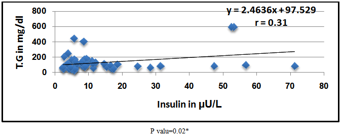
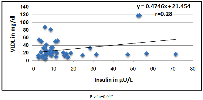
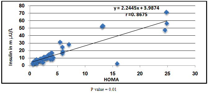
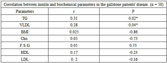
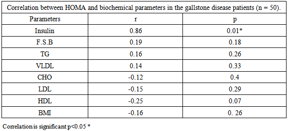
Moreover there was a significant positive correlation between serum insulin hormone levels and TG (r=0.31, p=0.02) and VLDL (r=0.28, p=0.04) in patients group as shown in Table (3) and Figures (3, 4), also there was a significant positive correlation between HOMA levels in patient group and insulin hormone (r=0.86, p=0.01) (Table 4) (Figure 5).
Table 3. Correlation between Insulin and other parameters in the gallstone disease patients.*Correlation is significant p< 0.05
     
Table 4. Correlation between HOMA and other parameters in the gallstone disease patients
     
Figure 3. The correlation between insulin and T.G
Figure 4. The correlation between insulin and VLDL
Figure 5. The correlation between HOMA and Insulin concentration

4. Conclusions

In conclusion: hyperinsulinemia, hyperglycaemia, insulin resistance, increase leptin level, abnormal changes of lipoprotein, abnormal changes in liver functions and increase in BMI, all of these parameters are associated with cholelithiasis.

References

[1]  Al-Allaf LI, Taher MT, Al-Mukhtar SBA, Al-Dulaimy LH: Histochemical study of alkaline phosphatase enzyme in gallbladder containing cholesterol stones. Ann Coll Med Mosul 2013, 39 (2):166-171.
[2]  Al-Obaidi SMM, Abdulla TS, Al-Alawi MS, Mohammed HMK: The prevalence of silent gall stones and Its relation to some risk factors in Iraq, The Iraqi postgraduate medical Journal 2006,5(2):146-150.
[3]  Atamer A, Övünc AOK, Yeşil AY, Atamar Y: Evalution of leptin and insulin resistance in patients with cholelithiasis, Indian Journal of biochemistry and biophysics 2013,50(4): 266-272.
[4]  Belousov Y. V. Pediatric Gastroenterology. Up-to-date guide. Moscow: Exama, 2006, p. 112.
[5]  Channa NA, Khand F, Ghanghro AB, Soomro AM: Quantitative analysis of Serum Lipid profile in Gallstone patients and Controls, Pakistan J Anal Environ Chem 2010, 11(1) 59.
[6]  Channa NA, Shaikh HR, Khand FD, Bhanger MI, Laghari MH: Association of gallstone daises risk with serum level of alkaline phosphatase, JLUMHS 2005, 4(1):18-22.
[7]  Chen LI, Qiao QH, Zhang SC, Chen YH, Chao GQ, Fang LZ: Metabolic syndrome and gallstone disease. World J Gastroenterol 2012, 18(31): 4215-4220.
[8]  Friedewald WT, Levy RI, Fredrickson DS. Estimation of the concentration of low-density lipoprotein cholesterol in plasma, without use of the preparative ultracentrifuge. ClinChem 1972, 18(6):499-502.
[9]  Kim SS, Lee JG, Kim DW, Kim BH, Jeon YK, Kim MR, Huh JE, Mok JY, Kim SJ, Kim YK, Kim IJ: Insulin resistance as a risk factor for gallbladder stone formation in Korean postmenopausal women, Korean J Intern Med 2011, 26(3): 285–293.
[10]  Kumari DJ, Krishna SHB: Role of body mass index, physical activity and nutrients in cholelithiasis in guntur, Andhra Pradesh. J Hum Ecol 2010, 31(3): 151-155.
[11]  Marschall HU, Einarsson C: Gallstone disease. J Inter Med 2007, 261:529-542.
[12]  Matthews DR, Hosker JP, Rudenski AS, Naylor BA, Treacher DF, Turner RC: Homeostasis model assessment: insulin resistance and beta-cell function from fasting plasma glucose and insulin concentrations in man. Diabetologia 1985, 28(7): 412-419.
[13]  Mendhez-Sanchez N, Ponciano-Rodrigoez G, Chavez –Tapia N, Uribe M: Effects of leptin on biliary lipids: potential consequences for gallstone formation and therapy in obesity. CurrDrug Targets Immune Endocrmetabol disorder 2005, 5(2): 203-208.
[14]  Misciagna G, Vguerra A, Leo D, Correale M, Trevisan M: Insulin and gallstone: apopulation case study in southern Italy.Gut2000, 47:144-147.
[15]  Reaven GM: Insulin resistance, the insulin resistance syndrome, and cardiovascular disease. Panminerva Med 2005, 47(4):201-210.
[16]  Reshetnyak, V.I. Concept of the pathogenesis and treatment of cholelithiasis, World J Hepatology 2012, 27; 4(2): 18-34.
[17]  Ruhl C. E. and Everhart JE: Relationship of serum leptin concentration and other measures of adiposity with gallbladder disease. Hepatology 2001, 34(5): 877-883.
[18]  Sharma R, Sachan SG, Sharma SR: Preponderance of gallstone in female. World Journal of Pharmacy and Pharmaceutical Sciences 2013, 2(6): 5871-5877.
[19]  Sharma R., Kumar A, Jha NK, Sacchan SG, Vidyarthi AS, Sharma SR: Probing into the prevalence and factors of gallstone formation in Jharkhand region in India, International journal of applied biology and pharmaceutical technology. 2012, 3(3):36-41.
[20]  Sun H, TangH, Jiang S, ZengL, ChenEQ, ZhouTY, Wang YJ: Gender and metabolic differences of gallstone diseases. World J Gastroenterol 2009, 15(15): 1886–1891.
[21]  Taher MA: Descriptive study of cholelithiasis with chemical constituents’ analysis of gallstones from patients living in Baghdad, Iraq, Int J Med Medical Sci2013, 5(1):19-23.
[22]  Tassaduqe K, Ali M, Salam A, Latif M, Afroze N, Masood S, Soban U: Studies on the chemical composition and presentation of gallstone in relation to sex and age among human population of Multan, Pakistan. J Bio Sci2004, 4(4): 470-473.
[23]  Tirziui S, Bel S, Bondor CI, Acalovschi M: Risk Factors for gallstone disease in patients with gallstones having gallstone heredity. A case-control study. Rom J InterMed 2008, 46 (3): 223–228.
[24]  Wittenburg H: Hereditary liver disease: Gallstones. Best Practice & Research Clinical Gastroenterology2010, 24:747–756.