-
Paper Information
- Previous Paper
- Paper Submission
-
Journal Information
- About This Journal
- Editorial Board
- Current Issue
- Archive
- Author Guidelines
- Contact Us
American Journal of Medicine and Medical Sciences
p-ISSN: 2165-901X e-ISSN: 2165-9036
2013; 3(5): 108-111
doi:10.5923/j.ajmms.20130305.03
Maximum Incidence of Appendicitis during Pubertal and Peri Pubertal Age Group Observed by Histological Study of Appendix
 Abstract
 Reference
 Full-Text PDF
 Full-text HTML
Hemanth Kommuru, Rajeswara Rao N., Anuradha S., Swayam Jothi S.
Department of Anatomy, Shri Sathya Sai Medical College & Research Institute, Tamil Nadu
Correspondence to: Hemanth Kommuru, Department of Anatomy, Shri Sathya Sai Medical College & Research Institute, Tamil Nadu.
| Email: |  |
Copyright © 2012 Scientific & Academic Publishing. All Rights Reserved.
The prime function of appendix in adults is the immune function. Inflammation of the appendix is known as appendicitis. Although its etiology is controversial, in the majority of patients, appendicitis is thought to be provoked by obstruction of the appendiceal lumen caused by fecalith impaction, muscular incoordination, lymphoid hyperplasia, or other processes. For the present work 318 appendix specimens from the patients who had been operated for appendicitis in General Surgery Department of Shri Sathya Sai Medical College Hospital were studied retrospectively and prospectively. 135 slides belonged to pubertal and peripubertal age groups. In that 71 cases showed obstruction due to fecalith in the lumen and 18 cases were due to lymphoid hyperplasia. Fecalith, associated with inflammation of mucosa, and lymphoid hyperplasia are believed to be the major cause of appendicitis.
Keywords: Fecalith, Lymphoid Hyperplasia, Appendicitis
Cite this paper: Hemanth Kommuru, Rajeswara Rao N., Anuradha S., Swayam Jothi S., Maximum Incidence of Appendicitis during Pubertal and Peri Pubertal Age Group Observed by Histological Study of Appendix, American Journal of Medicine and Medical Sciences, Vol. 3 No. 5, 2013, pp. 108-111. doi: 10.5923/j.ajmms.20130305.03.
Article Outline
1. Introduction
- Scientific studies reveal that the appendix plays a significant role in the development of fetus. Appendix starts developing in 11th week of pregnancy, in the form of endocrine cells. These cells produce number of biogenic amines and peptide hormones, essential for biological control mechanism. The prime function of appendix in adults is the immune function. Inflammation of the appendix is known as appendicitis. The life time risk of acute appendicitis is 7%[1, 2]. It is mostly seen between 20-40 years of age. Even today, fatalities due to appendicitis are significant[3, 4]. Appendicitis requires removal of the inflamed appendix, either by laprotomy or laparoscopy. Untreated, the appendix may rupture, leading to peritonitis, followed by shock, and, if still untreated, death. The incidence of appendicitis, which varies among different geographic populations, is less common in rural population. Despite the controversies and relative lack of knowledge regarding the function of this organ, surgical approaches have been followed. There are numerous articles and theories about the cause of appendicitis and many more experimental studies have been performed in order to reveal the pathophysiology of this disorder. Nowadays evacuation of luminal contents blocking is the generally accepted mechanism.
2. Aim
- To find the causative factor for the appendicitis in pubertal and peri pubertal aged (8 – 20 years) individuals by histological study.
3. Materials and Methods
- For the present work 318 appendix specimens from the patients who had been operated for appendicitis in General Surgery Department of Shri Sathya Sai Medical College Hospital were studied retrospectively and prospectively. Slides which were reported as appendicitis by the pathology department were made use for the study. Cases were charted out according to the type of infiltrating cells, age, sex, type of inflammation, and luminal contents. We included only 8 – 20 years aged individuals for the study and excluded the remaining age group.The slides will be studied for:- 1. Presence of fecalith or not in the lumen.2. Presence of lymphoid hyperplasia3. Eosinophilic or polymorphic or lymphocytic infiltration in the wall of the appendix4. Lumen obliteration5. Presence of submucosal fat
4. Observations
- A total of 318 slides of appendix specimen were studied retrospectively and prospectively. In that pubertal and peripubertal aged (8 – 20 years) individuals were 135 cases. Out of which 86 were males and 49 were females. Of the 135 cases 96 were chronic, 28 were acute and 11 cases were acute on chronic. Out of 135 slides in 71 cases lumen contained fecalith, (Fig - 1), necrosis and tissue debris, was present in 22 cases, lumen was empty in 38 cases and in 4 cases lumen was completely obliterated (Fig - 2). In those 71 cases which had fecalith, 57 cases were due to chronic appendicitis, 7cases were acute and 7 cases were acute on chronic. Of the 135 cases● Lymphoid hyperplasia (Fig - 3) was seen in18 cases. ● Submucosal fatty infiltration (Fig - 4) was seen in 77 cases.● In 23 cases hyperplastic epithelium was observed and was associated with fecalith in 5 cases, tissue debris and necrosis in 14 cases and, in 4 cases lumen was empty ● In 17 cases doesn’t have any special clinical features that what I have mentioned in the other cases and these cases showed edema, congestion, ulceration of mucosa.
 | Figure 1. Fecalith in the lumen of the appendix. F - Faecalith |
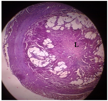 | Figure 2. Total obliteration of the lumen of appendix. L - Lumen |
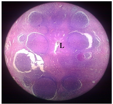 | Figure 3. Lymphoid hyperplasia narrowing the lumen. L - Lumen |
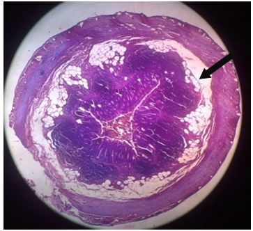 | Figure 4. Submucosa fat in the appendix |
5. Discussion
- Appendicitis is predominantly a disease of modern Western culture and communities and is influenced by it[5]. Abdominal pain, loss of appetite, nausea, vomiting, weakness and sometimes diarrhea are some well-known signs and symptoms. Pain traditionally starts from the epigastrium and descends to the right lower quadrant of the abdomen and settles down there. Rebound tenderness is generally absent when the pain first appears in the epigastrium, but is experienced later when the pain settles in the right lower quadrant of the abdomen. It suggests that the inflammation has reached the parietal peritoneum.[6]Appendicitis is most common between the ages of 10 and 20 years but can occur at any age and is more common in men[7]. In the present study out of 318 specimens 135 cases were between 8-20 years (42.4%) of age in that 86 were males (63.8%) and 49 were females (36.2%). According to Sing. JP[8] the number of specimens containing fecalith as a unique histopathologic finding was found in 44.9% in appendicitis. Mysorekar.VV[9] observed that the obstruction of lumen due to fecaliths was seen in only 20.1% of the inflamed appendices retrospectively. Fecaliths form a specific cause of appendicitis in about one third of specimens [10]. In the present study out of 135 cases the major causative factor for appendicitis was lumen obliteration by fecalith in 71 cases (52.6%). The lymphoid hyperplasia has been observed to occur in 25% of patients with appendicitis by Babekir AR[11]. In the present study it was observed only in 18 cases (13.3%).More over in the present study of the pubertal and peri pubertal age group individuals the major causative factor (52.6%) for appendicitis was obliteration of lumen by the fecalith. The fecalith itself is a rigid stool particle seen in the appendicular lumen. According to current consensus, luminal obstruction plays an important role in the pathophysiology of appendicitis. Ongoing secretion production from the mucosa and luminal exit obstruction secondarily cause increased intra luminal pressure. This pressure reduces appendix wall tissue perfusion and apparent congestion and edema appear. Faecalith fills and dilates the lumen causing erosions and irreparable damage to the mucosa. These breaks in the protective lining of the mucosa allow the entrance of bacteria into submucosa, thus initiating inflammation. Continued mounting pressure results in necrosis or even perforation. Perforation leads to intraluminal material scattering in the peritoneal cavity. Microorganisms, parasites, undigested food, plant seeds and foreign material are some components of appendix luminal content. An intra abdominal abscess formation can be the outcome of appendix perforation[12]. Besides the fecaliths, lymphoid hyperplasia is also a major causative factor in the pathophysiology of appendicitis in this age group. Fibro-obliterative changes in the lumen were found in a relatively older age group, with a mean age of 39.1 years in females and 33.6 years in males, respectively [13]. Following an attack of acute and subacute appendicitis which subsides by resolution, the mucosa is either partially or totally destroyed and the inflamed portions of the lumen now unprotected by lining of mucosa adhere together and later become solidly fused[14]. In the present study lumen obliteration was observed in 4 cases in that 3 cases were chronic and 1 case belonged to acute appendicitis. All the 4 cases were males with a mean age of 14.3 years. Following phagocytosis during inflammation, a lacework of reticular connective tissue alone remains in the place of mucosa and submucosa which were destroyed. The areas between the fibrils of this mesh sooner or later become filled with adipose tissue. In the present study submucosal fatty infiltration was seen in 77 cases (57.3%).
6. Conclusions
- Fecalith, associated with inflammation of mucosa, and lymphoid hyperplasia are believed to be the cause of appendicitis in most patients. The obstruction of the lumen is the dominant etiologic factor in appendicitis in the 8 – 20 years of age. Fecaliths are the most common cause of appendicular obstruction. Fecaliths are associated with complications like perforation and abscess. Peritonitis and intra abdominal abscess formation can be the outcome of appendix perforation. In case of fecalith demonstration on radiologic images of a patient with early complaints of appendicitis, appendectomy must be performed without waste of time and this will reduce morbidity and mortality. Mortality rate after perforation of appendix was 5.1 per 1000 individuals9. Fecalith prevalence is geographically distributed, as the incidence of appendicitis, being higher in Western than developing countries[15]. In the present study higher incidence of fecalith is observed in the rural population in Tamil Nadu of South India. Probably increased intake of fibre diet reduces the fecalith formation and there by appendicitis[12].