American Journal of Medicine and Medical Sciences

p-ISSN: 2165-901X    e-ISSN: 2165-9036

2013;  3(4): 61-67

doi:10.5923/j.ajmms.20130304.02

Red Blood Cell Alloimmunization among Sudanese Homozygous Sickle Cell Disease Patients

Mohammed Abbas1, Ahmed Bolad2, Nasreldin Jiefri3, Adil Mergani4

1College of Applied Medical Sciences, Medical Laboratory Department, Salman bin Abdulaziz University, Riyadh-Al Kharj, 11942, Saudi Arabia (KSA)

2Department of Microbiology, Faculty of Medical Laboratory Sciences, Al Neelain University, Khartoum, Sudan

3Hematology department, Faculty of Medicine , Al Neelain University , Khartoum, Sudan

4Department of Molecular Biology and Biotechnology, College of Applied Medical Sciences, Medical Laboratory Department, Taif University, –Tariba, Taif, Saudi Arabia (KSA)

Correspondence to: Mohammed Abbas, College of Applied Medical Sciences, Medical Laboratory Department, Salman bin Abdulaziz University, Riyadh-Al Kharj, 11942, Saudi Arabia (KSA).

Email:

Copyright © 2012 Scientific & Academic Publishing. All Rights Reserved.

Abstract

Background: Transfusion of RBCs is a major therapeutic option for anemia in homozygous SCD. Numerous studies conducted in the world to determine the frequency of alloimmunization in SCD patients, but there are no studies conducted in Sudan in open literature, to our knowledge. The objective of this study was to assess the incidence of RBC alloimmunization in SCD HbSS patients in Sudan. Methods: This study was conducted in Omdurman between August 2011 - Aril 2012, in 100 SCD HbSS patients of age between 6 month –17 years, who had received at least 2 transfusions with units of ABO and D matched RBCs, the alloimmunization was determined. Results: The mean No of blood units transfused per patient was 6.73 (SD, 4.461). 55% of the patients had been transfused from non relative donors, while 45% from relative donors. Only four patients was found to have antibodies 4% were detected among the 100 SCD (HbSS) patients who received transfusion.Conclusions: Although limited precompatibility testing is done in blood banking services in Sudanese hospital, low incidence of red cell alloimmunization (4%) was observed in this study. We recommend that to improve the blood bank services and polices to overcome the risk of alloimmunization.

Keywords: SCD, Transfusion, Alloimmunization

Cite this paper: Mohammed Abbas, Ahmed Bolad, Nasreldin Jiefri, Adil Mergani, Red Blood Cell Alloimmunization among Sudanese Homozygous Sickle Cell Disease Patients, American Journal of Medicine and Medical Sciences, Vol. 3 No. 4, 2013, pp. 61-67. doi: 10.5923/j.ajmms.20130304.02.

1. Introduction

Sickle cell disease (SCD) is an inherited disorder of hemoglobin (Hb), it is the most prevalent genetic disease in African region[1] with high mortality rate at age one to five years.[2] Since description of the first case of SCD in Sudan at 1926.[3] There was and still increasing research activities in this field. The highest prevalence of SCD in Sudanese is among the population from the western Sudan.[4-6]
SCD is caused by an autosomal recessive inherited of an abnormal beta globin gene (sickle cell gene) which leads to substitution of thymine by adenine in glutamic acid which in turn results in the substitution of valine to glutamic acid at the sixth position on the beta globin chain. Hemoglobin S (HbS), is the defective Hb that produced as a result of this defect, is a tetramer (alpha2/beta2) that is poorly soluble and polymerizes when deoxygenated.[7] The two characteristic features in the pathophysiology of SCD is the chronic hemolytic anemia and the vaso-occlusion.[8]
Transfusion of red blood cells is a major therapeutic option in SCD. It improve the oxygen-carrying capacity by increasing the total Hb level, decrease blood viscosity and increasing oxygen saturation by diluting the concentration of HbS; and suppressing endogenous production of sickle RBCs by increasing tissue oxygenation[9-11].
The repeated or chronic RBC transfusions and the exposure to different red cells antigen are often complicated by RBC alloimmunization, which results in difficulties in the selection of compatible blood, acute or delayed transfusion reactions.[12] A considerable number of studies have dealt with Alloimmunization in SCD, the range of RBCs alloimmunization has been reported to be in the range of 2.6% to 76%.[13-29,50].
The precompatibility testing procedures in the majority of the Sudanese hospital is limited to perform only ABO Rh grouping, and cross-matching by using saline and anti human globulin techniques by tube method. Although some major hospitals start to use the column technique in across-matching procedures, but they still use limited tests. There are no antibody screening , limited phenotyping, or even further antibody identification were done as a routine work far as we know.
Despite of the numerous studies conducted in the world to determine the frequency of red cells alloimmunization in SCD patients[13-29,50], unfortunately there are no enough studies conducted in Sudan in open literature, to our knowledge. The objective of this study was to assess the incidence of RBC alloimmunization in SCD patients in Sudan.

2. Material and Method

After appropriate ethical approval prior the commencement of the study, a cross-sectional study was carried out, random samples were analyzed. 5 ml of venous blood in K2 ethylenediaminetetraacetic acid (EDTA) for each sample. Questionnaires were used to collect demographic and clinical data include sex, age, tribe, number and indication for transfusion, transfusion reaction, and malarial attack . A total of hundred (100) homozygous sickle cell disease (HbSS) patients of age between 6 month –17 years, both males (57%) and females (43%) belonging to different tribes who had received at least 2 transfusions with units of ABO and D matched RBCs were selected from Albuluk pediatric teaching hospital and Omdurman pediatric hospital between August 2011 and Aril 2012. The number of blood transfusion ranged from 2 to 20 units per patient, the total number of transfusion to all patients was 673 units.

2.1. Laboratory Investigation

After ABO and Rh blood grouping by the standard tube method, the following tests were done for every sample:
Antibody screening. The collected plasma were tested for the presence of alloantibodies using a 3-cell panel with homozygous expression of the antigens (Panoscreen I,II and III, Immucor, Inc, USA) by using saline phase and indirect human antiglobulin test (indirect Coombs) in low-ionic-strength solution (LISS) by the tube method.
Antibody identification. Antibody specificity was determined with a standard panel of red cells reacting to known antigens using the same technique in the positive antibody screening (Panocell -10, Immucor, Inc, USA).
Patients were considered to be alloimmunized if antibodies to one or more RBC antigens were identified.

2.2. Data Analysis

Statistical software packages (Excel 5.0, Microsoft, Redmond, WA; and Statistical Package for the Social Sciences 20.0, SPSS, Inc., Chicago, IL) were used for data management and analysis, respectively.

3. Results

A total of 100 patients with SCD (HbSS) formed the study population. Fifty seven patients were males (57%), and forty three were female (43%) of age between 6 month – 17 years. (67%) of patients ages fall between 2 and 8 years, (25%) between 9 and 17 years and the lower proportion of patients aged less than two years (8%). Over (97%) of the patients involved in this study belong to tribes from the West of Sudan. Miserria, Marareet, Rizegat and Bargo constituted the largest proportions of the tribes, (28%, 22%, 12% and 8% respectively). Other tribes represents low percentage include: Tama, Habbania, Jawamaa, Zagawa, Masalma, Slihab, Bnehalba and Kwahla. (59%) of the patients parents are close relatives, (26%) are tribal, (5%) are far relation, while (10%) are not relatives. Only (2%) of the patients has had a history of malarial attack.
Figure 1 shows the rank order of blood group among subjects: O Rh + (n = 45 patients), A Rh + (n = 22), B Rh + (n = 20), AB Rh + (n = 8) , B Rh – (n = 2) ,A Rh – (n = 2) ,O Rh – (n = 1).
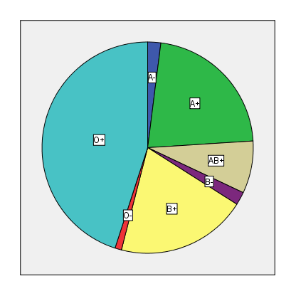
Figure 1. The frequency of ABO RH group among patients
Figure 2. The frequency of blood units taken by the patients
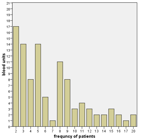
The patients were transfused with a total of 673 (SD, 4.461;median, 5; mean, 6.73; maximum, 20; minimum,2) units of blood. Figure 2 shows the frequency of blood units taken by the patients. (55%) of the patients were transfused from non related donor, while the residue (45%) from relative donor. Only one patient (1%) showed delayed transfusion reaction and another one showed immediate reaction.
Four antibodies (4%) were detected among the 100 SCD (HbSS) patients who received transfusion. Anti-K was detected in two patients, while anti-C and anti-E were detected in another patients, Table 1 shows the profile of the patients who developed alloantibodies in this study. Three of four patients (75%) showed no relation to donors. The patients who developed the alloantibodies received the higher rate of transfusion (between 15-20 units). There were no red cell antigens autoantibodies detected in patients samples.

4. Discussion

The alloimmunization is considered one of the major blood transfusion complications in SCD patients who received regular transfusion therapy, unfortunately, there are no published studies to determine the frequency of alloimmunization in SCD Sudanese patients as far as we know. In this study we try to establish the incidence of the alloimmunization in this category in Sudan.
The sickle cell anaemia (SCA) is the one of the major types of anaemia especially in Kordofan and Darfur states[4], western Sudan. Over (97%) of the patients involved in this study belong to tribes from that area, Elderdery, A[30] also noticed the high frequency of homozygous HbSS in his study among western Sudan tribes. Misseria which is a sub group of Baggara tribe, showed the highest percentage of the entire study group of patients in this study (28%). This tribe showed the prevalence of SCD to be (30%), (16%) among immigrants from the province of Blue Nile[31].
Blood group O (46%) was predominant among patients blood groups, this finding in agreement with three previous studies performed among Sudanese (52.7%) ,(51.5%) and (43.7% )[32-34], respectively. The result also consistent with other studies performed in African countries, shown an increase in group O percentage : Nairobi- Kenya (69%)[35], Nigeria (48.9%)[36]. The predominance of group O also observed in Western countries, in UK blood group O constitute (46.7%)[37], (47%) and (46%) of Caucasians and blacks in USA were group O respectively[38]. The increase percentage of group O in this study may be related to the low incidence of malarial attack (2%), which in agreement with some findings suggested the diverse relations between group O and different types of malaria[39-42]. Rowe JA et al; observed that group O was associated with a (66%) reduction in the odds of developing severe malaria compared with the non-O blood groups[43]. Furthermore, there is some convincing evidence to suggest that patients with SCD are protected from malaria[44-48]. The low prevalence of malaria observed in this study is matched with that reported by Makani J, et al.[49] the prevalence of malaria parasitemia was (0.72%) of 1808 SCA patients at the outpatient clinics and (3.04%) of 497 SCA patients during hospitalization.
Table 1. Profile of homozygous SCD patients who developed alloantibodies
     
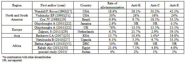
Table 2. review of alloimmunization in patients with homozygous SCD and the prevalence of anti-K, anti-C, and anti-E alloantibodies
     
The range of RBCs alloimmunization in a considerable number of previous studies has been reported to be in the range of 2.6% to 76%.[13-29,and 50]. Table 2 illustrate the frequency of RBCs alloimmunization in a number of studies conducted in different countries and the frequency of anti-K, anti-C and anti-E. This wide variation could be attributed to the transfusion load, closer matching of donor to recipient and the heterogeneity of the population in the community which may lead to increase the frequency of uncommon blood group antigens. The results of this study demonstrated that alloantibodies were developed in 4% of the patients, a frequency consider within the lower limit determined in literatures. The 4% rate of alloantibody formation in Sudanese SCD patients is incomparable with the 6.1% alloimmunization in neighboring regional study among Ugandan SCD patients reported by Natukunda B and colleagues[27] where there was less heterogeneity among donors and patients. Other studies carried out in African countries also reported low alloimmunization rate: (10%) in Democratic Republic of Congo (DRC)[51] and (7.75%) in Tunisia[52]. The low frequency of alloimmunization (2.6%) in Jamaica was anticipated by the author to the conservative use of transfusion there (range 1-22)[22], this finding matched with the range of blood unit taken by the patients in this study (2-20). The low frequency of alloimmunization in the current study may be attributed to the homogenicity of ethnic population since all donor although (55%) of them were not related to the recipient but there were Sudanese. The relatively low number of blood units taken (mean, 6.73) also may explain the low frequency. Young age of participants in this study consider an important factor in minimizing the alloantibodies frequency this finding in agreement with several studies[17,20,52,and 61]. The high rate of alloimmunization (21.4%) reported by Rabab Aly in 42 Egyptian SCA patients with mean age 12 year[50] is not matched neither with the result of alloimmunization in this study, nor with the results in other African countries[27,51and 52], the studied patients received from 4 to 12 units of blood per year. (11.3%) of a total 501 transfusion dependent Egyptian thalassemic patients of age between two and twenty six years were develop red cells alloantibodies[53]. The differences between the sample size in the previous studies together with the RBCs antigenic phenotypes variations between the patients and donors even they were from the same nationality especially they received approximately the same rate of blood transfusion per year (2 12 units/year), may explain the distinction in the rate of red cell alloimmunization among Egyptian patients receiving chronic transfusion. The rate of alloimmunization is higher in USA and UK compared to Uganda, DRC, and the present study. This may be explained by the assumed high phenotypic compatibility between blood donors and SCD patients in African countries, the most donors in the United States of America are Caucasian and SCA recipients are almost exclusively of African ancestry; this has been reported to be a cause for a high rate alloimmunization in these patients[54], Furthermore the greater red cell alloimmunization reported among UK SCD patients reflects the racial disparity between donor and recipient populations in the UK as well as greater use of transfusions[22].
The alloantibodies to the Kell system antigen K as well as to the Rhesus system antigens C and E, are comprise the percentage of the RBCs alloimmunization in this study, this finding in is in accordance with others[12,17,20, and 50]. The low frequency of K antigen among Sudanese (5.6%)[55] , together with unrelated donor to recipient may explain the present of anti-K alloantibody in two patients in this study. The frequency of antigen E and C is also relatively lower in percentage in comparison to antigen c, e and D among Sudanese[56]. The rhesus antigens are characterized by high immunogenicity[57] which intern lead to the formation of alloantibodies when transfused to the recipient lack such antigens. Anti-C anti-E appeared to be frequent alloantibodies in many studies[17,19,20,22,,27,29 and 51]. In this study the formation of Anti-C anti-E also may be attributed to the decrease their frequency in Sudanese population and to the unrelated donor. The phenotype matched donor RBCs viewed benefits to chronically transfused SCD, in one study the rate of alloimmunization dropped from 3% to 0.5%[21]
This study show that the rate of RBC alloimmunization was associated with the number of units of blood transfused and the number of transfusion episodes this finding in agreement with many studies observed that the RBCs alloimmunization is more likely in patients receiving multiple transfusion[13-20,27,50,54,58-60]. The patients who developed the alloantibodies received the higher rate of transfusion (between 15 -20 units) , their age range between 3 to10 years (Table 1) .
There were no red cell antigens autoantibodies detected in patients samples. The increase frequency of alloimmunization in females compared with male was observed in number of studies[13,50 and 54] the result in the current study not in harmony with that finding, there was no observable gender based differences in formation of alloantibodies.

5. Conclusions

The present study is the first study to determine the incidence of alloimmunization among homozygous SCD Sudanese patients. Although the limited tests were performed as a routine precompatibility testing in blood banks in Sudan hospitals, but fortunately low incidence of alloimmunization was detected (4%) which can be compared with other studies with low incidence of alloimmunization [22,27, and 29]. We recommend that to improve the blood bank services and polices in the way of RBCs compatibility tests, limited phenotype matching in Kell and Rhesus blood group systems specially in K, C, and E antigens, and improve the laboratory techniques to involve the alloantibody detection and identification. After making these improvement the alloantibodies formation in chronically transfused patients can easily be prevented.

ACKNOWLEDGEMENTS

The authors thank the staff of the haematology department at Albuluk pediatric teaching hospital and Omdurman pediatric hospital , Sudan, specially: dr. Areej Aljizoli and Ms. Israa Abdul Mahmoud for the help in recruiting patients and taking of blood samples and the Research Center in the medicine faculty in Alneelain University , Sudan, for the storage of samples during the period of the study. They also extend their appreciation to the dr. Abdelmoneim A. Sulieman and dr. Omar H. Aldirdeery for they help and advices.

Conflict of Interest

The authors certify that they have no affiliation with or financial involvement in any organization or entity with a direct financial interest in the subject matter or materials discussed in this manuscript.

References

[1]  Cook GC, Zumla AI (eds). Manson’s tropical diseases, 21st edition. London, WL Saunders, 2003.
[2]  Weatherall DJ et al. Inherited disorders of hemoglobin. In: Disease Control Priorities in Developing Countries. Jamison D et al. New York, Oxford University Press and the World Bank, 2006, 663-80.
[3]  Archibald RG (1926) A case of sickle cell anaemia in the Sudan. Trans R Soc Trop Med Hyg19:389–393
[4]  Attalla B, Mohammed AO, Bashir FM, Ahmed FE, El Hassan AM, Ibnauf G, Jiang W, Cavalli-Sforza LL, Karrar ZA, Ibrahim ME (2006). Relationship of the sickle cell gene to the ethnic and geographic groups populating the Sudan. Community Genet., 9(2): 113-120.
[5]  Vella F (1964) Sickling in the Western Sudan. Sudan Med J 1:16–17.
[6]  Lauder IR, Ibrahim SA (1970) Sickling in south-west Kordofan. Sudan Med J 8:206–214
[7]  Bunn HF. Pathogenesis and treatment of sickle cell disease. New Engl J Med 1997.
[8]  Clarice D. Reid, Samuel Charache, Bertram Lubin: MANAGEMENT AND THERAPY OF SICKLE CELL DISEASE. NATIONAL INSTITUTES OF HEALTH. National Heart, Lung, and Blood Institute. NIH PUBLICATION NO. 96-2117 DECEMBER 1995. Pp 1-2.
[9]  ReedW, Vichinsky E: New considerations in the treatment of sickle cell disease. Annu Rev Med 49:461 - 474, 1998
[10]  Nifong T, Domen R: Oxygen saturation and hemoglobin A content in patients with sickle cell disease undergoing erythrocytapheresis. Ther Apher 6:390 - 393, 2002
[11]  Thurston G, Henderson N, Jeng M: Effects of erythrocytapheresis transfusion on the viscoelasticity of sickle cell blood. Clin Hemorheol Microcirc 30:83 - 97, 2004
[12]  Moreira Júnior G, Bordin JO, Kuroda A, Kerbauy J. Red blood cell alloimmunization in sickle cell disease: the influence of racial and antigenic pattern differences between donors and recipients in Brazil. Am J Hematol. 1996 Jul;52(3):197-200.
[13]  Orlina AR, Unger PJ, Koshy M. Post-transfusion Alloimmunization in patients with sickle cell disease. Am J Hematol 1978;5:101-6.
[14]  Coles SM, Klein HG, Holland PV. Alloimmunization in two multitransfused populations. Transfusion 1981;21:462-6.
[15]  Ambruso DR, Githens JH, Alcorn R, Dixon DJ, Brown LJ, Vaughn WM, Hays T. Experience with donors matched for minor blood group antigens in patients with sickle cell anemia who are receiving chronic transfusion therapy. Transfusion 1987;27:94-8.
[16]  Luban NL. Variability in rates of alloimmunization in different groups of children with sickle cell disease: effect of ethnic background. Am J Pediatr Hematol Oncol 1989;11: 314-9.
[17]  Rosse WF, Gallagher D, Kinney TR, Castro O, Dosik H, Moohr J, Wrang W, Levy PS. Transfusion and Alloimmunization in sickle cell disease: the Cooperative Study of Sickle Cell Disease. Blood 1990;76:143-7.
[18]  Davies SC, McWilliam AC, Hewitt PE, Devenish A, Brozovic M. Red cell alloimmunization in sickle cell disease. Br J Haematol 1986;63:241-5.
[19]  Cox JV, Steanne E, Cunningham G, Frenkel EP. Risk of alloimmunization and delayed hemolytic transfusion reactions in patients with sickle cell disease. Arch Intern Med 1988;148:2485-9.
[20]  Sarnaik S, Schornack J, Lusher JM. The incidence of development of irregular red cell antibodies in patients with sickle cell anemia. Transfusion 1986;26:249-52.
[21]  Castro O, Sandler G, Houston-Yu P, Rana S. Predicting the effect of transfusing only phenotype-matched RBCs to patients with sickle cell disease: theoretical and practical implications. Transfusion 2002;42:684-90.
[22]  Olujohungbe A, Hambleton I, Stephens L, Serjeant B, Serjeant G. Red cell antibodies in patients with homozygous sickle cell disease: a comparison of patients in Jamaica and the United Kingdom. Br J Haematol 2001;113:661-5.
[23]  Bashawri LA. Red cell alloimmunization in sickle cell anemia patients. East Med Health J 2007;13:1181-9.
[24]  Vichinsky EP. Current issues with blood transfusion in sickle cell disease. Semin Hematol 2001;38 Suppl: 14-22.
[25]  Aygun, B., Padmanabhan, S., Paley, C. and Chandrasekaran, V. (2002), Clinical significance of RBC alloantibodies and autoantibodies in sickle cell patients who received transfusions. Transfusion, 42: 37–43.
[26]  Ameen, R., Al Shemmari, S. and Al-Bashir, A. (2009), Red blood cell alloimmunization among sickle cell Kuwaiti Arab patients who received red blood cell transfusion. Transfusion, 49: 1649–1654.
[27]  Natukunda B, Schonewille H, Ndugwa C, Brand A. Red blood cell alloimmunization in sickle cell disease patients in Uganda. 2010 Jan;50(1):20-5. Epub 2009 Oct 10.
[28]  Vichinsky EP, Luban NL, Wright E, et al. Prospective RBC phenotype matching in a stroke-prevention trial in sickle cell anemia: a multicenter transfusion trial. Transfusion. 2001;41(9):1086-1092.
[29]  Zalpuri, S., Zwaginga, J. J., le Cessie, S., Elshuis, J., Schonewille, H. and van der Bom, J. G. (2012), Red-blood-cell alloimmunization and number of red-blood-cell transfusions. Vox Sanguinis, 102: 144–149. doi: 10.1111/j.1423-0410.2011.01517.x
[30]  Elderdery, A., Mohamed, B., Cooper, Alan, Knight, Gavin and Mills, Jeremy (2011) Tribal distribution of haemoglobinopathies in a Sudanese patient population. Journal of Medical Laboratory and Diagnosis, 2 (4). pp. 31-37. ISSN 2141-2618.
[31]  Ahmed HA, Baker EA (1986). Sickling in the Sudan. Result of surveys in Blue Nile Province. East Afr. Med. J., 63(6): 395-399.
[32]  Fathelrahman Mahdi Hassan, Frequency of ABO, Subgroup ABO and Rh(D) , Pak J Med Res. Vol. 49, No. 1, 2010.
[33]  Waleid M. Shahata, Hiba B. Khalil , Awad-Elkareem Abass, Ishag Adam, Shahad M. Hussien. Blood group and Rhesus antigens among Blood donors attending the Central Blood Bank, Sudan. Sudan JMS Vol. 7, No.4. December 2012. Pp. 245-248.
[34]  Adam I, Babiker S, Mohmmed AA, Salih MM, Prins MH, Zaki ZM: ABO blood group system and placental malaria in an area of unstable malaria transmission in eastern Sudan. Malar J 2007, 6:110.
[35]  Lyko J, Gaertner H, Kaviti JN, Karithi MW, Akoto B. The blood group antigens ABO and Rh in Kenyans. Hamdard Medicus 1992;35:59–67.
[36]  Falusi AG, Ademowo OG, Latunji CA, Okeke AC, Olatunji PO, Onyekwere TO, et al.. Distribution of ABO and Rh genes in Nigeria. Afr J Med Sci 2000;29:23–6.
[37]  Talib, V.H. Hand book of Medical Laboratory Technology, 2nd ed. CBS Publishers, New Delhi, India. p. 205-210 (1991).
[38]  Seeley RR, Stephens TD, Tate P. Anatomy and Physiology. 4th edition. The McGraw Hill Companies, Inc.1998; USA p. 1098.
[39]  Fischer PR, Boone P. Short report: severe malaria associated with blood group. Am J Trop Med Hyg 1998; 58: 122-123.
[40]  Lell B, May J, Schmidt-Ott RJ, Lehman LG, Luckner D, Greve B, et al. The role of red blood cell polymorphisms in resistance
[41]  Pathirana SL, Alles HK, Bandara S, Phone-Kyaw M, Perera MK, Wickremasinghe AR, et al. ABO-blood-group types and protection against severe, Plasmodium falciparum malaria. Ann Trop Med Parasitol 2005; 99: 119-124.
[42]  Singh N, Shukla MM, Uniyal VP, Sharma VP. ABO blood groups among malaria cases from district Mandla, Madhya Pradesh. Indian J Malariol 1995; 32: 59-63.
[43]  Rowe JA, Handel IG, Thera MA, et al. Blood group O protects against severe Plasmodium falciparum malaria through the mechanism of reduced rosetting. Proc Natl Acad Sci U S A. 2007; 104(44):17471-17476.
[44]  Okuonghae HO, Nwankwo MU, Offor E. Malarial parasitaemia in febrile children with sickle cell anaemia. J Trop Pediatr. 1992;38(2):83-85.
[45]  Aluoch JR. Higher resistance to Plasmodium falciparum infection in patients with homozygous sickle cell disease in western Kenya. Trop Med Int Health. 1997;2(6):568-571.
[46]  Kotila R, Okesola A, Makanjuola O. Asymptomatic malaria parasitaemia in sickle-cell disease patients: how effective is chemoprophylaxis? J Vector Borne Dis. 2007;44(1):52-55.
[47]  Awotua-Efebo O, Alikor EA, Nkanginieme KE. Malaria parasite density and splenic status by ultrasonography in stable sickle-cell anaemia (HbSS) children. Niger J Med. 2004;13(1):40-43.
[48]  KombaAN, Makani J, Sadarangani M, et al. Malaria as a cause of morbidity and mortality in children with homozygous sickle cell disease on the coast of Kenya. Clin Infect Dis. 2009;49(2):216-222.
[49]  Makani J, Komba AN, Cox SE, Oruo J, Mwamtemi K, Kitundu J, Magesa P, Rwezaula S, Meda E, Mgaya J, et al. 2010. Malaria in patients with sickle cell anemia: Burden, risk factors, and outcome at the outpatient clinic and during hospitalization. Blood 115: 215–220.
[50]  Rabab Aly, Mohamed R. El-sharnoby, Adel A. Hagag, Frequency of red cell alloimmunization in patients with sickle cell anemia in an Egyptian referral hospital, Transfusion and Apheresis Science, Volume 47, Issue 3, December 2012, Pages 253-257, ISSN 1473-0502, 10.1016/j.transci. 2012.07.014.
[51]  Batina-Agasa S, Dupont E, Kayembe T, et al.: Multiple transfusions for sickle cell disease in the Democratic Republic of Congo: the importance of the hepatitis C virus. Transfus Clin Biol 2010; 17:254–259
[52]  S. Hmida, N. Mojaat, M. Maamar, M. Bejaoui, M. Mediouni, K. Boukef. Red cell alloantibodies in patients with haemoglobinopathies. Nouv Rev Fr Hematol, 36 (1994), pp. 363–366.
[53]  Ahmed AM, Hasan NS, Ragab SH, Habib SA, Emara NA, Aly AA. Red cell alloimmunization and autoantibodies in Egyptian transfusion-dependent thalassaemia patients. Arch Med Sci. 2010 Aug 30;6(4):592-8.
[54]  Vichinsky EP, Earles APNP, Johnson RA, Hoag MS, Williams A & Lubin B (1990). Alloimmunization in sickle cell anemia and transfusion of racially unmatched blood. New England Journal of Medicine, 322: 1617-1621.
[55]  Akasha, Ahmed. "The Frequency of Kell Red Cell Antigens (K,k) Among The Major Sudanese Tribes" Recent Research in Science and Technology[Online], 4 26 Feb 2012
[56]  Waleid M. Shahata, Hiba B. Khalil , Awad-Elkareem Abass, Ishag Adam, Shahad M. Hussien. Blood group and Rhesus antigens among Blood donors attending the Central Blood Bank, Sudan. Volume 7, Number 4 (December 2012) , 245-248
[57]  P.L. Mollison, C.P. Engelfriet, M. Contreras. Blood transfusion in clinical medicine (10th ed.)Blackwell Science, Oxford, UK (1997)
[58]  Hoeltge GA, Domen RE, Rybicki LA, et al.: Multiple red cell transfusions and alloimmunization. Experience with 6996 antibodies detected in a total of 159,262 patients from 1985 to 1993. Arch Pathol Lab Med 1995; 119:42–45
[59]  Reisner EG, Kostyu DD, Phillips G, Walker C and Dawson V (1987). Alloantibody responses in multiply transfused sickle cell patients. Tissue Antigens, 30: 161-166.
[60]  Fluit CR, Kunst VA, Drenthe-Schonk AM: Incidence of red cell antibodies after multiple blood transfusion. Transfusion 1990; 30:532–535.
[61]  Murao M., Viana M.B. Risk factors for alloimmunization by patients with sickle cell disease. Brazilian J Med Biol Res. 2005;38:675–682.