American Journal of Dermatology and Venereology

p-ISSN: 2332-8479    e-ISSN: 2332-8487

2022;  11(2): 21-26

doi:10.5923/j.ajdv.20221102.01

Received: Oct. 13, 2022; Accepted: Oct. 31, 2022; Published: Nov. 14, 2022


Role of Biomarker and Personalized Medicine in Atopic Dermatitis Management

Helmina Robiyatul Umami

Faculty of Medicine Muhammadiyah Surakarta University, Surakarta, Indonesia

Correspondence to: Helmina Robiyatul Umami, Faculty of Medicine Muhammadiyah Surakarta University, Surakarta, Indonesia.

Email:

Copyright © 2022 The Author(s). Published by Scientific & Academic Publishing.

This work is licensed under the Creative Commons Attribution International License (CC BY).
http://creativecommons.org/licenses/by/4.0/

Abstract

Atopic dermatitis (AD) is closely related to the immunological disorder. One of the main therapeutic strategies for atopic dermatitis is to control the immune system. Many biomarkers were identified to have some role in atopic dermatitis progression. Disease biomarkers are important and helpful in the treatment of atopic dermatitis. Biomarkers may identify the best-personalized treatment option for atopic dermatitis management which is known as personalized medicine. Here, we discuss some biomarkers that may relate to a treatment option for atopic dermatitis in different conditions and different patients. Biomarker profile differences in some people may correlate to a different treatment option. Here we discuss the best treatment option for atopic dermatitis based on biomarkers. This study design as a review and was conducted following PRISMA guidelines. Publications were searched in PubMed. 15 studies related to AD biomarkers were found that met the inclusion criteria. Th1 (CXCL9), Th2 (IL-13, CCL17/CCL22), Th17/Th22, IgE and epidermal thickness change significantly in clinical trial treatments. Biomarkers play role as severity assessment and personalized medicine.

Keywords: Atopic dermatitis, Biomarker, Personalized medicine, Management

Cite this paper: Helmina Robiyatul Umami, Role of Biomarker and Personalized Medicine in Atopic Dermatitis Management, American Journal of Dermatology and Venereology, Vol. 11 No. 2, 2022, pp. 21-26. doi: 10.5923/j.ajdv.20221102.01.

1. Introduction

The prevalence of AD is estimated to be 15-20% in children and 1-3% in adults, and the incidence has increased by 2 to 3 fold during the past decades in industrialized countries [1]. Atopic dermatitis (AD) is caused by a complex interaction of immune dysregulation, epidermal gene mutations, and environmental factors that disrupts the epidermis causing intensely pruritic skin lesions. Repeated scratching triggers a self-perpetuating itch-scratch cycle, which can have a significant impact on the patient's quality of life [2]. In a review of cohort studies, 38% of adults with AD had symptom onset in childhood. Many describe pain, stinging and embarrasing from their AD impacting their choice of clothing. The burden increases with increasing severity of disease [3]. The diagnose tool that use widely worldwide is Hanif and Rajka’s criteria that based on clinical appearance. The severity of AD also identified by tools based clinical appearance, such as SCORing of Atopic Dermatitis (SCORAD), Eczema Area and Severity Index (EASI) and Investigator’s Global Assessment (IGA) that’s rely on the observer’s subjective assessment [4] But it is also known that in the AD patient's skin lesions there is found infiltration of T-helper type 2 (Th2) cells and increased dendritic cells. Dendritic cells induce cytotoxins that activate eosinophils, and Natural Killer (NK) cells [5]. In addition, Th2 cells produce various pro-inflammatory cytokines, including Interleukin (IL)-4, IL-5, and IL-13. IL-4 induced the production of immunoglobulin (Ig) E to develop hypersensitivity reaction [6]. It is clear that the pathogenesis of AD is not only associated with Th2 but also Th1 and Th17. The heterogeneity of Th22 cytokine it is unlikely that novel molecular therapies targeting specific immunologic pathways will be equally effective in all patients with AD, which makes stratification of subtypes of patients with AD of increasingly important [7]. The specific pathway of immunological mechanism in AD lead many study to found specific biomarker. Some biomarkers may relate to a treatment option for atopic dermatitis in different conditions and different patients.

2. Method

This study design as a review and was conducted following PRISMA guidelines. Publications were searched in PubMed. The following inclusion criteria of the articles were: (a) Articles published in 2017 until 22 September 2022; (b) Keyword used “atopic dermatitis biomarker”; (c) Publication defined as Clinical trials; (d) Publication defined as a Free-full text article; (e) Publication correlate with diagnosis and management of atopic dermatitis disease; (f) English language. Additionally, we conducted a manual search in the references to search the meaning or terms that used in publication.

3. Biomarkers to Diagnostic in DA

Biomarker comes from the word "biological marker" which means a broad subcategory of medical signs that can be measured objectively. Biomarker difference from clinical signs and could play a significant role in the diagnosis, prognosis and management of AD. WHO defined biomarker as “any substance, structure or process that can be measured in the body or its products and influence or predict the incidence of outcome or disease. Biomarkers; can be classified into markers of exposure, effect, and susceptibility” [4]. The biologic origin of a biomarker could be genomic information, transcriptomic profiles obtained by analysis of mRNA and miRNA, proteins such as cytokines and other mediators from body fluids (whole blood, serum, plasma, tissue fluids) or tape stripping, and morphological information [8].
The elevated of serum IL-19 level is known relevance in AD and psoriasis severity [9]. In a cohort study involving Asians, African-American and Caucasians, serum IgE levels were elevated in All Asian patients (>480ng/ml) while among Caucasian and African-American patients the relative distribution for elevated IgE levels was 80%. Asian patients were predominately associated with the low inflammatory subgroup, whereas African-American associated with the high inflammatory subgroup, and Caucasian distributed evenly. There is association to baseline EASI Score, SCORAD Index, Itch Numeric Rating Scale (NRS) and Dermatology Life Quality Index (DLQI). Specifically, the high inflammatory subgroup was associated with higher disease score as compared to the low inflammatory subgroup. The study also found that Th2/Th22-related biomarkers, including IL-13, IL-22, Thymus and activation-regulated chemokine (TARC)/CCL17, Macrophage-derived chemokine (MDC)/CCL22, Monocyte chemo-attractant protein (MCP)-4/CCL13 and MCP-3/CCL17 correlate with disease severity. Furthermore IL-13, MCP-3 and TNFβ were found to be variable in both high inflammatory and low inflammatory subgroup and can be potential biomarker to detect severity of AD [10].
With the skin tape strip (STS) method, IL4R, CCL22, CCR4 also found correlate with severity of AD [11]. In European-Caucasian patients, a study using skin tape strip found that concentration of inflammatory cytokine in stratum corneum such as IL-18 CCL17, TARC/CCL17, MDC/CCL22, Cutaneous T-cell attracting chemokine (CTACK)/CCL27, IL-8/CXCL8 are high significantly in AD with skin lesions rather than in AD non lesions [12] Another study that using skin tape strip, found a group with 45 proteins as principal component 1 (PCI1) highest in AD with food allergy (FA). It has correlation with elevated trans-epidermal water loss (TEWL), specifically serine protease inhibitors b12 (SERPINB12) and gelsolin (GSN). 45 PCI1 proteins also significantly high in AD with FA rather than AD without FA and lowest in control group [13]. Studies found that there is characteristics in AD sample both blood or skin and can be identify the severity of AD (Table 1).
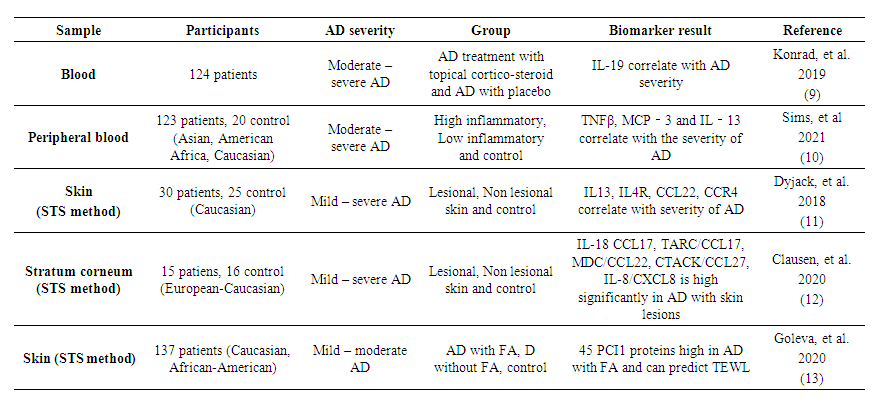
Table 1. Characteristics and Severity of Atopic Dermatitis
Treatments for AD based on Biomarker and Personalized Medicine:
Study found that Th2 cytokines play a key role in allergic disorders, include AD. Specifically; IL-4 and IL-13 and the heterodimeric IL-4 receptor (IL-4R). That’s way the potential treatment of AD may correlate with that markers. Dupilumab is a humanized IgG4 monoclonal antibody that targets the IL-4 receptor alpha chain (IL-4Rα) complex. The potential mechanism of action of Dupilumab in targeting the IL-4R complex is may inhibit IL-4 binding to IL-4Rα, inhibit the recruitment of γc to IL-4Rα chain, and inhibit the recruitment of the IL-R4α to IL-13Rα1. Dupilumab has been approved by the food and drug administration (FDA) in the United States for the treatment of moderate-to-severe atopic dermatitis in uncontrolled patients [14]. In a clinical trial, Dupilumab significantly reduce the inflammatory markers by inhibit the IL-4Rα rather than the placebo treatment. The biomarkers that notably the type 2 chemokines such as CCL17, CCL18 and the periostin after 16 week of treatment. TARC and pulmonary and activation-regulated chemokine (PARC) decrease and higher microbial diversity is correlate with the staphylococcus aureus colonization [15-16]. Tralokinumab, a fully human monoclonal antibody, specifically neutralizes IL-13, a key cytokine driving peripheral inflammation in AD. Its known to reduce daily pruritus through NRS [17]. Tralokinumab 300mg proven affect the clinical severity of AD through SCORAD index and Itchy NRS, the biomarker that found decrease with the severity is TARC, PARC and serum IgE, reversely dipeptidyl peptidase-4 (DPP-4) which the protein biomarker-induced by IL-13, increase in patients without Tralokinumab 300mg treatment [18].
OX40 is one of the co-stimulatory molecules expressed on T cells, and it is engaged by OX40L, primarily expressed on professional antigen-presenting cells such as dendritic cells [19]. GBR 830 is a humanized mAb against OX40. The clinical study found that the GBR 830 treatment with dose 10mg/kg can reduce significantly the clinical sign through Itchy NRS in patients with moderate to severe AD [20].
Tezepelumab is fully human monoclonal antibody that binds to thymic stromal lymphopoietin (TSLP), thereby preventing its reaction to TSLP receptor complex. TSLP is a innate cytokine that activates dendritic cells (DC) [21]. Tezepelumab 280 mg subcutan every 2 weeks with the class 3 topical corticosteroids (TCS) can reduce the clinical symptom of AD and reduce biomarkers such as IgE and DPP-4 that are high in AD with TCS treatment. The other biomarkers found was serum Periostin, and TARC/CCL17 which already low with TCS treatment, and being lower with Tezepelumab treatment although not significantly [22]. Crisaborole ointment 2% is a nonsteroidal phosphodiesterase 4 inhibitor for the treatment of mild to moderate AD. Inhibition of phosphodiesterase 4 by Crisaborole increase levels pf 3’4’-cyclic adenosine monophosphate in inflammatory cells, leading to activation of nuclear factor κB and nuclear factor of activated T-cell lead pathways and subsequent suppression of inflammatory cytokine release [23]. Crisaborole ointment 2% induced significant changes compared with vehicle in several pathogenic processes associated with AD, including IL-13, Interferon (IFN)γ, IL-22, IL-1 and/or TNF, T-cell and DC genes and Th17/th22 [24].
Phosphodiesterase-4 (PDE4), mainly present in immune cells, epithelial cells and brain cells, manifest as an intracellular non receptor enzyme that modulates inflammation and epithelial intact. Inhibition of PDE4 is predicted to have diverse effect via the elevation of the level of cyclic adenosine monophosphate (cAMP) and the subsequent regulation of a wide array of genes and proteins [25]. Apremilast is an oral PDE-4 inhibitor, approved for use in the management of psoriasis and psoriatic arthritis as well AD [26]. Apremilast 40 mg (APR40) give to patients with moderate – severe AD twice a daily and its improved the clinical symptom through EASI score significantly than apremilast 30 mg (APR30) and more from placebo. APR40 also significantly reduce the biomarkers, epidermal hyperplasia and inflammatory marker associated with Th17/Th22 pathways [27].
Study found that the symbiotic mixture intervention in infants with high IgE-associated AD can improvement significantly the SCORAD score than in the placebo, but not significant in infant with no appearance of IgE-associated AD. The symbiotic mixture is hydrolysed formula with Bifidobacterium breve M-16V and galacto-/fructooligosaccharide mixture [28]. Recent clinical trial study also did intervention with the symbiotic mixture of prebiotic short-chain galacto-oligosaccharides (scGOS), long-chain fructo-oligosaccharides (lcFOS), and a probiotic strain Bifidobacterium breve M-16V together with class 7 TCS treatment in infants with IgE-associated AD. After 4 month dietary intervention, SCORAD index improved with Th2/Th1 chemokine ratios for CCL17/CXCL9, CCL22/CXCL9, CCL20/CXCL10 and CCL20/CXCL11 was detected associated with the active intervention after 4 month dietary [29].
Gusacitinib (ASN002) is an oral dual inhibitor of (Janus kinase) JAK and tyrosine-protein kinase spleen tyrosine kinase (SYK) [30]. In a clinical trial study, ASN002 is given in three dose of 20 mg, 40 mg and 80 mg. There is significant improvement in EASI score and decreasing biomarkers (Th1, Th2 and Th17/Th22) after the treatment of 40 mg and 80 mg ASN002 [31].
The TCS treatment was broadly use worldwide in a decade and known to be effective as anti-inflammatory drugs. A study order the potency of TCS including pimecrolimus, betamethasone dipropionate, and clobetasol propionate versus emollient as a vehicle to moderate-severe AD patients. Betamethasone dipropionate, and clobetasol propionate significantly improved the clinical symptom through total sign score (TSSs), TEWL level, tissue biomarkers (Th1, Th2 and Th17/Th22), and epidermal hyperplasia rather pimecrolimus, same as vehicle that has no significant improvement [32]. The role of biomarker in management of AD recently developed across many study (Table 2).
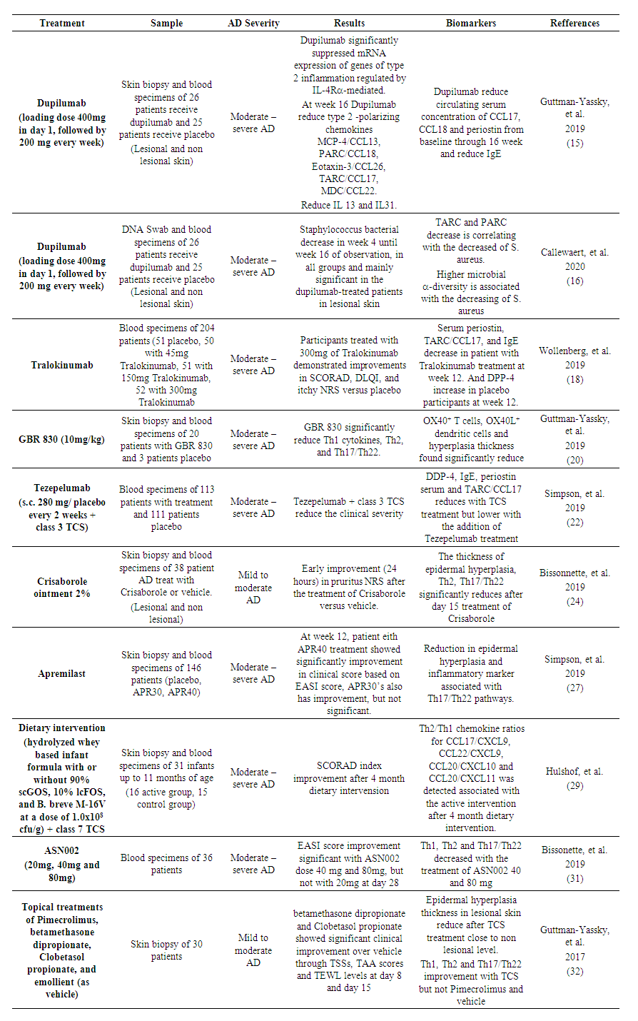
Table 2. The role of biomarker in management of Atopic Dermatitis
From 2017 to September 2022, many clinical trial studies of AD biomarkers were published. In PubMed search, 15 studies related to AD biomarkers were found that met the inclusion criteria. Most visible in these 15 studies was the role of biomarkers in the objective assessment of AD severity and drug efficacy in both adult patients and dietary interventions in infants. The Th1 chemokines (CXCL9), Th2 (IL-13, CCL17/CCL22) and Th17/Th22 appeared to be significantly decreased which was consistent with the improvement of clinical symptoms. IgE and epidermal thickness are also often used as biomarkers to assess drug effectiveness.
In the management of AD, biomarkers also play role to be a part of personalized medicine. In infant patients who suffers moderate – severe DA, biomarkers help to breakdown that dietary intervention only improved in infant with IgE-associated AD. In Dupilumab treatment of moderate to severe AD, study also found that the decrease of S. aureus only found in lesional skin, but not in non lesional skin.

4. Conclusions

In AD, clinical symptom assessment such as SCORAD index, EASI score, pruritus NRS, widely used worldwide. But the assessment is subjective. Biomarkers in AD play important role as objective assessment. Biomarkers also can help to breakdown the best personalized medicine for DA. It is still necessary to study the role of biomarkers to detect the safety and possibility of appropriate therapy in several conditions.

References

[1]  Nutten, Sophie. Atopic dermatitis: global epidemiology and risk factors. Ann Nutr Metab. 2015; 66 Suppl 1: 8-16. doi: 10.1159/000370220.
[2]  Frazier, Winfred., Bhardwaj, Namita. Atopic Dermatitis: Diagnosis and Treatment. Am Fam Physician. 2020; 101(10): 590-598.
[3]  Ali, Faraz., Vyas, Jul., Finlay, Andrew Y. Counting the Burden: Atopic Dermatitis and Health-related Quality of Life. Acta Derm Venereol. 2020 Jun 9; 100(12): adv00161. doi: 10.2340/00015555-3511.
[4]  Mastraftsi, Styliani., Vrioni, Georgia., Bakakis, Michail., Nicolaidou, Electra., Rigopoulos, Dimitrios., Stratigos, Alexender J., et al. Atopic Dermatitis: Striving for Reliable Biomarker. J Clin Med. 2022 Aug 9; 11(16): 4639. doi: 10.3390/jcm11164639.
[5]  Banchereau J, Briere F, Caux C, Davoust J, Lebecque S, Liu YJ, dkk. Immunobiology of dendritic cells. Annu Rev Immunol. 2000. doi:10.1146/annurev.immunol.18.1.767.
[6]  Kusumawati, Dewi., Prakoeswa, Cita Rosita Sigit., Rahmadewi. Profil Kadar Interleukin-31 Serum pada Pasien Dermatitis Atopik. Berkala Ilmu Kesehatan Kulit dan Kelamin. Agustus 2017. Vol 29. No 2.
[7]  Bakker, Daohne S., Nierkens, Stefan., Knol Edward F., Giovannone, Barbara., Delemarre, Eveline M., Schaft, Jorien van der., et al. Confirmation of multiple endotypes in atopic dermatitis based on serum biomarkers. J Allergy Clin Immunol. 2021 Jan; 147(1): 189-198. doi: 10.1016/j.jaci.2020.04.062. Epub 2020 Jun 8.
[8]  Renert-Yuval, Yael., Thyssen. Jacob P., Bissonnette, Robert., Bieber, Thomas., Kabashima, Kenji., Hijnen, DirkJan., et al. Biomarkers in atopic dermatitis - a review on behalf of the International Eczema Council. J Allergy Clin Immunol. 2021 Apr; 147(4): 1174-1190. e1. doi: 10.1016/j.jaci.2021.01.013.Epub 2021 Jan 28.
[9]  Konarad, Robert J., Higgs, Richard E., Rodgers, George H., Ming, Wenyu., Qian, Yue-Wei., Bivi, Nicoletta., et al. Assesment and Clinical Relevance of Serum IL-19 Levels in Psoriasis and Atopic Dermatitis Using Sensitive and Specific Novel Immunoassay. Sci Rep. 2019; 9: 5211. Doi: 10.1038/s41598-019-41609-z.
[10]  Sims, Jonathan T., Chang, Ching-Yun., Higgs, Richard E., Engle, Sarah M., Liu, Yushi., Sissons, Sean E., et al. Insights into adult atopic dermatitis heterogeneity derived from circulating biomarker profiling in patients with moderate-to-severe disease. Exp Dermatol. 2021 Nov; 30(11): 1650–1661. doi: 10.1111/exd.14389.
[11]  Dyjack, Nathan., Goleva, Elena., Rios, Cydney., Kim, Byung Ei., Bin, Lianghua., Taylor, Patricia., et al. J Allergy Clin Immunol. 2018 Apr: 141(4): 1298-1309. Doi: 10.1016/j.jaci.2017.10.046.
[12]  Clausen, Maja-Lisa., Kezic, S., Olesen, C.M., Agner, T. Cytokine concentration across the stratum corneum in atopic dermatitis and healthy controls. Sci Rep. 2020; 10: 21895. Doi: 10.1038/s42598-020-78943-6.
[13]  Goleva, Elena., Calatroni, Agustin., LeBeau, Petra., Berdyshev, Evgeny., Taylor, Patricia., Kreimer, Simion., et al. Skin tape proteomics identifies pathways associated with transepidermal water loss and allergen polysensitization in atopic dermatitis. J Allergy Clin Immunol. 2020 Dec: 146(6): 1367-1378. Doi: 10.1016/j.jaci.2020.04.002.
[14]  Harb, Hani., Chatila, Talal. Mechanisms of Dupilumab. Clin Exp Allergy. 2020 Jan: 50(1): 5-14. Doi: 10.1111/cea.13491.
[15]  Guttman-Yassky, Emma., Bissonette, Robet., Ungar, Benjamin., Suarez-Farinas, Mayte., Ardeleanu, Marius., Esaki, Hitokazu., et al. Dupilumab progressively improves systemic and cutaneous abnormalities in patients with atopic dermatitis. J Allergy Clin Immunol. 2019 Jan; 143(1): 155-172. Doi: 10.1016/j.jaci.2018.08.022.
[16]  Callewaert, Chris., Nakatsuji, Teruaki., Knight, Rob., Kosciolek, Tomasz., Vrbanac, Alison., Kotol, Paul., et al. IL-4Rα blockade by Dupilumab Decreases Staphylococcus aureus Colonization and Increase Microbial Diversity in Atopic Dermatitis. J Invest Dermatol. 2020 Jan: 140(1): 191-202.e7.
[17]  Wollenberg, Andreas., Blauvelt, A., Gutman-Yassky, Emma., Worm, M., Lynde, C., Lacour, J.P., et al. Tralokinumab for moderate to severe atopic dermatitis: results from two 52 week, rnadomized, double-blind, multicentre, placebo-controlled phase III trials (ECZTRA). Br J Dermatol. 2021 Mar, 184(3): 437 – 449. Doi: 10.111/bjd.19674.
[18]  Wollenberg, Andreas. Howell, Michael D., Gutman-Yassky, Emma., Silverberg, Jonathan I., Kell, Christopher., Ranade, Koustubh., et al. Treatment of atopic dermatitis with tralokinumab, an anti-IL-13 mAb. J Allergy Clin Immunol. 2019 Jan; 143(1): 135-141. Doi: 10.1016/j.jaci.2018.05.029.
[19]  Furue, Masutaka., Furue, Mihoko. Ox40L-OX40 signaling in atopic dermatitis. J Clin Med. 2021 Jun 11; 10{12}: 2578. Doi: 10.2290/jcm10122578.
[20]  Gutman-Yassky, Emma., Pavel, Ana B., Zhou, Lisa., Estrada, Yeriel D., Zhang, Ning., Xu, Hui., et al. GBR 830, an anti-OX40, improves skin gene signatures and clinical scores in oatients with atopic dermatitis. J Allergy Clin Immunol. 2019 Aug; 144(2): 482-493.e7. doi: 10.1016/j.jaci.2018.11.053.
[21]  Pelaia, Corrado., Pelaia, Giulia., Crimi, Claudia., Maglio, Angelantonio., Gallelli, Luca., Terracciano, Rosa., et al. Tezepelumab: A potential new biological theraphy for severe refractory Asthma. Int J Mol Sci. 2021 May; 22(9): 4369.
[22]  Simpson, Eric L., Parnes, Jane R., She, Dewei., Crouch, Sarah., Rees, William., Mo, May., et al. Tezepelumab, an anti-thymic stromal lymphopoietin monoclonal antibody, in the treatment of moderate to severe atopic dermatitis: A randomized phase 2a clinical trial. J Am Acad Dermatol. 2019 Apr; 80(4): 1013-1021. Doi: 10.1016/j.jaad.2018.11.059.
[23]  Callender, Valerie D., Alexis, Andrew F., Gold, Linda F. Stein., Lebwohl, Mark G., Paller, Amy S., Desai, Seemal R., et al. Efficacy and safety of crisaborole ointment 2% for the treatment of mild to moderate atopic dermatitis across racial and ethnic groups. Am J Clin Dermatol. 2019 Oct; 20(5): 711-723. Doi: 10.1007/s40257-019-00450-w.
[24]  Bissonnette, Robert., Pavel B., Diaz, Aisleen., Werth, John L., Zang, Chuanbo., Vranic, Ivana., Purohit, Vivek S., et al. Crisaborole and atopic dermatitis skin biomarkers: An intrapatient randomized trial. J Allergy Clin Immunol. 2019 Nov; 144(5): 1274-1298. Doi: 10.1016/j.jaci.2019.06.047.
[25]  Li, Heng., Zuo, Jianping., Tang, Wei. Phosphodiesterase-4 inhibitors for the treatment of inflammatory disease. Front Pharmacol. 2018 Oct 17; 9: 1048. Doi: 10.3389/fphar.2018.01048.
[26]  Rajagopalan, Murlidhar., Dogra, Sunil., Saraswat, Abir., Varma, Sachin., Banodkar, Pravin. The use of apremilast in psoriasis: An Indian perspective on Real-world scenarios. Psoriasis (Auckl). 2021 Aug 14; 11: 109-122. Doi: 10.2147/PTT.S320810.
[27]  Simpson, Eric L., Imafuku, Shinichi., Poulin, Yves., Ungar, Benjamin., Zhou, Lisa., Malik, Kunal., et al. A phase 2 randomized trial of apremilast in patients with atopic dermatitis. J Invest Dermatol. 2019 May; 139(5): 1063-1072. Doi: 10.1016/j.jid.2018.10.043.
[28]  Aa, L B van der., Heymans, H. S., Aalderen, W M van., Smitt, J H Sillevis., Knol, J., Amor, K Ben., et al. Effect of a new symbiotic mixture on atopic dermatitis in infants: a randomized-controlled trial. Clin Exp Allergy. 2010 May; 40(5): 795-804. Doi: 10.1111/j.1365-2222.2010.03465.x.
[29]  Hulshof, Lies., Overbeek, Saskia A., Wyllie, Anne., Chu, Mei Ling J N., Bogaert, Debby., de Jader, Wilco., et al. Exploring immune development in infants with moderate to severe atopic dermatitis. Front Immunol. 2018 Mar 29; 9: 630. Doi: 10.3389/fimmu.2018.00630.
[30]  Le, Michelle., Berman-Rosa, Melissa., Ghazawi, Feras M., Bourcier, Marc., Fiorillo, Loretta., Gooderham, Melinda., et al. Systematic Review on the efiicacy and safety of oral janus kinase inhibitors for the treatment of atopic dermatitis. Front Med (Lausanne). 2021; 8: 682547. Doi: 10.3389/fmed.2021.682547.
[31]  Bissonette, R., Maari, C., Forman, S., Bhatia, N., Lee, M., Fowler, J., Trying, S., et al. The oral janus kinase/spleen tyrosine kinase inhibitor ASN002 demonstrates efficacy and improves associated systemic inflammation in patients with moderate-to-severe atopic dermatitis: results from randomized double-blind placebo-controlled study. Br J Dermatol. 2019 Oct; 181(4): 733-742. Doi: 10.1111/bjd.17932.
[32]  Gutman-Yassky, Emma., Ungar, Benjamin., Malik, Kunal., Labuda, Tord., Suarez-Farinas, Mayte., Bissonette, Robert. Molecular signatures order the potency of topically applied anti-inflammatory drugs in patients with atopic dermatitis. J Allergy Clin Immunol. 2017 Oct; 140(4): 1032-1042. Doi: 10.1016/j.jaci.2017.01.027.