American Journal of Dermatology and Venereology

2016;  5(3): 39-45

doi:10.5923/j.ajdv.20160503.01


Clinicoepidemiological Study Prevalence and Types of Alopecia Areata in Khartoum Dermatology Teaching Hospital, Outpatient Clinic, Khartoum, Sudan

AO Khogali 1, M. El-Kordofani 1, Anilkumar Mithani 1, Abdel Rahman M. Ramadan 2, Gamal O. Elhassan 3, Abdel Khalig Muddathir 4, Khalid O. Alfarouk 5, Adil H. H. Bashir 6

1Faculty of Medicine, University of Bahri, Bahri, Sudan

2Faculty of Dentistry, Taibah University, AlL-Madinah Al-Munawarah, KSA

3Faculty of Pharmacy, Omdurman Islamic University, Omdurman, Sudan

4Faculty of Pharmacy, University of Khartoum, Khartoum Sudan

5Faculty of Pharmacy, AL-Neelain University, Khartoum, Sudan

6Institute of Endemic Diseases, University of Khartoum, Khartoum, Sudan

Correspondence to: Adil H. H. Bashir , Institute of Endemic Diseases, University of Khartoum, Khartoum, Sudan.

Email:

Copyright © 2016 Scientific & Academic Publishing. All Rights Reserved.

This work is licensed under the Creative Commons Attribution International License (CC BY).
http://creativecommons.org/licenses/by/4.0/

Abstract

Introduction: This study was compiled at Khartoum Dermatology Teaching Hospital (KDTH), Khartoum, Sudan, with Juba University Department of Dermatology and Venereology diseases on 170 patients, during the period May 1st, 2005- May 1st, 2006. Objective: this study was conducted to identify the prevalence of the common Alopecia Areata (AA) and different types, attending outpatient clinics of KDTH in Khartoum city. Patients & Methodology: The design was based on clinical studies, i.e. descriptive. A 170 case of Alopecia Areata (AA) and different types was considered as the study population. Conclusions: This study revealed that % of the cases attending the out-patient clinic at KDTH, presented with Alopecia Areata (AA). The results showed that most of the Alopecia Areata (AA) cases were males (70%), and %80 of AA are in the age group 14-40 yr- old, and 74% of them are single. Most of the AA patients are of Northern Sudanese origin (40%), the Southerners, the Easterners, and the Westerners showed lower percentages i.e. 6, 26, and 24%, respectively. AA is common among school children, as they represent approximately 58%. Those with primary school educational background represent 26% AA population, illiterates 12%, and as high as 30% among holders of higher degrees. Most of the patients are cases of AA (98%), for months duration (%). Thyroid and DM were among the most commonly associated diseases where they represented ca. 96%. The results also demonstrated that most of the patient’s complaints of localized loss of hair (90%). In the examination, and to according to the distribution of the lesions, it was noticed that most of the lesions are localized (82%), in a cluster (8%), to specific part-systematized (3%). Morphologically, 94% of the lesions were macules, patches (4%), papules (2%). The observed lesions colors were the skin colored (96%), followed by the white (4%). Most of the lesions represented in palpation were soft (94%) and firm (6%). No doughy lesions were encountered. Investigations in all of the cases of the study population (50) were presumed to confirm or to exclude a diagnosis. Skin tests of different types and other investigations were performed in the study population. Skin scraping for fungal infection represented 100%. Consequently, it was concluded that: the AA are detectable in % of the cases attending the out-patient clinic at KDTH. Alopecia Areata (90%) were the most common manifestations.

Keywords: Alopecia Areata, Prevalence, Variations, Sudan

Cite this paper: AO Khogali , M. El-Kordofani , Anilkumar Mithani , Abdel Rahman M. Ramadan , Gamal O. Elhassan , Abdel Khalig Muddathir , Khalid O. Alfarouk , Adil H. H. Bashir , Clinicoepidemiological Study Prevalence and Types of Alopecia Areata in Khartoum Dermatology Teaching Hospital, Outpatient Clinic, Khartoum, Sudan, American Journal of Dermatology and Venereology, Vol. 5 No. 3, 2016, pp. 39-45. doi: 10.5923/j.ajdv.20160503.01.

1. Introductions

Alopecia areata (AA) is a recurrent non-scarring type of hair loss that can affect any hair-bearing area. Clinically, characterized by patchy hair loss on the scalp or any hair-bearing area that can progress to cover the entire scalp (Alopecia totalis). Eventually the whole body (Alopecia universalis). It is a common dermatological problem with a worldwide occurrence. It is also a frequent cause of non-scarring hair loss usually diagnosed by the history and physical findings alone. Alopecia areata affects both sexes, children, and adults. In newborns and young infants, it is thought to be extremely rare. Alopecia areata can also evolutes alone or in association with a variety of other disorders. It is thought to be an autoimmune disease with evidence of association with other autoimmune conditions with a high incidence of association of autoimmune thyroiditis, a genetic predisposition where interaction with environmental factors results in episodes of terminal hair loss. Characteristic nail changes may accompany hair loss; the pathologic inflammatory process is proved on histopathology examination. While the exact etiology of this common condition has not been elucidated, a substantial body of evidence suggests that alopecia areata is an organ specific, autoimmune disease targeted to hair follicles. However, the antigenic targets, mechanisms, and consequences of auto-immune attack in alopecia areata have yet to be determined. While most patients have a good prognosis and can be effectively treated with available medications, treatment can be prolonged, and emotionally challenging, Early recognition, and intervention involving topical or intralesional treatment and education can afford patients with comforting reassurance for an eventual recovery. AA can present with many different patterns. Even though medically benign, AA can produce immense emotional and psychosocial stress in the affected patients and their families [1]. Alopecia areata is typified by a reversible form of patchy or complete loss of hair that is due to infiltration of hair follicles by T cells. The lifetime disease risk is approximately 1.4% of the general population is increased by more than 30% in the autoimmune polyendocrinopathy candidiasis ectodermal dysplasia syndrome (APECED), a recessive condition developing from a mutation in the autoimmune regulator (AIRE) gene on chromosome 21q22.3 [2].

2. Patients and Methods

The present study was conducted at HDTH, outpatient clinic.
It is estimated that the number of visitors to out-patient clinic 50 patients/ day (adults and children).

3. Methodology and Justification

Saturdays, Mondays, and Wednesdays were chosen for data collection, due to the availability of the Dermatologist specialties during these particular days, for revision and consultation. Every AA case was assigned a file number by the KDTH. As mentioned earlier, the data collection period extended from May 1st, 2005- May 1sth, 2006. A 230 case of AA patients was considered as the study population. For all cases, questionnaires (below) were filled out, and patients were cross- examined by the researcher personally. The highlight questionnaire ts were: The patient’s serial number, name, sex, age. The patients were categorized as those under 13yr, over 14, over 21, and those over 61 yr. The marital status was single, married, divorced, or widow). The residence, tribe, occupation, educational background, associated diseases (hypertension, thyroid swelling, renal, and others)was also recorded., The symptoms (type and duration), signs (distribution and configuration of cutaneous lesions, the morphology and color of the I individual lesions, and palpation of cutaneous lesions), professional diagnosis, confirmatory investigations, final diagnosis, and comments.
Lesions diagnostic criteria that have been followed in this study to define AA:
1. Aopecia Areata (AA).
2. Alopecia Marginalis- Ophiasis.
3. Alopecia Sub Totalis.
4. Alopecia Totalis
5. Alopecia Universalis.

4. Results

Table (1). Show the prevalence of Alopecia Areata (AA) according to sex in KDTH, Khartoum, for the period 1.5.2005- 1.3, 2006
     
Table (2). Showing the prevalence of Alopecia Areata (AA) according to age in KDTH, Khartoum, for the period 1.5.2005- 1.3, 2006
     
Table (3). Showing the prevalence of Alopecia Areata (AA) according to Marital in KDTH, Khartoum, for the period 1.5.2005- 1.3, 2006
     
Table (4). Showing the prevalence of Alopecia Areata (AA) according to a residence in KDTH, Khartoum, for the period 1.5.2005- 1.3, 2006
     
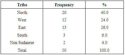
Table (5). Showing the prevalence of Alopecia Areata (AA) according to tribe in KDTH, Khartoum, for the period 1.5.2005- 1.3, 2006
     
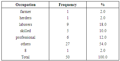
Table (6). Showing the prevalence of Alopecia Areata (AA) according to the occupation in KDTH, Khartoum, for the period 1.5.2005- 1.3, 2006
     
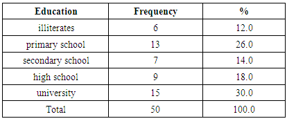
Table (7). Showing the prevalence of Alopecia Areata (AA) according to education in KDTH, Khartoum, for the period 1.5.2005- 1.3, 2006
     
Table (8). Showing the prevalence of Alopecia Areata (AA) according to associated diseases in KDTH, Khartoum, for the period 1.5.2005- 1.3, 2006
     
Table (9). Showing the prevalence of Alopecia Areata (AA) according to a number of relapses in KDTH, Khartoum, for the period 1.5.2005- 1.3, 2006
     
Table (10). Showing the prevalence of Alopecia Areata (AA) according to symptoms in KDTH, Khartoum, for the period 1.5.2005- 1.3, 2006
     
Table (11). Showing the prevalence of Alopecia Areata (AA) according to the duration of symptoms in KDTH, Khartoum, for the period 1.5.2005- 1.3, 2006
     
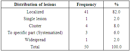
Table (12). Showing the prevalence of Alopecia Areata (AA) according to the distribution of lesions in KDTH, Khartoum, for the period 1.5.2005- 1.3, 2006
     
Table (13). Showing the prevalence of Alopecia Areata (AA) according to the configuration of lesions in KDTH, Khartoum, for the period 1.5.2005- 1.3, 2006
     
Table (14). Showing the prevalence of Alopecia Areata (AA) according to the morphology of lesions in KDTH, Khartoum, for the period 1.5.2005- 1.3, 2006
     
Table (15). Showing the prevalence of Alopecia Areata (AA) according to the color of lesions in KDTH, Khartoum, for the period 1.5.2005- 1.3, 2006
     
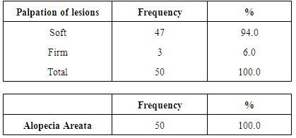
Table (16). Showing the prevalence of Alopecia Areata (AA) according to palpation of lesions in KDTH, Khartoum, for the period 1.5.2005- 1.3, 2006
     
Table (17). Showing the prevalence of Alopecia Areata (AA) according to confirmatory investigations in KDTH, Khartoum, for the period 1.5.2005- 1.3, 2006
     
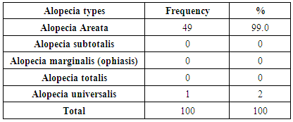
Table (18). Showing the prevalence of Alopecia Areata (AA) according to alopecia types in KDTH, Khartoum, for the period 1.5.2005- 1.3, 2006
     

5. Discussion

Two hundred patients with renal failure of different causes were seen at the Khartoum renal center in the period from June -3 to October-28- 2001. Fifty patients are pre-dialysis while hundred were on regular hemodialysis, and fifty were transplanted (recipients).
Those patients with cutaneous manifestations were found to be 167 out of the 200 (83.5%) (table 1). The highest percentage of cutaneous manifestations was seen in patients on regular hemodialysis (89%), followed by transplanted (recipients) (84%) and (72%) in pre–dialysis patients. These findings showed that the cutaneous manifestations increase when the renal failure progress and after transplantation the skin manifestations decrease to some extent but still more than in the pre–dialysis stage. That means the difficulty of management of cutaneous manifestations even after renal transplantation.
The frequency of the cutaneous manifestations according to the sex showed that 97 out of 167 (58%) were males, and 70 (42%) were females. This result showed that renal failure in Sudanese patients is more prevalent in males than females, the ratio about 1.5: 1. (Table 2), the rate of cutaneous manifestations in patients above 45 years was 51.5% and in the 26- 45 years age group it was 40.7%. These figures mean that the skin manifestations became more manifested as renal failure progresses.
The frequency of the cutaneous manifestations according to the duration of renal failure showed that 86.2% appeared after years while 13.8% appeared after months (Table 4). Moreover, the frequency of the skin manifestations according to the duration of the cutaneous manifestations, showed that 64.7% for years and 34.1% for the duration of months (Table 5). The findings in tables 4 & 5 revealed that the cutaneous manifestations go simultaneously with the chronicity of the renal failure.
The common symptoms observed are pruritus, skin dryness and hyperpigmentation; (tables 7, 8 and 9. The pruritus is more perceived in the pre-dialysis patients (83.3%) and decreases in patients on hemodialysis to (76.4%). Whereas, in transplanted patients it is only (47.6%). These findings indicate that renal transplantation improves the relief of this troublesome symptom. In this study, it was found that the percentage of patients with pruritus was similar to that found in the international literature which was 80% [66]. Dimkovic (1992). Barbara et al. (1980) stated that patients with renal failure experienced exacerbation of their symptoms during the summer due to the raising of skin temperature that may reduce the threshold for perception of uremic pruritus just as it does in other forms of pruritus. Maybe, the weather in Sudan, which is hot, most of the year round plays a role in the exacerbation of the condition in the Sudanese patients. The same findings also observed for the skin dryness (xerosis) which was 88.8% in pre-dialysis patients, 48.3% in patients on hemodialysis and 23.8% in transplanted patients and usually the pruritus is accompanied by xerosis that is considered to be one of the leading causes of the itching.
The hyperpigmentation was high in the three categories of patients, 77.7% in pre-dialysis patients, 84.2% in patients on hemodialysis and 85.7% in transplanted recipients. These findings pointed to that the hyperpigmentation is unaffected by either the hemodialysis or the renal transplantation. The skin discoloration depends on the pre-morbid color, and perhaps the type of skin plays a role. Most of the Sudanese patients belong to skin type four.
For the morphology of the cutaneous lesions, (tables 10, 11 and 12). The most common are hyperpigmented macules and patches observed in the three categories of patients with high percentages. Seventy-seven percents are macules, and 59.8 % are patches in pre-dialysis patients in the patients on hemodialysis, 80.9% are macules, and 47.8% are patches whereas, in the transplanted recipients 85.7% are macules and 59.5% are patches. These findings showed that the morphological signs became evident as renal failure progresses while they are unaffected by hemodialysis or even renal transplantation.
The next most common morphological lesions are nail changes that were found as follows; 16.6% of the pre-dialysis patients, 26.9% in patients on hemodialysis and 19.5% in the transplanted recipients. These nail changes ranged from Beaue’s lines, the half and half nail, to nail dystrophy.
Scales that were found in 16.6% of the pre-dialysis patients, 14.5% of the patients on hemodialysis and 38.9% in the transplanted recipients. There were few or minimal signs of papules, vesicles, cysts and bullae seen in a small number of patients in the different categories of renal failure patients.
Table (6): showed that the most common associated disease is hypertension, and followed by diabetes mellitus followed by gout. There are some patients associated with more than one concomitant disease, e.g., hypertension plus diabetes mellitus and hypertension plus gout. For the parathyroid diseases, only three patients were found and for the thyroid diseases, only two patients were recorded.
Regarding the effect of hemodialysis on the skin manifestations, 50.6% showed no effect while 38.2% showed improvement and 11.2% deteriorated after the hemodialysis (Table 13).
In this study, three transplant recipients developed nodular and granulomatous lesions that were confirmed by histopathology to be Kaposi sarcoma. Seven patients developed fungal nail infections, four patients developed Pityriasis Versicolor, and two patients developed herpes zoster infection. Most likely the low immune status of those patients due to immunosuppressive therapy played a significant role in these conditions. There are numerous other cutaneous manifestations mentioned in the literature e.g. bullous dermatosis of dialysis, cutaneous calcification, and calciphylaxis. Conditions such as perforating dermatosis, –were not encountered in this study. In this study the patients were seen were from different tribes and various social, economic and education classes. These findings pointed to that the socioeconomic factors do not play any role in the development of renal failure and associated cutaneous manifestations.
The drugs used in the management of renal failure that included calcium, Arabic gum, ferrous, folic acid, erythropoietin, have no effect on the associated cutaneous manifestations. Also, neither the antihypertensives nor the antidiabetic drugs showed any effect. The drugs used in hemodialysis and those used in sterilization of the renal dialysis machine tubes that mentioned in the literature to cause allergic reactions did not display any effect and no allergic reactions were recorded in the study.

6. Conclusions

Consequently, it was concluded that: the AA are detectable in %????? of the cases attending the out-patient clinic at KDTH. Alopecia Areata (90%) were the most common manifestations.

7. Recommendations

From the results of the present work, the followings are recommended:
1. Every patient of AA must be well clerked, with a full history being taken as well as a proper examination, as the diagnosis can be established by the characteristic clinical features of alopecia areata including its severe forms alopecia area Tato Alis and universalis.
2. As the unpredictable course of the disease is a major handicap, the patient must be given a full explanation of the course and prognosis of the condition.
3. As no similar study has been done in Sudan before, so recommended to consider this study as an initial step for further research.

References

[1]  Tan E, Tay YK, Giam YC. A clinical study of childhood alopecia areata in Singapore. Pediatr Dermatol. 2002 Jul-Aug; 19(4):298-301.
[2]  Tazi-Ahnini R, Cork MJ, Gawkrodger DJ, Birch MP, Wengraf D, McDonagh AJ, and Messenger AG. Role of the autoimmune regulator (AIRE) gene in alopecia areata: strong association of a potentially functional AIRE polymorphism with alopecia universalis. Tissue Antigens. 2002 Dec; 60(6): 489-95.
[3]  Green J, Sinclair RD. Genetics of alopecia areata. Australas J Dermatol. 2000 Nov; 41(4): 213-8.
[4]  Tazi-Ahnini R, di Giovine FS, McDonagh AJ, Messenger AG, Amadou C, Cox A, Duff GW, Cork MJ. Structure and polymorphism of the human gene for the interferon-induced p78 protein (MX1): evidence of association with alopecia areata in the Down syndrome region. Hum Genet. 2000 Jun; 106(6): 639-45.).
[5]  Tazi-Ahnini R, McDonagh AJ, Cox A, Messenger AG, Britton JE, Ward SJ, Bavik CO, Duff GW, Cork MJ. Association analysis of IL1A and IL1B variants in alopecia areata. Heredity. 2001 Aug; 87(Pt 2):215-9.
[6]  Yosefy C, Ronnen M, Edelstein D. Pseudo alopecia areata caused by skull-caps with metal pin fasteners used by Orthodox Jews in Israel. Clin Dev Immunol. 2003 Jun-Dec; 10(2-4):193-5.
[7]  McDonagh AJ, Messenger AG: The pathogenesis of alopecia areata. Dermatol Clin 1996 Oct; 14(4): 661-70.
[8]  Perini GI, Veller Fornasa C, Cipriani R, et al.: Life events and alopecia areata. Psychother Psychosom 1984; 41(1): 48-52.
[9]  Kalish RS, Johnson KL, Hordinsky MK. Alopecia areata. Autoreactive T cells are variably enriched in scalp lesions relative to peripheral blood. Arch Dermatol. 1992 Aug; 128(8): 1072-7.
[10]  Tazi-Ahnini R, Cork MJ, Gawkrodger DJ, Birch MP, Wengraf D, McDonagh AJ, and Messenger AG. Role of the autoimmune regulator (AIRE) gene in alopecia areata: strong association of a potentially functional AIRE polymorphism with alopecia universalis. Tissue Antigens. 2002 Dec; 60(6): 489-95.
[11]  Rokhsar CK, Shupack JL, Vafai JJ, Washenik K: Efficacy of topical sensitizers in the treatment of alopecia areata. J Am Acad Dermatol 1998 Nov; 39(5 Pt 1): 751-61).
[12]  Mainardi E, Montanelli A, Dotti M, Nano R, Moscato G. Thyroid-related autoantibodies and celiac disease: a role for a gluten-free diet?J Clin Gastroenterol. 2002 Sep; 35(3):245-8.
[13]  Galbraith GM, Thiers BH, Pandey JP. Gm allotype associated resistance and susceptibility to alopecia areata. Clin Exp Immunol. 1984 Apr; 56(1):149-52.
[14]  Price VH, Colombe BW: Heritable factors distinguish two types of alopecia areata. Dermatol Clin 1996 Oct; 14(4): 679-89.
[15]  Colombe BW, Price VH, Khoury EL, Garovoy MR, Lou CD. HLA class II antigen associations help to define two types of alopecia areata. J Am Acad Dermatol. 1995 Nov; 33(5 Pt 1): 757-64.
[16]  Kavak A, Baykal C, Ozarmagan G, Akar U. HLA in alopecia areata. Int J Dermatol. 2000 Aug; 39(8):589-92.
[17]  Tazi-Ahnini R, McDonagh AJ, Cox A, Messenger AG, Britton JE, Ward SJ, Bavik CO, Duff GW, Cork MJ. Association analysis of IL1A and IL1B variants in alopecia areata. Heredity. 2001 Aug; 87(Pt 2):215-9.
[18]  Yang S, Yang J, Liu JB, Wang HY, Yang Q, Gao M, Liang YH, Lin GS, Lin D, Hu XL, Fan L, Zhang XJ. The genetic epidemiology of alopecia areata in China. Br J Dermatol. 2004 Jul; 151(1):16-23.
[19]  Tazi-Ahnini R, di Giovine FS, McDonagh AJ, Messenger AG, Amadou C, Cox A, Duff GW, Cork MJ. Structure and polymorphism of the human gene for the interferon-induced p78 protein (MX1): evidence of association with alopecia areata in the Down syndrome region. Hum Genet. 2000 Jun; 106(6):639-45.
[20]  van der Steen P, Traupe H, Happle R, Boezeman J, Strater R, Hamm H. The genetic risk for alopecia areata in first-degree relatives of severely affected patients. An estimate. Acta Derm Venereol. 1992 Sep; 72(5):373-5.
[21]  Shellow WV, Edwards JE, Koo JY. Profile of alopecia areata: a questionnaire analysis of patient and family. Int J Dermatol. 1992 Mar; 31(3):186-9.
[22]  McElwee KJ, Tobin DJ, Bystryn JC, et al.: Alopecia areata: an autoimmune disease? Exp Dermatol 1999 Oct; 8(5): 371-9).
[23]  Colombe BW, Price VH, Khoury EL, et al.: HLA class II antigen associations help to define two types of alopecia areata. J Am Acad Dermatol 1995 Nov; 33(5 Pt 1): 757-64.
[24]  Ruiz-Doblado S, Carrizosa A, Garcia-Hernandez MJ. Alopecia areata: psychiatric comorbidity and adjustment to illness. Int J Dermatol. 2003 Jun; 42(6):434-7.
[25]  Muller, SA: Alopecia areata: an evaluation of 736 patients. Arch Dermatol 1963; 88: 106-13.
[26]  Nanda A, Al-Fouzan AS, Al-Hasawi F. Alopecia areata in children: a clinical profile. Pediatr Dermatol. 2002 Nov-Dec; 19(6):482-5.
[27]  Tan E, Tay YK, Giam YC. A clinical study of childhood alopecia areata in Singapore. Pediatr Dermatol. 2002 Jul-Aug; 19(4):298-301.
[28]  Sharma VK, Dawn G, Kumar B. Profile of alopecia areata in Northern India. Int J Dermatol. 1996 Jan; 35(1):22-7.
[29]  Ro BI. Alopecia areata in Korea (1982-1994).J Dermatol. 1995 Nov; 22(11):858-64.
[30]  Shellow WV, Edwards JE, Koo JY. Profile of alopecia areata: a questionnaire analysis of patient and family. Int J Dermatol. 1992 Mar; 31(3):186-9.
[31]  Safavi KH, Muller SA, Suman VJ, Moshell AN, Melton LJ 3rd. Incidence of alopecia areata in Olmsted County, Minnesota, 1975 through 1989. Mayo Clin Proc. 1995 Jul; 70(7):628-33.
[32]  Tan E, Tay YK, Goh CL, Chin Giam Y. The pattern and profile of alopecia areata in Singapore--a study of 219 Asians. Int J Dermatol. 2002 Nov; 41(11):748-53.
[33]  Nanda A, Al-Fouzan AS, Al-Hasawi F. Alopecia areata in children: a clinical profile. Pediatr Dermatol. 2002 Nov-Dec; 19(6):482-5.
[34]  Sharma VK, Dawn G, Kumar B. Profile of alopecia areata in Northern India.Int J Dermatol. 1996 Jan; 35(1): 22-7.
[35]  Ro BI. Alopecia areata in Korea (1982-1994). J Dermatol. 1995 Nov; 22(11):858-64.
[36]  Shellow WV, Edwards JE, Koo JY. Profile of alopecia areata: a questionnaire analysis of patient and family. Int J Dermatol. 1992 Mar; 31(3):186-9.
[37]  Bertolino AP. Alopecia areata. A clinical overview. Postgrad Med. 2000 Jun; 107(7):81-5, 89-90.
[38]  Taylor CR, Hawk JL: PUVA treatment of alopecia areata partialis, totalis, and universalis: audit of 10 years' experience at St John's Institute of Dermatology. Br J Dermatol 1995 Dec; 133(6): 914-8.
[39]  Ro BI. Alopecia areata in Korea (1982-1994). J Dermatol. 1995 Nov; 22(11):858-64.
[40]  Ro BI. Alopecia areata in Korea (1982-1994). J Dermatol. 1995 Nov; 22(11):858-64.
[41]  Ro BI. Alopecia areata in Korea (1982-1994). J Dermatol. 1995 Nov; 22(11):858-64.
[42]  Shellow WV, Edwards JE, Koo JY. Profile of alopecia areata: a questionnaire analysis of patient and family. Int J Dermatol. 1992 Mar; 31(3):186-9.
[43]  Hoffmann R, Happle R: Topical immunotherapy in alopecia areata. What, how, and why? Dermatol Clin 1996 Oct; 14(4): 739-44.
[44]  Ro BI. Alopecia areata in Korea (1982-1994). J Dermatol. 1995 Nov; 22(11):858-64.
[45]  Ro BI. Alopecia areata in Korea (1982-1994). J Dermatol. 1995 Nov; 22(11):858-64.
[46]  Dominguez Ortega L. Narcolepsy and alopecia areata: a new association? An Med Interna. 1992 May; 9(5):234-6.
[47]  Shellow WV, Edwards JE, Koo JY. Profile of alopecia areata: a questionnaire analysis of patient and family. Int J Dermatol. 1992 Mar; 31(3):186-9.
[48]  Werth VP, White WL, Sanchez MR, Franks AG. Incidence of alopecia areata in lupus erythematosus. Arch Dermatol. 1992 Mar; 128(3):368-71.
[49]  Isa AR, Noor M. Non-ionizing radiation exposure causing ill-health and alopecia areata. Med J Malaysia. 1991 Sep; 46(3):235-8.
[50]  No authors listed. Epidemiological evidence of the association between lichen planus and two immune-related diseases. Alopecia areata and ulcerative colitis. Gruppo Italiano Studi Epidemiologici in Dermatologia. Arch Dermatol. 1991 May; 127(5):688-91.
[51]  Somos Z, Horvath T. Alopecia areata and bis albuminaemia (a casual coincidence). Acta Med Hung. 1989; 46(2-3):163-7.
[52]  Hatzis J, Kostakis P, Tosca A, Parissis N, Nicolis G, Varelzidis A, Stratigos J. Nuchal nevus flammeus as a skin marker of prognosis in alopecia areata. Dermatologica. 1988; 177(3): 149-51.
[53]  Puavilai S, Puavilai G, Charuwichitratana S, Sakuntabhai A, Sriprachya-Anunt S. Prevalence of thyroid diseases in patients with alopecia areata. Int J Dermatol. 1994 Sep; 33(9):632-3.
[54]  Puavilai S, Puavilai G, Charuwichitratana S, et al.: Prevalence of thyroid diseases in patients with alopecia areata. Int J Dermatol 1994 Sep; 33(9): 632-3.
[55]  Broniarczyk-Dyla G, Lewinski A, Zerek-Melen G, Sewerynek E, Wozniacki A. Abnormalities of structure and function of the thyroid in patients with alopecia areata. Przegl Dermatol. 1989 Sep-Dec; 76(5-6):416-21.
[56]  Tosti A, De Padova MP, Minghetti G, Veronesi S: Therapies versus placebo in the treatment of patchy alopecia areata. J Am Acad Dermatol 1986 Aug; 15(2 Pt 1): 209-10.
[57]  Taylor CR, Hawk JL: PUVA treatment of alopecia areata partialis, totalis and universalis: audit of 10 years' experience at St John's Institute of Dermatology. Br J Dermatol 1995 Dec; 133(6): 914-8.
[58]  Wang SJ, Shohat T, Vadheim C, Shellow W, Edwards J, Rotter JI. Increased risk for type I (insulin-dependent) diabetes in relatives of patients with alopecia areata (AA). Am J Med Genet. 1994 Jul 1; 51(3):234-9.
[59]  Tazi-Ahnini R, di Giovine FS, McDonagh AJ, Messenger AG, Amadou C, Cox A, Duff GW, Cork MJ. Structure and polymorphism of the human gene for the interferon-induced p78 protein (MX1): evidence of association with alopecia areata in the Down syndrome region. Hum Genet. 2000 Jun; 106(6):639-45.
[60]  Koo JY, Shellow WV, Hallman CP, Edwards JE. Alopecia areata and increased prevalence of psychiatric disorders. Int J Dermatol. 1994 Dec; 33(12):849-50.
[61]  Wygledowska-Kania M, Bogdanowski T. Testing the significance of psychic factors in the etiology of alopecia areata. II. Examination of personality by means of Eysenck's Personality Inventory (MPI) adapted by Choynowski. Przegl Lek. 1995; 52(11): 562-4.
[62]  Liakopoulou M, Alifieraki T, Katideniou A, Kakourou T, Tselalidou E, Tsiantis J, Stratigos J. Children with alopecia areata: psychiatric symptomatology and life events. J Am Acad Child Adolesc Psychiatry. 1997 May; 36(5):678-84.
[63]  Jackow C, Puffer N, Hordinsky M, et al.: Alopecia areata and cytomegalovirus infection in twins: genes versus environment? J Am Acad Dermatol 1998 Mar; 38(3): 418-25.
[64]  Suzuki S, Shimoda M, Kawamura M, Sato H, Nogawa S, Tanaka K, Suzuki N, Kuwana M. Myasthenia gravis accompanied by alopecia areata: clinical and immunogenetic aspects. Eur J Neurol. 2005 Jul; 12(7): 566-70.
[65]  Corazza GR, Andreani ML, Venturo N, Bernardi M, Tosti A, Gasbarrini G. Celiac disease and alopecia areata: report of a new association. Gastroenterology. 1995 Oct; 109(4):1333-7.
[66]  Kubota A, Komiyama A, Hasegawa O. Myasthenia gravis, and alopecia areata. Neurology. 1997 Mar; 48(3):774-5.
[67]  Roselino AM, Almeida AM, Hippolito MA, Cerqueira BC, Maffei CM, Menezes JB, Vieira RE, Assis SL, Ali SA. Clinical-epidemiologic study of alopecia areata. Int J Dermatol. 1996 Mar; 35(3):181-4.
[68]  Tosti A, Morelli R, Bardazzi F, Peluso AM. Prevalence of nail abnormalities in children with alopecia areata. Pediatr Dermatol. 1994 Jun; 11(2):112-5.
[69]  Garcia-Hernandez MJ, Rodriguez-Pichardo A, Camacho F. Congenital triangular alopecia (Brauer nevus). Pediatr Dermatol. 1995 Dec; 12(4):301-3.
[70]  Trakimas C, Sperling LC, Skelton HG 3rd, Smith KJ, Buker JL. Clinical and histologic findings in temporal triangular alopecia. J Am Acad Dermatol. 1994 Aug; 31(2 Pt 1):205-9.
[71]  van der Steen P, Traupe H, Happle R, et al.: The genetic risk for alopecia areata in first-degree relatives of severely affected patients. An estimate. Acta Derm Venereol 1992 Sep; 72(5): 373-5.
[72]  Bystryn JC, Orentreich N, Stengel F. Direct immunofluorescence studies in alopecia areata and male pattern alopecia. J Invest Dermatol. 1979 Nov; 73(5):317-20.
[73]  MacDonald N: Alopecia areata: identification and current treatment approaches. Dermatol Nurs 1999 Oct; 11(5): 356-9, 363-6.
[74]  Safavi KH, Muller SA, Suman VJ, Moshell AN, Melton LJ 3rd. Incidence of alopecia areata in Olmsted County, Minnesota, 1975 through 1989. Mayo Clin Proc. 1995 Jul; 70(7): 628-33.