American Journal of Dermatology and Venereology

2016;  5(2): 17-24

doi:10.5923/j.ajdv.20160502.01


Cutaneous Manifestations of Renal Failure in Khartoum Renal Centre in Khartoum, Sudan

Salah EH Elsheikh 1, M. El-Kordofani 1, Anilkumar Mithani 1, Gamal O. Elhassan 2, 3, Ali B. Sidahmed 4, Abdel Khalig Muddathir 5, Khalid O. Alfarouk 6, Intisar A. Bashir 6, Abdel Rahman Murtada Ramadan 7, Adil H. H. Bashir 8

1Faculty of Medicine, University of Bahri, Bahri, Sudan

2Faculty of Pharmacy, Omdurman Islamic University, Khartoum, Sudan

3Uneizah Pharmacy College, Qassim University, KSA

4Doctors Clinic, Khartoum, Sudan

5Faculty of Pharmacy, University of Khartoum, Sudan

6Faculty of Pharmacy, AL-Neelain University, Khartoum, Sudan

7Faculty of Dentistry, Taibah University, Al-Madinah Al-Munawarah, KSA

8Institute of Endemic Diseases, University of Khartoum, Khartoum, Sudan

Correspondence to: Adil H. H. Bashir , Institute of Endemic Diseases, University of Khartoum, Khartoum, Sudan.

Email:

Copyright © 2016 Scientific & Academic Publishing. All Rights Reserved.

This work is licensed under the Creative Commons Attribution International License (CC BY).
http://creativecommons.org/licenses/by/4.0/

Abstract

Introduction: This study was done at Khartoum renal Centre (KRC), Khartoum, with Bahri University, Department of Dermatology and Venereology in 200 patients with renal failure, during the period from June 3 - October 28, 2001. The Objective: this study was to identify the frequency of the common cutaneous manifestations in patients with renal failure attending KRC. Patients & methodology: The design was based on the clinical study, i.e., descriptive. 200 cases of renal failure from different causes were seen, 50 out them were pre-dialysis, 100 were on regular hemodialysis and 50 were transplant recipients. Conclusions: This study revealed that 83.5% of RF patients examined have Cutaneous Manifestations (CMs). The showed that most of the CMs seen in patients on regular haemodialysis 89%, transplant recipients 84%, and then pre-dialysis patients 72%. This study showed that males represent 58%, and 51.5% of CMs are in the age group above 45 years. The common symptoms represented in this study, hyperpigmentations 83.2%, pruritus 70.6%, and xerosis 50.9% respectively. In the examination according to lesions morphology, hyperpigmented macules 81.4%, patches 52.1%, while plaques 1.2%, and bullae 0.6% represents in frequency. The most commonly associated diseases with RF were hypertension 64.1%, DM 7.2%, and gout 2.4% respectively. In this study, fungal infections of the nails, pityriasis versicoloured, Kaposi, s sarcoma, and Herpes zoster are observed mainly in transplant recipients patients. This study showed that socioeconomic factors have no prominent role affecting CM of RF, as well as drugs used in the treatment of RF. Thus, it was concluded that; CMs were detected in this study, 83.5% out of study population, hyperpigmentation represents 83.2%, hyperpigmented macules show 81.4%, fungal nail infections represent the highest presentation 22.8%, and then Kaposi’s sarcoma and Herpes zoster represents 1.8 & 1.2% respectively. Some of known CM of RF as dermatosis of dialysis, perforating collagenous, and cutaneous calcification not encountered in this study.

Keywords: Cutaneous manifestations, Renal Failure, Renal dialysis, Sudan

Cite this paper: Salah EH Elsheikh , M. El-Kordofani , Anilkumar Mithani , Gamal O. Elhassan , Ali B. Sidahmed , Abdel Khalig Muddathir , Khalid O. Alfarouk , Intisar A. Bashir , Abdel Rahman Murtada Ramadan , Adil H. H. Bashir , Cutaneous Manifestations of Renal Failure in Khartoum Renal Centre in Khartoum, Sudan, American Journal of Dermatology and Venereology, Vol. 5 No. 2, 2016, pp. 17-24. doi: 10.5923/j.ajdv.20160502.01.

1. Introductions

Uremia is the term applied to the clinical syndrome that results from profound loss of renal function [1]. Although the causes of the syndrome remain unknown, the term uremia was adopted initially because of the presumption that the abnormalities result from retention in the blood of urea, and other end products of metabolism, normally excreted in urine. However, the term uremia represents more than the renal excretory failure alone. A host of metabolic, endocrinologic, haematology and dermatologic alterations usually accompany uremia. Therefore, uremia refers to the constellation of signs and symptoms associated with chronic renal failure (CRF) regardless of cause.
The progression of (CRF) leads, the majority of instances, to end-stage renal disease (ESRD) at which point renal-replacement therapy is required. Since the mid-1980s, there has been a marked increase in the incidence of ESRD in the world. In the UK, the incidence of ESRD varies between 45 and 85 new patients per million of the population (pmp) per year. The corresponding figure in the USA is much higher and was around 268 pmp in 1996. The prevalence of patients on replacement therapy in Europe was around 462 ppm in 1996 and USA, 1041 pp in 1996. In general, the incidence of ESRD increases with age, reaching around 1000 ppm per year in patients over the age of 65 [2].
Renal failure is a known medical problem all over the world and associated with many cutaneous manifestations, e.g.,- pruritus, xerosis, hyperpigmentation, hyperkeratosis penetrations, purpura, bullous dermatosis, calciphylaxis and nail changes, etc. These skin manifestations may be seen in pre-dialysis patients, patients on dialysis and transplanted patients [3].
There are also some diseases characterised by distinctive cutaneous and renal manifestations that reciprocally can lead to renal failure, e.g., - systemic lupus erythematosus, scleroderma, polyarteritis nodosa, thrombocytopenic purpura, leukemia, lymphoma, diabetes mellitus, gout, amyloidosis and sarcoidosis [4].
Although renal failure is a worldwide problem no much has been written about its cutaneous manifestations in the world and this framework of renal failure in Sudan can be compared with international studies. This frame aimed to study the cutaneous manifestations Sudanese patients with renal failure. Thus, this can help to compare the cutaneous manifestations in Sudanese patients with renal failure with the international literature.

2. Patients and Methods

Study area: -
The was done at Khartoum renal center that is localised in Khartoum East. This centre is the first specialised centre in Sudan and serves patients with renal failure from all parts of Sudan with their different tribes. It is nicely organized and well equipped. It contains 20 hemodialysis machines, modern laboratory and a small theatre. In the future, a big theatre and wards will be open and renal transplantation operations will be carried on. The centre conduct hemodialysis six days per week from Saturday to Thursday in two shifts. It has organized referred clinics for the different categories of renal failure patients and nice filing with computer registrations. The staff consist of consultant nephrologists, registrars, sisters and well-trained nurses plus the administrative staff and labour workers.
Study population: -
The centre contains files. The average number of patients seen at the centre about daily and per month.
Study design and sample size: -
Random 200 patients have seen 50 out of them are pre-dialysis, and 100 are on regular hemodialysis, and the last 50 were transplanted recipients. The patients already investigated and diagnosed. For every patient, a detailed history and proper clinical examination were done, and a questionnaire was filled. From these random samples, those patients with cutaneous manifestations were identified.
Tools: -
A comprehensive questionnaire including the demographic data, type and duration of renal failure, duration of cutaneous manifestations, symptoms and morphology of the cutaneous lesions filled by every patient.
The analysis: -
Was done by manual and computer through tables, percentages, histograms and charts.

3. Results

Table (1). Frequency of Pts. with C.M. out of total R.F cases (200) attending K.R.C in period from (6-10-2001)
     
Table (2). Frequency of C.M. in Pts. With R.F attending K.R.C in period from (6-10-2001) according to sex
     
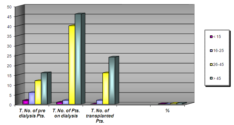
Figure 1
Table (3). Frequency of C.M. in Pts. With R.F attending K.R.C in period from (6-10-2001) according to age group
     
Figure 2
Table (4). Frequency of C.M. in Pts. With R.F attending K.R.C in period from (6-10-2001) according to the duration of R.F
     
Table (5). Frequency of C.M. in Pts. With R.F attending K.R.C in period from (6-10-2001) according to the duration of C.M
     
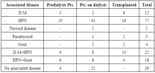
Table (6). Frequency of C.M. in Pts. With R.F attending K.R.C in period from (6-10-2001) according to the associated diseases
     
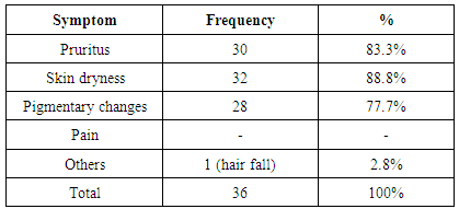
Table (7). Frequency of cutaneous symptoms in pre-dialysis Pts. Attending K.R.C in period from (6-10-2001)
     
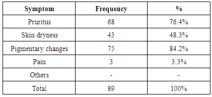
Table (8). Frequency of cutaneous symptoms in Pts. On haemodialysis attending K.R.C in period from (6-10-2001)
     
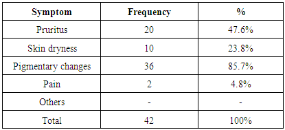
Table (9). Frequency of cutaneous symptoms in transplanted Pts. Attending K.R.C in period from (6-10-2001)
     
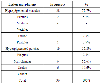
Table (10). Frequency of C.M in pre-dialysis Pts. Attending K.R.C in period from (6-10-2001) according to their lesions morphology
     
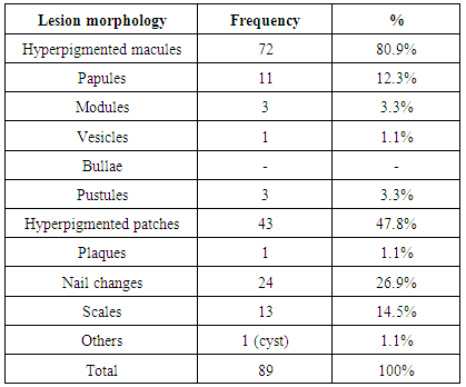
Table (11). Frequency of C.M in Pts. On haemodialysis attending K.R.C in period from (6-10-2001) according to their lesions morphology
     
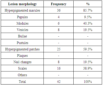
Table (12). Frequency of C.M in transplanted Pts. On haemodialysis attending K.R.C in period from (6-10-2001) according to their lesions morphology
     
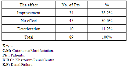
Table (13). The effect of haemodialysis on the C.M in Pts. Attending K.R.C in period from (6-10-2001)
     

4. Discussion

Two hundred patients with renal failure of different causes were seen at Khartoum renal centre in the period from June -3 to October-28- 2001. Fifty patients are pre-dialysis; hundred were on regular hemodialysis, and fifty were transplanted (recipients).
Those patients with cutaneous manifestations were found167 out of the 200 (83.5%) according to the table (1). The highest percentage of cutaneous manifestations seen in patients on regular hemodialysis (89%), then transplanted (recipients) (84%) and (72%) in pre–dialysis patients. These findings showed that the cutaneous manifestations increase when the renal failure progress and after transplantation the skin manifestations decrease to some extent but still more than in the pre–dialysis stage. That means the difficulty of management of cutaneous manifestations even after renal transplantation.
The frequency of cutaneous manifestations according to the sex showed that 97 out of 167 (58%) were males, and 70 (42%) were females. This result showed that the renal failure in Sudanese patients is more frequent in males than females, the ratio about 1.5: 1. Table (2).
According to the age, table (3) the rate of cutaneous manifestations in patients above 45 years were 51.5% and in age group (26-45 years) were 40.7%, which again means that the skin manifestations became more as renal failure progress.
The frequency of cutaneous manifestations according to the duration of renal failure showed that 86.2% appeared after years while 13.8% appeared after months [Table 4]. Moreover, the frequency of the skin manifestations according to the duration of the cutaneous manifestations, showed that 64.7% for years and 34.1 % for the duration of months [Table 5]. These findings in tables (4) & (5) revealed that the cutaneous manifestations go simultaneously with the chronicity of the renal failure.
The common symptoms observed are pruritus, skin dryness and hyperpigmentation; tables (7), (8) and (9). The pruritus is more noticed in the pre-dialysis patients (83.3%) and decreased in patients on hemodialysis to (76.4%) while in transplanted patients only (47.6%).
These findings pointed to that renal transplantation helps in the relief of this troublesome symptom. In this study found that the percentage of pruritus similar to that found international literature 80% [66]. Dimkovic N.1992. Barbara et al. 1980 mentioned that patients with renal failure experienced exacerbation of their symptoms during summer due to the raising of skin temperature that may reduce the threshold for perception of uremic pruritus just as it does in other forms of pruritus. Maybe the weather in Sudan, which is hot, most of the year play a role in exacerbation in Sudanese patients. The same findings also observed for the skin dryness (xerosis) 88.8% in pre-dialysis patients, 48.3% in patients on hemodialysis and 23.8% in transplanted patients and usually the pruritus accompanied by xerosis that is considered as one of the main causes of the itching.
The hyperpigmentation is high in the three categories of patients, 77.7% of pre-dialysis patients, 84.2% in patients on hemodialysis and 85.7% in transplanted recipients. These findings pointed to that the hyperpigmentation not affected by hemodialysis or renal transplantation. The skin discoloration depends on the pre-morbid colour and may be the type of skin play a role, and most of the Sudanese patients belongs to skin type four.
For the cutaneous lesions morphology, tables (10), (11) and (12) the common are hyperpigmented macules and patches observed in the three categories of patients with high percentages 77.7% macules & 59.8% patches in pre-dialysis patients, 80.9% macules & 47.8% patches in patients on hemodialysis and 85.7% macules & 59.5% in transplanted recipients. These findings showed that the morphological signs became more while renal failure progress and they are not affected by hemodialysis or even renal transplantation.
The next common morphological lesions are nail changes that found as follows 16.6% in pre-dialysis patients, 26.9% of patients on hemodialysis and 19.5% in transplanted recipients. These nail changes ranged from Beau's lines, the half and half nail, to nail dystrophy.
Then scales that were found as follows 16.6% in pre-dialysis patients, 14.5% in patients on hemodialysis and 38.9% in transplanted recipients. There were few or minimal signs of papules, vesicles, cysts and bullae seen in a small number of patients in the different categories of renal failure patients.
Table (6): showed that the commonest associated disease is hypertension and next common is diabetes mellitus then gout. There are some patients associated with more than one disease, e.g., hypertension plus diabetes mellitus and hypertension plus gout. For the parathyroid diseases, only three patients were found and for the thyroid diseases only two patients recorded.
About the effect of hemodialysis on the skin manifestations, 50.6% showed no effect while 38.2% showed improvement and 11.2% deteriorate after hemodialysis [table 13].
In this study, three transplant recipients developed nodular and granulomatous's lesions that were confirmed by histopathology to be Kaposi sarcoma. Seven patients developed fungal nail infections, four patients developed Pityriasis Versicolor and two patients developed herpes zoster infection. Most likely the low immune status of those patients due to immunosuppressive therapy play a major role in these conditions.
There are many other cutaneous manifestations mentioned in the literature like bullous dermatosis of dialysis, cutaneous calcification and calciphylaxis, perforating dermatosis, etc. - not encountered in this study.
In this study patients, seen were from different tribes and different social, economic and education classes. These findings pointed to that the socioeconomic factors do not play any role in the development of renal failure and associated cutaneous manifestations.
The drugs used in the management of renal failure that include calcium, Arabic gum, ferrous, folic acid, erythropoietin, etc. do not any effect on associated cutaneous manifestations. Also, neither the antihypertensives nor the antidiabetes drugs showed any effect. The drugs used in hemodialysis and those used in sterilization of the machine tubes that mentioned in the literature to cause allergic reactions did not show the effect and no allergic reactions recorded in the study.

5. Conclusions

Out of 200, known cases of renal failure examined for cutaneous findings, 167 (83.5%) showed cutaneous manifestations. Patients on regular hemodialysis showed the highest skin manifestations (89%), and then transplant recipients (84%) and pre- dialysis patients (72%). The most common symptoms observed were pigmentary changes (82.5%), itching (69.1%) and skin dryness (53.6%). The cutaneous signs seen were.

6. Recommendations

(1) Renal failure is a real problem, and the cases is increasing there for efforts must be done to develop this Khartoum renal centre to accommodate the increasing number of patients and to put a plan for new centres.
(2) There are a lot of cutaneous manifestations associated with renal failures like pruritus, hyperpigmentation, fungal infections and skin tumours so a dermatologist needed to be available at the renal centre to supervise these cases and manage them correctly or to be in contact if this not possible.
(3) There are many cutaneous manifestations been found in other studies like skin calcification and calciphylaxis, bullous dermatosis, etc. which were not found in this study, so further studies recommended being conducted.

References

[1]  Anderson S, and Brenner BM. Progessive renal disease: A disorder of adaptation :QJ Med 1989; 70:185, 1989.
[2]  Meguid EL Nahas. Progression of chronic renal failure in: Richard Johnes and John Feehly text book of comperhension clinical nephrology (first edition) 2001; 14:67.1.
[3]  Gilchrest BA et al. Clinical and histological cutaneous findings in uremia: Evedence from a dialysis - resistant, transplant – responsive micro angiopithy. Lancet 1980; 2: 1271.
[4]  Joseph L, Jorizzo and Elizabith F. Shevertz. Cutaneous changes related to chronic renal failure in Fitz Patrick text book of dermatology in general medicine 2000; 165 : 2058.
[5]  Maxwell MH et al. Clinical disorders of fluid and electrolyte metabolism, 4th ed 1987; New York, Mc Graw – Hill.
[6]  Brezis M et al. Acute renal failure, in the kidney, 4th ed, BM Brenner, FC Rector Jr (eds). Philadelphia, Saunders 1991; 993.
[7]  Brenner BM, Lazarus JM (eds). Acute Renal Failure, 3rd ed 1993; New York, churchil Living stone.
[8]  Glassock RJ. Nutrition, immunlogy and renal disease. Kidney Int 1983; 24: S 194.
[9]  Dewaurdner, H.E. The kidney. An outline of normal and abnormal structure and function 1973; P: 432.
[10]  Brickman A.S. P.H, Massvy S.G and Norman A.W. Impaired calcium absorption in uremic man, J. Lab .clin. Med 1982; 48 ,791.
[11]  Clarkson E.M, Eastwad J.B., koutsaimanis K.G and Dewaurdener H.E.. Net intestinal absorption of calcium – patient with CRF Kidney Int 1973; 3:258.
[12]  Parker R.F, Vergne – Marini P, Hull A.R, and pake C.Y. Jejenal absorption and secretion of calcium in patients with CRF on haemodialysis. J. Clin . investigation 1974; 54:359 [quoted from Andreucci , V.E.1982].
[13]  Avioli L.V. Renal osteodystrophy and v.t.D . pt . on dialysis transplanted patient 1978; 7:244.
[14]  Atkius D. (1976). A possible role of 24,25 (oh)2 1)3 in bone resorption. Journal Eudocrinol, 69:28.
[15]  Stern P. Bone resorbing of 25 – oh stereoisomens of 24 – oh at D3 and 24, 25 (oh)2 D3. Bio.chem. Biophys. Res common 1975; 67, 965.
[16]  Lee S.W., Russell J., and Avioli L.V. 25 – oh 1,3 to 1,25 oh2 1,3: conversion impaired by systemic metabolic acidosis. Science 1977; 194, 994.
[17]  Lumb G.A, Mawer and Stanbury S.W. The apparent vitamin D resistance of CRF. Am . J . Med 1971; 50, 421.
[18]  Kanis, J.A, Oliver D., Ledingham J.G.G, and Russel R.G.G. Evidence that endogenous calcitorin protects against renal bone disease . Lancet 1976; 2, 1322.
[19]  Tranzer F.S, and Naria J.M. Calcitonin inhibition of intestinal phosphate absorption. Nature, london, 1973; 242, 221.
[20]  Haussler M.R., and Mc cain T.H. Basic and clinical concepts related to vitamin D metabolism and action N.E ng .J. Med 1977; 297, 974.
[21]  Habener J.F. and Potts J.T. Biosynthesis of parathyroid hormone, N. Eng. J. Med 1978; 299, 635.
[22]  John W. Rowe, and Barbara A.Gilchrest. Cutaneous aspects of renal disease. ed. 2 New York Mc Graw - Hill Bookco pp 1979; 1408 –1411.
[23]  Barbara A. Gilchrest, John W., Rowe and Martin C.M. Clinical and histological skin changes in chronic renal failure. Lancet 1980; 2.1271-1275.
[24]  Cawly E.P. The eccrine sweat glands of patients in uremia. Arch. Dermatol 1961; 84:51 –58.
[25]  Bartman I. and Landing. 13.14: (1966). Morphology of sweat apparatus in cystic fibrosis. Amer. J . Clin. Path. 54: 455 - 459.
[26]  Stanburry S.W., (1971). Calcium and phosphorus metabolism in renal failure. Diseases of the Kidney. 2nd.ed. 305 -333.
[27]  Arnaud C.D. (1973). Hyperparathyroidism and renal failure . Kidney. Int. 4: 89.
[28]  Vosik W. M. Successful medical mangement of osteitis fibrosa due to tertiary hyperparathyroidism. Mayoclin. Proc 1972; 47, 110.
[29]  Massry S.G., Broutbar N., and Goldstein D.A. Ca + P in health and CRF. Nephrology an approach to the patient with renal disease. J.B., Lippincott company 1982; P.154 –179.
[30]  Muligan R. M. Metastatic calcification. Arch. Path 1947. 43: 177–230.
[31]  Putkonen T and Wangel G. A. Renal hyperparathyroidism with metastatic calcification of the skin. Dermatologica,118: 127-144.
[32]  Parfitt A. M. (1969). Soft tissue calcification in uremia. Arch . intern. Med 1959. 124: 544 –556.
[33]  Eisenberg E. and Bartholow P. V. Reversible calcinosis cutis: calciphylaxis in man. New. Eng. J. Med 1963; 268: 1216-1220.
[34]  Massry S.G. Intractable pruritus as a manifestation of secondary hyperparathyroidism in uremia: Disappearance of itching often subtotal parathyroidectomy. New. Eng. J. Med 1968; 279: 697 –700.
[35]  Gayler B.W. and Brogdon B.G. Soft – tissue calcification in the extremities in systemic disease. Amer. J. Med. Sci 1965; 249: 590 –605.
[36]  Longan M.A. Advances in the chemistry of calcification. Physiol. Rev 1940; 20: 522 – 560.
[37]  Walser M. The chronic composition of uremic plasma. Abstracted, J. Clin. Investigation 1959; 38: 1052.
[38]  Herbert F. K., Miller H. G. and Richardvon G. O. Chronic renal disease, secondary parathyroid hyperplosia, Decalcification of bone and metabolic calcification. J. Path. Bact 1941; 53: 161–182.
[39]  Ress J. K. and Coles G. A. Calciphyloxis in man. British Med. J. 1969; 2: 670– 672.
[40]  Selye H. Tuchweber B and Gabbiani G. Acta. Endocrinologica, suppl 1964; 90: 203.
[41]  Anderson D.G. Stewart W. K. and Pievcy D. M. Lancet 1968; 2: 323.
[42]  Copp D. H. Calcium and phosphorus metabolism. Amer. J. Med 1957; 22: 275–285.
[43]  Urist M. R., Moss M. J., and Adams J. M. Calcification of terdon: A triphosic local mechanism. Arch. Path 1964; 77: 594 – 608.
[44]  David G. Adesonm William K. Stewart, and David M. Pievry. Calcifying panniculitis with fat and skin necrosis in a case of uremia with hyperparathyroids. Lancet 10 August 1968; 323 –325.
[45]  Friedman M., Marshall – Jones P. and Ross E. J. Q. J. Med 1966; 35: 193.
[46]  Olmostead E. G. , and Lunseth J. H. Skin manifestations of chronic acidosis. Arch. Dermatol 1958; 77: 304 – 314.
[47]  Worth R.L. Calciphylaxis: Pathogenesis and therapy. Cutan Med Sung Apr 1998; 2 (4): 245 – 8.
[48]  Roe – SM, Graham – L.D. Brock – W.B., and Barker – D.E. Calciphylaxis: Early recognition and mangement. Am – Sung 1994 Feb; 60 (2): 81 – 6.
[49]  Hohl D. Necrotizing vasculan calcinosis. MEDLINE Dermatology 1994; 189: 432 – 4.
[50]  Benkalfate L., Zein K., Le – Gall F., Chevvant – Breton J., Rivalan J, and Le – Pogamp P. Calcified intertrigo; a vare cause of cutaneous calcinosis. Ann–Dermatology–Venereol 1995; 122: 789 – 92.
[51]  Tan H .H., and Cheong W. K. Cutaneos gangvene secondary to metastatic calcification in end stage renal failure. Singapore – Med – J 1996 Aug.; 37: 438 – 40.
[52]  Uchida M., Sakemi T., Ikeda Y., and Maeda T. Acute progressive and extensive metastatic calcification in a nephrotic patient following chronic hemodialysis. Am. J. Nephrol 1995; 15: 427–30.
[53]  Gilkes J.H., Eady R. A., and Lesley H. Ress. Plasma immunoreactive melanotrophic hormones in patients on mainterarce haemodialysis. British Medical Journal 1 1975; 656 – 658.
[54]  Smith A. G., Sam Shouster and A. J.Thody. Role of the kidney in regulation plasma immunoreactive beta – melanocyte stimulating hormone. British medical journal 1 1976; 874 – 876.
[55]  Abe K. Levels of immunoreactive B. MSH in CRF. British J. of clinical investigation 1969; 48: 1580.
[56]  Shuster S., and Thody A. J. Immunoreactive B. MSH. Journal of investigation Dermatology 1974; 62: 172.
[57]  Hmida M. B., Turki H., Hachicha J., Reygagne P., Rabier D., Zahaf A., and Jarraya A. Hypopigmentation in hemodialysis. MEDLINE Dermatology 1996; 192:148 – 52.
[58]  Kalman Keczkes and Malcolm Farr. Bullouss dermatosis of chronic renal failure. British journal of Dermatoloy 1976; 95: 541–546.
[59]  Maureen Poh -Fitzpatrick. Bullous disease of dialysis: Topic Last Updated 11/3/ 2000; 11: 54: 20.
[60]  Mantoux F., Bahadovan P., Perrin C., Bermon C., Lacour J.P., and Ortonne J. P. Flutamide –induced late cutaneous pseudoporphyra. Ann–Dermatology–Venereol 1999 Feb.; 126:150 – 2.
[61]  Glynne P., Deacon A., Goldsmith D.; Pusey C., and Clutterbuck E. Bullous dermatosis in E .S .R .O: Porphyria on pseudoporphyria . Am. J. kidney. Dis 1999 Jul.; 34:155 – 60.
[62]  Kenedy A. C. , and Lyell A. Accquived epydermolysis bullosa due to high dose of frusemide. British medical journal 1 1976; 1509 -1510.
[63]  Kalman Keczkes and Malcolm Farr. Cutaneous bullous and frusemide in CRF. British medical journal 1972; 2: 236.
[64]  Shaul G. Massry, Mordecai M., Poportzen and Jake W. Coburn. Intractable pruritus as a manifestation of secondary hyperparathyroidism in uremia. New England Journal of Medicine 1968; 279: 697 –703.
[65]  Berson S. A. and Yallow R. S. Parathyroid hormonein plasma in adenomatous hyperparathyroidism. Science 1966; 154 : 907.
[66]  Dimkovic N., Djukanovic L., Radmilovic A., Bojic P., and Juloski T. Uremic pruritus and skin most cells. Nephron 1992; 61: 5 – 9.
[67]  David Maclachlan and Alexander L. Forrest: (1974). Itching in renal failure. Lancet, 2: 355.
[68]  Barbara A. Gilchrest, John W. Rowe and Robert S.Brown. Relief of uremic pruritus with ultraviolet phototherapy.Lancet 1977; 297:136–138.
[69]  Eugene L. Saltzer. Relief from uremic pruritus: A therapeutic approach. Cutis 1975; 16: 298 –299.
[70]  Yatzidis H., Digenis P., and Tountas C. Heparin treatment of uremic itching. Jama 1972; 222:1183.
[71]  Silverberg D. S., Iaina A., and Reisin E. Cholestyramine in uremic pruritus. British medical journal 1 1977; 752 – 753.
[72]  Luis Tapia, Jhoong S. Cheigh, and David S. David. Pruritus in dialysis patients treated with parentral lidocaine. New Enngland Journal 1977; 296: 261–262.
[73]  Constantine L. Hampers, Adrian I. Katz and Richard E.Wilson. Disappearance of uremic itching after subtotal parathyroidectomy. New England Journal 1968; 279: 695–697.
[74]  Robertson K.E. et al. Uraemic pruritus, Am. J. Health . Syst. Pharm 1996. 53: 2159.
[75]  Muchrcke R. C. The finger nails in chronic hypoalbuminemia. British Medical Journal 1 1956; 1327–1328.
[76]  Samuel L. Moschella, Donald M. Pillsbury and Harvy J. Hurty. Diseases of nutrition and metabolism. Dermatology vol II. W. B. Saunders company. Philadelphia, London, Toronto P 1975; 1237 – 1322.
[77]  James B. Hudson and Allen J. Dennis. Transverse white lines in the finger nails after. Acute and chronic renal failure. Arch. Intern. Med. Vol 1966.117: 276–279.
[78]  Philip G. Lindsay. The half and half nail. Arch. Intern. Med 1967; 119: 583–587.
[79]  James J. Leyden and Margared Gray Wood.“ The half–and –half nail ``A uremic onychopathy. Arch. Derm. Vol 1972; 105: 591–592.
[80]  Lindsay P. G. The half and half nail. J. Lab. Clin. Med. 1965; 66: 82.
[81]  Lubach D., Strubbe J. and Schmitt J. The half and half nail phenomenon in chronic hemodialysis patients. Dermatologica 1982; 164: 350–353.
[82]  Constantine V. S., and Carter V. H. Kyrle’s disease: Histopathologica Findings in five cases and review of the literatune. Arch. Dermatol 1968; 97: 633–639.
[83]  Antoinette F., Gray L. Hardegen and Alfredo R. Zarate. Kyrle’s disease in patients with CRF. Arch. Dermatol 1982; 118: 85–88.
[84]  Patterson J.W. The perforating disorders. J. Am. Acad. Dermatol 1984; 10: 561.
[85]  Mehta R. L. Skin necrosis associated with acquired protein C deficiency in patients with renal failure and calciphylaxis. Am J. Med 1990; 88.
[86]  Goldblum O. M. Pseudo – kaposi sarcoma of the hand associated with an acquired, iatrogenic arteriovenous fistula. Arch. Dermatol 1985; 121: 1038.
[87]  Fuchs E. Dialysis acne. J. Am. Acad. Dermatology 1990; 23:125.
[88]  Rudlinger R. Human papilloma virus infections in a group of renal transplant recipients. Br. J. Dermatology 1986; 115: 681.
[89]  Gupta A.K. Cutaneous malignant neoplasms in patients with renal transplants. Arch. Dermatology 1986; 122: 1288.