-
Paper Information
- Next Paper
- Paper Submission
-
Journal Information
- About This Journal
- Editorial Board
- Current Issue
- Archive
- Author Guidelines
- Contact Us
American Journal of Dermatology and Venereology
2015; 4(1): 5-8
doi:10.5923/j.ajdv.20150401.02
Hemangiopericytoma (HPC), in Adolescent Sudanese Patient
 Abstract
 Reference
 Full-Text PDF
 Full-text HTML
Adil H. H. Bashir1, Lamyaa A. M. Elhassan2, Abdel Khalig Muddathir3, Khalid O. Alfarouk1, 4, Rehab O. Mohammed4, Gamal O. Elhassan5, 6, Anilkumar Mithani7, Ahmed M. El Hassan1
1Institute of Endemic Diseases, University of Khartoum, Khartoum, Sudan
2Ahfad University for Women, Omdurman, Sudan
3Faculty of Pharmacy, University of Khartoum, Khartoum, Sudan
4Alfarouk Biomedical Research LLC, San Antonio, Texas, USA
5Unizah Pharmacy Collage, Qassim University, Qassim, KSA
6Faculty of Pharmacy, Omdurman Islamic University, Omdurman, Sudan
7Faculy of Medicine, University of Bahri, Khartoum, Sudan
Correspondence to: Adil H. H. Bashir, Institute of Endemic Diseases, University of Khartoum, Khartoum, Sudan.
| Email: |  |
Copyright © 2015 Scientific & Academic Publishing. All Rights Reserved.
Hemangiopericytoma (HPC) is a tumor with malignant potential composed with vascular channels and proliferating pericyte-like spindle cells. Itg rows in the body’s soft tissue, which includes fat, muscles, tendons, nerves, blood vessels and another fibrous tissue. We reported rare case, of a male adolescent, 13 years old, presented with insidious onset of right leg, easily bleed, scabbed, well-demarcated skin ulcer, with offensive necrotic floor, and crusts. The case was diagnosed and confirmed histopathologically as Hemangiopericytoma, considered to be the first casebeen reported in Sudan.
Keywords: Hemangiopericytoma
Cite this paper: Adil H. H. Bashir, Lamyaa A. M. Elhassan, Abdel Khalig Muddathir, Khalid O. Alfarouk, Rehab O. Mohammed, Gamal O. Elhassan, Anilkumar Mithani, Ahmed M. El Hassan, Hemangiopericytoma (HPC), in Adolescent Sudanese Patient, American Journal of Dermatology and Venereology, Vol. 4 No. 1, 2015, pp. 5-8. doi: 10.5923/j.ajdv.20150401.02.
1. Background
- Hemangiopericytoma is a rare mesenchymal neoplasm, accounting for about 1% of vascular tumours [1] Hemangiopericytoma is known to be derived from the vascular pericyte and was first reported by Stout and Murray in 1942 [2]. It has been reported at all ages and sexes are equally affected. Some cases had been present at birth. Haemangiopericytoma usual situation is in the subcutaneous or muscular tissues, and the lower trunk, pelvis, head and neck or thigh. The tumor is flesh colored, firm, often circumscribed nodular, mass up to 8 cm varying in size. [3]. The tumour occurs most commonly in the skin, subcutaneous soft tissues, muscles of the extremities, retroperitoneum but rarely in the lung, trachea or mediastinum [4]. Herein, a surgical case of primary mediastinal hemangiopericytoma is presented.Hemangiopericytoma often is painless masses and may not have any associated symptoms. These tumors can originate anywhere in the body where there are capillaries. They can be either benign or malignant, and can metastasize or spread to other areas of the body, primarily the lungs and bones. Though rare, hemangiopericytomas can be located in the nasal cavity and paranasal sinuses. Their prognosis is better because they tend to be less aggressive and do not metastasize.
2. Case Report
- A 13-year-old adolescent male, descent from first-degree relative parents, resident in Khartoum, was referred to our hospital complaining of massive right leg non-healing wound, for the last two months. Physical examination indicated large punch-out sloughing ulcer affecting almost all anterior, lateral and medial aspects of left leg (Figure 1). Some of the floors showed with adherent scabs, and some areas were necrotic while others are granulating. A nodular lesion at same leg popliteal area is noticed, 3cm in diameter, skin colored, and slightly tender. No palpable regional lymph nodes. The condition was associatedlow-grade fever, lassitude and loss of appetite. Laboratory studies other and chest X-ray were essentially within normal limits. Culture of the wound was also negative. The patient was negative for both HIV and VDRL (Venereal disease research laboratory) using ICT; Helicobacter pylori infection was non-reactive. The complete Blood Count showed Hb 10.2 RBCs 3.65; platelets 590 TWBc Lymph 33.5Neutr. 50.4Eosino. 2 Mono. 8 Baso.0 with mild hypochromia and ESR 105.
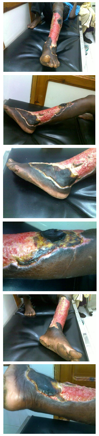 | Figure 1. Left leg ulcer along anterior, lateral and medial aspects |
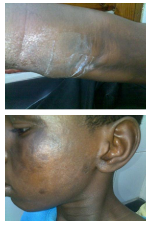 | Figure 2. Left leg popluteal fossa nodular lesion and another pigmented patches at right side face |
3. Discussion
- Hemangiopericytoma is an uncommon, potentially malignant tumour originating from pericytes in the small vessels [4]. Our case was a cutaneous Hemangiopericytoma, which is rare. Only a few isolated case reports are available in the literature [5]. Hemangiopericytoma has no uniform clinical or radiographic features, usually affects older individuals, and mostly presents as an asymptomatic. These tumours are composed of closely-packed spindle cells and prominent vascular channels. The histological differential diagnosis includes many mesenchymal tumours, such as the solitary fibrous tumour and the synovial sarcoma [4]. No single clinical or histological feature including histological type or DNA ploidy allows prediction of biologic aggressiveness [6]. Malignant Hemangiopericytoma is recognized by its increased mitotic rate, tumour size and foci of haemorrhage and necrosis [4]. Immunohistochemically, hemangiopericytomas are known to show a positive response to antibodies against vimentin and type IV collagen and a negative response to VIII-related antigen, S-100 protein, neuron specific enolase, carcinoembryonic antigen, desmins, laminin and cytokeratins [7].Surgical radical excision is the treatment of choice for hemangiopericytoma, although the criteria for determining the area of resection have not been established. Hansen and colleagues stated that it was necessary to consider all hemangiopericytomas as malignant and perform extended surgery [8]. During the resection, it is important to look for invasion of the surrounding tissue and to avoid the spread of tumour cells by manual examination. With respect to adjuvant therapy, chemotherapy or radiotherapy have been recommended but is considered to be almost ineffective [4]. On the other hand Rusch et al., reported that combination therapy or single therapy with Adriamycin was effective against metastases [9].The 5-year survival of patients with hemangiopericytomasoriginating in any organ has been reported to be 85%, whereas the survival of patients with a tumour of pulmonary origin is 30–35%. Approximately 50% of hemangiopericytoma have been reported to recur within five years [4, 8]. It has been demonstrated that recurrent diseaseusually occurs within two years after initial treatmentand recurrences are commonly found in the thorax, either in the pulmonary parenchyma or the pleura. Distant metastases to liver, brain and bone have also been reported [8].
4. Conclusions
- Hemangiopericytoma is an uncommon, potentially malignant tumour originating from pericytes in the small vessels, and surgical radical excision is the treatment of choice, although the criteria for determining the area of resection have not been established. International literature has demonstrated that recurrent disease usually occurs within 2 years and therefore a long-term careful follow-up is required.