-
Paper Information
- Next Paper
- Paper Submission
-
Journal Information
- About This Journal
- Editorial Board
- Current Issue
- Archive
- Author Guidelines
- Contact Us
American Journal of Dermatology and Venereology
2014; 3(4): 72-74
doi:10.5923/j.ajdv.20140304.02
Herpes Simplex Dermatitis (Keratitis): Two Cases Report
 Abstract
 Reference
 Full-Text PDF
 Full-text HTML
Adil HH Bashir1, Anil Mitani2, Gamal O. Elhassan3, 4, Lamyaa AM Elhassan5, Abdel Khalig Muddathir6, Khalid O. Alfarouk1, Ahmed M. Elhassan1
1Institute of Endemic Diseases, University of Khartoum, Khartoum, Sudan
2Faculty of Medicine, University of Bahri, Khartoum North, Sudan
3Uneizah Pharmacy College, Qassim University, Alqassim, KSA
4Faculty of Pharmacy, Omdurman Islamic University, Khartoum, Sudan
5School of Medicine, Ahfad University for Women, Omdurman, Sudan
6Faculty of Pharmacy, University of Khartoum, Khartoum, Sudan
Correspondence to: Adil HH Bashir, Institute of Endemic Diseases, University of Khartoum, Khartoum, Sudan.
| Email: |  |
Copyright © 2014 Scientific & Academic Publishing. All Rights Reserved.
Several cutaneous disorders may occur in organ transplant recipients. The immunosuppressive regimen consisted of combinations including cyclosporine, systemic corticosteroids, azathioprine, tacrolimus, Mycophenolate mofetil, antiviral, and antibiotics. Ninety-one cutaneous manifestations were identified in 55% kidney transplant patients. 17.5% cutaneous viral infections, 10.0% included verruca vulgaris, herpes zoster and herpes simplex. We reported two rare cases of adult Herpes Simplex Dermatitis (Keratitis) (HSD) occurred in a 30 year old Sudanese young adult with renal transplant 5 months duration, presented with erythematous widespread diffuse and discrete vesicle with few typical herpetiform groups in upper back for 3 days duration, and 33 years old female patient presented with small blisters progressed to Toxic Epidermal Necrolysis (TEN). The cases were diagnosed and confirmed histopathologically as HSD, considered to be the first two cases of HSD been reported in Sudan.
Keywords: Dermatitis, Renal transplant
Cite this paper: Adil HH Bashir, Anil Mitani, Gamal O. Elhassan, Lamyaa AM Elhassan, Abdel Khalig Muddathir, Khalid O. Alfarouk, Ahmed M. Elhassan, Herpes Simplex Dermatitis (Keratitis): Two Cases Report, American Journal of Dermatology and Venereology, Vol. 3 No. 4, 2014, pp. 72-74. doi: 10.5923/j.ajdv.20140304.02.
1. Background
- Several cutaneous disorders may occur in organ transplant recipients. The immunosuppressive regimen consisted of combinations including cyclosporine, systemic corticosteroids, azathioprine, tacrolimus, Mycophenolate mofetil, antivirals, and antibiotics. Ninety-one cutaneous manifestations were identified in 55.0% kidney transplant patients. 17.5% cutaneous viral infections, 10.0% included verruca vulgaris, herpes zoster and herpes simplex (1). Renal transplant patients were submitted to dermatological check-up investigations at regular intervals over a mean period of 26.2 months after surgery. 98% of the case material showed dermatological complications. In an analysis of the findings in 55 patients with infectious complications, viral infections occurred in 40, bacterial in 30 and mycotic infections in 20 patients. Dermatological manifestations of non-dermatological complications were mainly due to immunosuppressive therapy. The data stress the necessity of optimum cooperation between dermatologists and the attendant physician in the case of renal transplant patients [1].Threats of bioterrorism have renewed efforts to better understand poxvirus pathogenesis and to develop a safer vaccine against smallpox. Individuals with atopic dermatitis are excluded from smallpox vaccination because of their propensity to develop eczema vaccinatum, a disseminated vaccinia virus (VACV) infection [2, 3].
2. Case I
- A male patient, single, 30 years old, civil employee, descent from second degree relative parents, resident in Taief, Gaalei tribe.The condition started 3 days ago with insidious onset and progressive course, with erythematous widespread diffuse and discrete mildly itchy, vesicles, mainly at face, upper trunk, and extremities. Patient is a case of renal transplant 5 months ago and on immunosuppressive Cellcept 500mg (Mycophenolate mofetil) and Prednisolne medication. The condition associated with low grade fever, malaise and lassitude.On general examination: The general condition is well, not pale, not icteric, as well no palpable spleen, and liver. No palpable lymph nodes.On dermatological examination, wide spread. diffuse symmetrically discrete, erythematous vesicular lesions; tense, clear, variable sized 1-3 mm, some arranged at herpetiform groups involving mainly face, upper back, upper arms, upper chest, upper and thighs. No erosions and ulcers seen.The blisters are small, tense, clear; some are eroded and crusted on erythematous base.Palms and soles: Similar lesions at both soles and palms.Nails: No nails dystrophy has been noticed. Ears: No Abnormality Detected. Hair: No Abnormality Detected. Oral cavity: No oral blisters or erosions are seen.Investigations done: Skin biopsy:Sections show an intraepidermal bulla. It shows two types of changes: ballooning and reticular degeneration. The former involves basal cells which are swollen and contain single or multiple nuclei. The nuclei have condensed chromatin beneath the nuclear membrane with microabscess formation. The dermis contains a mild peri- vascular lymphocytic infiltration.Diagnosis: Dermatitis Herpetiformis.
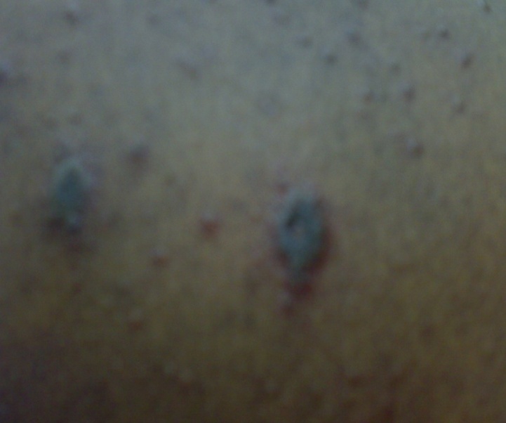 | Figure 1a. Shows erythematous widespread diffuse and discrete vesicles at the back-case 1 |
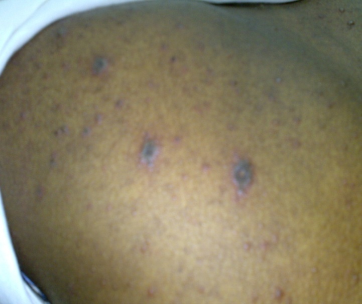 | Figure 1b. case 1 |
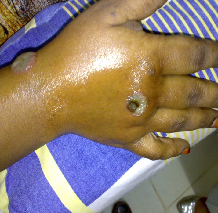 | Figure 2a. Shows discrete, yellow brownish, bullous impetigo-like lesions at dorsum of right hand-case 2 |
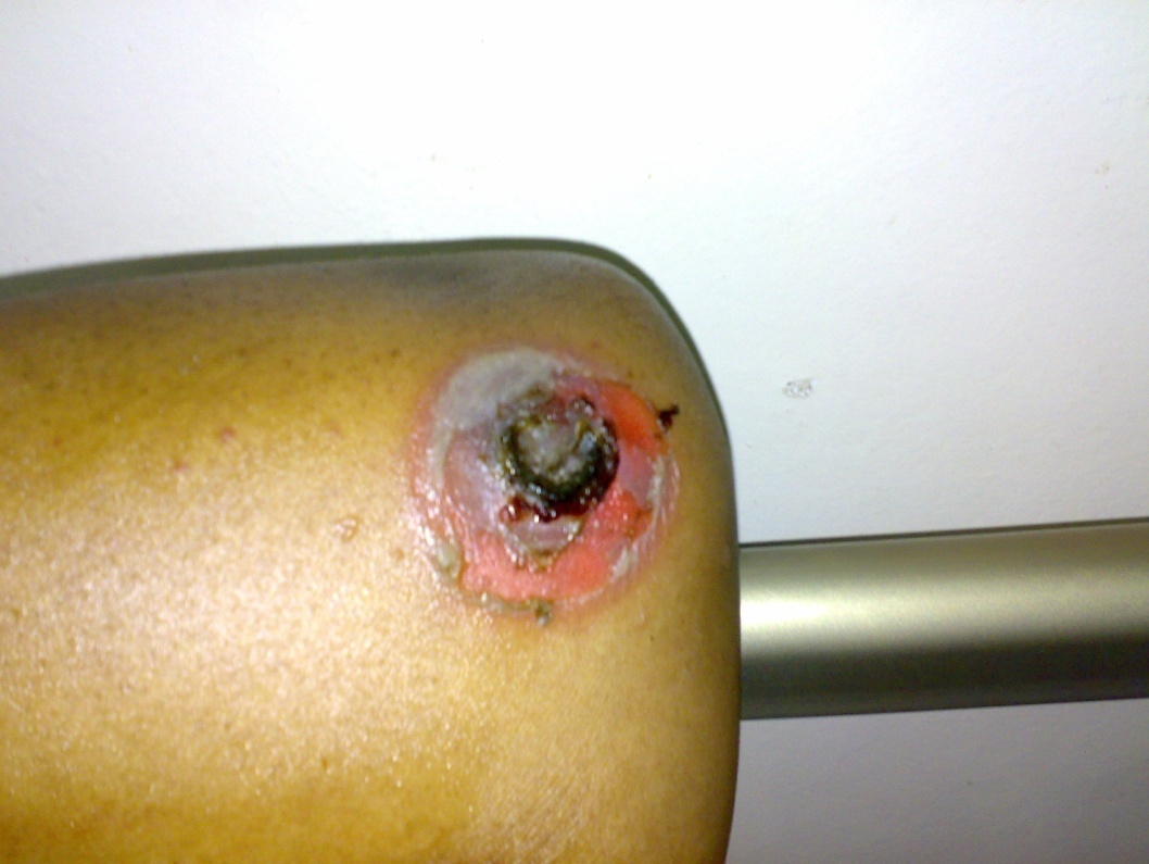 | Figure 2b. Shows vesiculo bullous target lesion (Herpes iris of Battman) at left elbow. (Closer view-case 2) |
 | Figure 2c. Closer view |
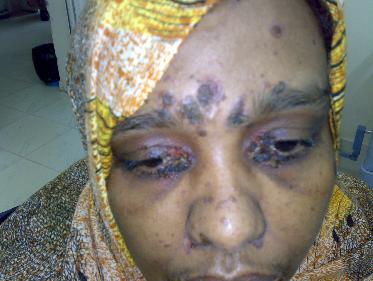
3. Case II
- A female patient 31 years old, presented with turbid blistering lesions mainly at face, periorifical a month ago, a diagnosis of bullous impetigo is raised where systemic and topical antibiotics prescribed with poor response.A new widespread diffuse individual blistering lesions appeared, blisters were small, tense and clear where a suspicion of viral infection raised as patient was in follow up with a dentist for oral thrush and systemic steroids given before flaring of the condition, a biopsy is taken to confirm the diagnosis Herpes Simplex Keratitis In Erythema Multiformi Major- Sever type ? Toxic Epidermal Necrolysis (TEN).Patient put on Antiviral regimen oral therapy, then parenteral, with other conservative treatments, then methyl-prednisolne 250 mg daily injection for 5 days, followed with prednisolne 40 mg daily dose and topical Mebo ointment.General condition is stable, and skin healing is slow.Skin biopsy:Sections show an intraepidermal bulla. It shows two types of changes: ballooning and reticular degeneration. The former involves basal cells which are swollen and contain single or multiple nuclei. The nuclei have condensed chromatin beneath the nuclear membrane with microabscess formation. The dermis contains a mild peri- vascular lymphocytic infiltration.Diagnosis: Herpes Simplex Dermatitis.
4. Discussion
- The two are typical rare HSD cases, in a male, 30 years old, with renal transplant, and 31 years female both had an adult onset of presentation. Clinical features and histopathology are typical, as well as responded dramatically to systemic Antiviral therapy. In case 1 the response was dramatic.
ACKNOWLEDGMENTS
- This work has been supported by Alfarouk Biomedical Research LLC.