-
Paper Information
- Paper Submission
-
Journal Information
- About This Journal
- Editorial Board
- Current Issue
- Archive
- Author Guidelines
- Contact Us
American Journal of Dermatology and Venereology
2014; 3(1): 5-8
doi:10.5923/j.ajdv.20140301.02
Papillon-Lefèvre Syndrome: A Report of Three Siblings
 Abstract
 Reference
 Full-Text PDF
 Full-text HTML
Adil H. H. Bashir1, Shaza Mohammed Yousif2, Anilkumar Mitani3, Abdel Khalig Muddathir4, Gamal O. Elhassan5, Abdel Rahman M. A. I. Ramadan6, Hanadi Abd Allah Elamin7, Khalid O. Alfarouk1, Lamya A. Elhassan8, Ahmed M. Elhassan1
1Institute of Endemic Diseases, University of Khartoum, Khartoum, Sudan
2International University of Africa, Khartoum, Sudan
3Faculty of Medicine, University of Bahri, Sudan
4Faculty of Pharmacy, University of Khartoum, Khartoum, Sudan
5Uneizah Pharmacy College, Qassim University, KSA
6College of Dentistry, Taibah University, Al-Madinah Al-Munawarah, KSA
7Khartoum Skin Hospital, Khartoum, Sudan
8Ahfad University for women, Omdurman, Sudan
Correspondence to: Adil H. H. Bashir, Institute of Endemic Diseases, University of Khartoum, Khartoum, Sudan.
| Email: |  |
Copyright © 2012 Scientific & Academic Publishing. All Rights Reserved.
We reported an 18 year old Sudanese female patient, Gaali tribe, and another 2 males, brothers ages 10 and 8 years old, presented with diffuse trangrediens palmoplantar keratosis with scaly, non-erythematous, psoriasiform-like transgradient plaque in elbows and knees, and periodontosis, the female patient has been misdiagnosed before as a case of Psoriasis and given phototherapy in the form of PUVA with satisfactory results and the 2 brothers has been also missed as a cases of eczemas with poor response to treatment and frequent relapses. The female case is associated with thyroid swelling and increased Thyroid Stimulating Hormone TSH, and considered as a first cases record at Sudan, as Papillon-Lefèvre syndrome is known as a rare presentation globally and in Sudan specifically.
Keywords: Syn. Papillon-Lefevre Syndrome, Disease, Papillon-Lefevre, Papillon Lefevre Disease, Papillon Lefevre Syndrome, Keratosis palmoplantar-periodontopathy
Cite this paper: Adil H. H. Bashir, Shaza Mohammed Yousif, Anilkumar Mitani, Abdel Khalig Muddathir, Gamal O. Elhassan, Abdel Rahman M. A. I. Ramadan, Hanadi Abd Allah Elamin, Khalid O. Alfarouk, Lamya A. Elhassan, Ahmed M. Elhassan, Papillon-Lefèvre Syndrome: A Report of Three Siblings, American Journal of Dermatology and Venereology, Vol. 3 No. 1, 2014, pp. 5-8. doi: 10.5923/j.ajdv.20140301.02.
Article Outline
1. Background
- The Papillon-Lefevre syndrome is psoriasiform hyperkeratosis of the palms and soles that can overflow onto the dorsal surfaces of the hands and feet. Psoriasiform lesions can also be seen on the limbs. This keratoderma associates, as early as infancy, intense gingivitis with alveolar bone lysis and early loss of baby teeth. During childhood, this phenomenon of periodontal disease recurs with rapid loss of adult teeth. This syndrome is accompanied, in half the patients, by enhanced susceptibility to infections (furunculosis, skin abscesses, hidradenitis suppurativa). Anomalies of chemotaxis and phagocytosis by polymorphonuclear leukocytes have been observed (Itin and Fistarol).This condition is inherited in an autosomal recessive fashion. The frequency of PPK with periodontitis is 4 cases per 1 million population. The male-to-female ratio is equal. Onset occurs between the first and fifth years of life. A variant, Haim-Munk syndrome features, in addition to PPK and periodontitis, arachnodactyly, acroosteolysis, and onychogryphosis (Ratnavel and Griffiths, 1997).Clinically, diffuse transgredient PPK may be observed, typically developing within the first 3 years of life. Punctiform accentuation, particularly along the palmoplantar creases, may be seen. Unless treated, periodontosis results in severe gingivitis and loss of teeth by age 5 years. No significant correlation has been demonstrated between the level of periodontal infection and the severity of skin affections, which supports the concept that these major components of this syndrome are unrelated to each other. Patient’s exhibit increased susceptibility to cutaneous and systemic infections. Scaly, psoriasiform lesions are often observed over the knees, elbows, and interphalangeal joints. Finally, patients may have malodorous hyperhidrosis (Griffiths et al., 1998).
 anodontia/oligodontia (Very frequent sign).
 autosomal recessive inheritance (Very frequent sign)
 dysplastic/thick/grooved fingernails (Very frequent sign)
 hamartoma/tumours of the mouth (Very frequent sign)
 palmoplantar hyperkeratosis (Very frequent sign)
 teeth anomalies (Very frequent sign)
 arachnodactyly (Frequent sign)
 intracranial calcifications (Frequent sign)
 thin/hypoplastic/hyperconvex fingernails (Frequent sign)
 decreased body hair (Occasional sign)
 increased body hair (Occasional sign)
 skin tumours/lumps (Occasional sign)
 sparse/absent scalp hair(generalized) (Occasional sign)Histologic findings include hyperkeratosis with irregular parakeratosis and moderate perivascular infiltration. Electron microscopic features include lipid like vacuoles in corneocytes and granulocytes, a reduction in tonofilaments, and irregular keratohyalin granules (Griffiths et al., 1998). Molecular biology findings include mutations in the Cathepsin C gene, mapping to 11q14-q21, which are responsible for this syndrome. Cathepsin C is a lysosomal protease known to activate enzymes that are vital to the body's defenses (Hart et al., 1998; Kimyai-Asadi et al., 2002). Treatment includes oral retinoids for the PPK. Elective extraction of involved teeth may prevent excess bone resorption. Appropriate antibiotic therapy may be required for periodontitis and recurrent cutaneous and systemic infections. Treatment with acitretin starting at an early age shows promise towards allowing patients to have normal adult dentition (Saywell C and WAD, 1994). Retinoids slow the alveolar bone lysis and attenuate the palmoplantar keratoderma. This genetic disease is transmitted by autosomal recessive inheritance. It is linked to an almost total loss of Cathepsin C activity (Hart et al., 1999; Toomes et al., 1999).
2. The Case 1
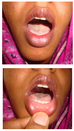
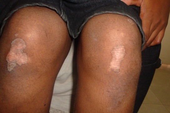
- A female patient, student, 18 years old, single, resident in Al-Dqba, north of Sudan. The patient is complaining of dry painful fissured, non-itchy, soles of feet with difficulty on walking since age of 4 months, loss of teeth with swollen painful gum since the age of 15 years old. Patient’s general condition looks well with average intelligence, a thyroid swelling is noticed, uniformly diffuse, moves with deglutition, no regional lymphadenopathy. The condition started at infancy, age of 4 months with peeling of soles of feet, with gradual onset and progressive course, where thickening of both soles ensues, fissured and worse with age and focal thickening in both palms. Patient noticed inability to move some of her hand fingers since childhood. Patient has been diagnosed as a case of Psoriasis and received topical treatment in the form of PUVA with more or less satisfactory result.Family history: Parents are first degree relatives, no similar condition in family, and heel fissurings in her cousins is noticed. On examination: Patient has normal facial features with diffuse transgradient palmoplantar keratoderma, and distant psoriasiform plaques-like at both knees and elbows. Hyperkeratotic knuckle pads mainly at proximal interphalangeal joints, with joint block at both little fingers, where patient can’t approximate palms of both hands together.Plaques are well defined, demarcated scaly, no fissures or erythema, 1-2 cms in diameter. No hyperhidrosis is seen. Lesions are skin colored.The lesions are Firm on palpation.Palms & soles: Diffuse transgradient palmoplantar keratoderma. No Palmar or plantar pits or erythema, as well as arachnodactyly or pseudoainhum of the digits, not detected. Nails: Nail dystrophy with hyperconvexity, transverse and longitudinal ridging is noticed at most of dysplastic nails. Hair: No Abnormality Detected, with normal body and scalp hair. Eye: No abnormality seen.Oral mucosa &Lips: Periodontosis with loss of teeth-Oligodontia. No Leukokeratosis.
3. Investigations Done
- 1. Skin biopsy:Macro: 2 small pieces of soft tissue, submitted all in one block.Micro: Sections show tumor cells form cords and clusters of cells with basophilic ovoid nuclei and little cytoplasm.The cells at the periphery of the cords show palisading.There are a melanin pigments and mitotic figures.Diagnosis: Basal Cell Carcinoma, BCC.
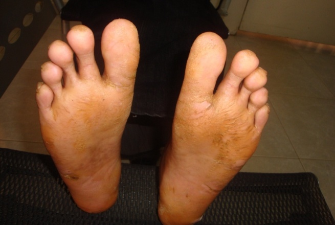 | Figure 1. Diffuse plantar keratoderma both feet |
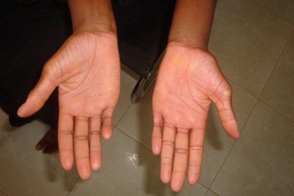 | Figure 2. Focal palmar keratoderma both hands |
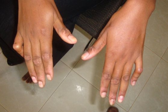 | Figure 3. Blocked joints little fingers of both hands |
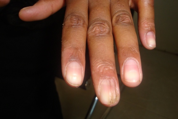 | Figure 4. Longitudinal nail ridging of middle finger with ragged nail cuticle |
 | Figure 5. Psoriasis like lesions both knees |
 | Figure 6. Complete denture |
4. The Case 2
- A male patient, student, 10 years old, resident in Al-Duba, north of Sudan. The patient is complaining of dry painful fissured, non-itchy, soles of feet with difficulty on walking since age of 4 months, loss of teeth with swollen painful gum since the age of 15 years old. Patient’s general condition looks well with average intelligence.The condition started at infancy, age of 4 months with peeling of soles of feet, with gradual onset and progressive course, where thickening of both soles ensues, fissured and worse with age and focal thickening in both palms. Patient noticed inability to move some of her hand fingers since childhood. Patient has been diagnosed as a case of Psoriasis and received topical treatment in the form of PUVA with more or less satisfactory result.Family history: Parents are first degree relatives, 2 similar condition in family, oldest sister and a younger brother. On examination: Patient has normal facial features with diffuse transgradient palmoplantar keratoderma, and distant psoriasiform plaques-like at both knees and elbows. Hyperkeratotic knuckle pads mainly at proximal interphalangeal joints, with joint block at both little fingers, where patient can’t approximate palms of both hands together. Plaques are well defined, demarcated scaly, no fissures or erythema, 1-2 cms in diameter. No hyperhidrosis is seen. Lesions are skin colored.The lesions are Firm on palpation.Palms & soles: Diffuse transgradient palmoplantar keratoderma. No Palmar or plantar pits or erythema, as well as arachnodactyly or pseudoainhum of the digits, not detected. Nails: Nail dystrophy with hyperconvexity, transverse and longitudinal ridging is noticed at most of dysplastic nails. Hair: No Abnormality Detected, with normal body and scalp hair. Eye: No abnormality seen.Oral mucosa &Lips: Periodontosis with loss of teeth-Oligodontia. No Leukokeratosis.
5. The Case 3
- A male patient, student, 8 years old, mixed race, Gaali tribe, resident in Al-Duba, north of Sudan. The patient is complaining of dry painful fissured, non-itchy, soles of feet with difficulty on walking since age of 4 months, loss of teeth with swollen painful gum since the age of 15 years old.Patient’s general condition looks well with average intelligence. The condition started at infancy, age of 4 months with peeling of soles of feet, with gradual onset and progressive course, where thickening of both soles ensues, fissured and worse with age and focal thickening in both palms. Patient noticed inability to move some of her hand fingers since childhood. Patient has been diagnosed as a case of Psoriasis and received topical treatment in the form of PUVA with more or less satisfactory result.Family history: Parents are first degree relatives, the youngest brother of case 2. On examination: Patient has normal facial features with diffuse transgradient palmoplantar keratoderma, and distant psoriasiform plaques-like at both knees and elbows. Hyperkeratotic knuckle pads mainly at proximal interphalangeal joints, with joint block at both little fingers, where patient can’t approximate palms of both hands together. Plaques are well defined, demarcated scaly, no fissures or erythema, 1-2 cms in diameter. No hyperhidrosis is seen. Lesions are skin colored.The lesions are Firm on palpation.Palms & soles: Diffuse transgradient palmoplantar keratoderma. No Palmar or plantar pits or erythema, as well as arachnodactyly or pseudoainhum of the digits, not detected. Nails: Nail dystrophy with hyperconvexity, transverse and longitudinal ridging is noticed at most of dysplastic nails. Hair: No Abnormality Detected, with normal body and scalp hair. Eye: No abnormality seen.Oral mucosa & Lips: Periodontosis with loss of teeth-Oligodontia. No Leukokeratosis.
6. Discussion
- ● Papillon-Lefevre syndrome is a rare disease, especially in Africans.● In our cases of discussion here, the patients are a Sudanese from northern part of Sudan. One of them associated with Toxic Goiter and has been missed as a case of Psoriasis where given PUVA therapy.● The condition has been put in due dental care where oral medicine and periodontal departments take care of the oral manifestations. The cases diagnosed provisionally as: Papillon-Lefevre syndrome, as mutilating palmoplantar keratoderma and periodontosis, where considered as the first cases been reported in Sudan. In conclusion, Papillon-Lefevre syndrome is a rare disease, with solid diagnostic major and minor features, supposes never missed if features were followed properly. In our case, oral and dental care is essential, as the patient has got satisfied cosmetically and psychologically.