-
Paper Information
- Paper Submission
-
Journal Information
- About This Journal
- Editorial Board
- Current Issue
- Archive
- Author Guidelines
- Contact Us
American Journal of Biomedical Engineering
p-ISSN: 2163-1050 e-ISSN: 2163-1077
2014; 4(2): 25-32
doi:10.5923/j.ajbe.20140402.01
Exploitation of Differential Pulse Code Modulation for Compression of EMG Signals by a Combination of DWT and DCT
 Abstract
 Reference
 Full-Text PDF
 Full-text HTML
Welba Colince 1, Ntsama Eloundou Pascal 1, Pierre Ele 2
1Physics Department, Faculty of Sciences, University of Ngaoundéré, Ngaoundéré, Cameroon
2Electrical Engineering and Telecommunications Department, National Advanced School of Engineering, University of Yaoundé 1, Cameroon and IUT of the University of Douala, Douala, Cameroon
Correspondence to: Ntsama Eloundou Pascal , Physics Department, Faculty of Sciences, University of Ngaoundéré, Ngaoundéré, Cameroon.
| Email: |  |
Copyright © 2014 Scientific & Academic Publishing. All Rights Reserved.
Storage and transmission of medical information are for more than a decade a subject of great importance, particularly because of exponential growth experienced by telemedicine. This paper presents a compression method of EMG signals, based on Differential Pulse Code Modulation (DPCM) encoder as pre-processing. The method consists to process a signal by the DPCM coder and transform signal into 2D, arrange this signal by correlation sorting function and cut it into blocks of pixels. Finally, we apply Discrete Cosine Transform (DCT) and Discrete Wavelet Transform (DWT). The coefficients thus obtained are coded by the SPIHT coding. The results obtained on actual signals are evaluated by the compression ratio (CR), the signal to noise ratio (SNR), percentage root mean square difference (PRD) and quality of reconstruction EMG signal. This method offers encouraging results in terms of Compression Ratio (CR) and PRD.
Keywords: Compression, EMG, DCT, DPCM, SPIHT, DWT
Cite this paper: Welba Colince , Ntsama Eloundou Pascal , Pierre Ele , Exploitation of Differential Pulse Code Modulation for Compression of EMG Signals by a Combination of DWT and DCT, American Journal of Biomedical Engineering, Vol. 4 No. 2, 2014, pp. 25-32. doi: 10.5923/j.ajbe.20140402.01.
Article Outline
1. Introduction
- Images, as the electrophysiological signals contain redundant information. The aim of compression is to minimize or eliminate such redundancy. The compression of electrophysiological signals is the subject of many studies that focus on improvement of compression algorithms and the development of new techniques and compression formats. Predictive coding also called Differential Pulse Code Modulation (DPCM) is a simple method for reduction of redundancy [1, 2]. The hidden theory of predictive coding is to predict the sample values of a signal based on previous values, and coding the prediction error. That is why we proposed to significantly reduce signal distortion by the predictive coding in order to obtain a low value of the percentage root mean square difference in compression.Compression of electrophysiological signals in 2D began with the electrocardiogram (ECG) signals, before extending to others as EMG. In this review, just a few works will be mentioned to show the exponential growth of this technique. Lee and Buckley in 1996 [3, 4], and Uyar et al. 2001 [5] proposed compression methods of electrocardiogram (ECG) signals based on discrete cosine transform 2D.Moghaddam and Nayebi [6], Bilgin et al. [7] presented another approach for the compression of ECG signal based on the discrete wavelet transform.The methods based on 2D coding on the discrete wavelet transform and SPIHT coding have been previously proposed [8, 9, 10]. The literature shows some works on the compression of EMG signal in 2D [11]. Most of works on compression of EMG signals are based on 1D model. Norris et al. [12] used Adaptive Differential Pulse Code Modulation. Guerrero et al. [13] compared different methods of EMG compression. Welling et al. [14] and Norris et al. [15] used Embedded Zerotree Wavelet (EZW) for compression of EMG signal. Berger et al. [16] proposed an algorithm for compression of EMG based on discrete wavelet transform. Paiva et al. [17] proposed adaptive compression of EMG using optimization wavelet filters. The work of Filho et al.[18] are based on compression of EMG signals using recurring models; Ntsama et al [19, 20] used fractals, associated Discrete Wavelet Transform (DWT) and Discrete Cosine Transform (DCT) SPIHT coding to compress EMG.The original EMG signal is transformed into an image (matrix). The obtained image is treated as an ordinary image. This compression technique has proven itself in terms of good results [11, 19, 20, 21]. Discrete Cosine Transform (DCT) has a great concentration of energy for highly correlated data [22]. Discrete Wavelet Transform is a widely used tool in signal processing. One of its main applications is image compression, because of its capacity to compact the energy in a small number of coefficients which leads to an efficient coding of image. Because of these advantages, we used DWT or DCT by taking the parameters which best adapt to algorithm so as to compress the EMG signal. SPIHT coding is finally applied. The obtained results are encouraging and provide many perspectives. The paper is organized as follows: in section 2, we describe Differential Pulse Code Modulation encoder (DPCM), Discrete Cosine Transform and Discrete Wavelet Transform and Set partitioning In Hierarchical Trees coding (SPIHT). In section 3, compression approach is described. Obtained results are given in section 4, and followed by a conclusion.
2. Background
2.1. Differential Pulse Code Modulation (DPCM)
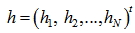
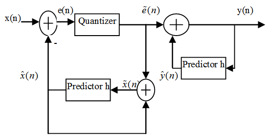
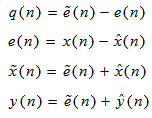
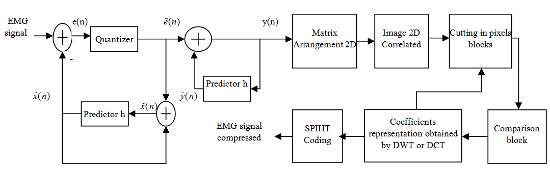
- The principle of DPCM (Differential Pulse Code Modulation) coding is to make a numerical prediction of the signal to be code. The difference between each sample and the respective estimated value for this sample is coded. The predictive coding theory is to predict the values of samples of a signal and to encode prediction error. Figure 1 illustrates a block DPCM coder, where the difference between input samples and those predicted, is quantized and encoded for transmission. As for decoder, the error signal received is added to predicted signal. There are several forms of predictors. In this paper, we use the linear predictor of order N on 1D signal.If
 is predicted signal and
  the original signal, linear predictor is following form:
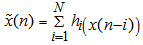 | (1) |
 | Figure 1. Diagram block of DPCM coding |
 | (2) |
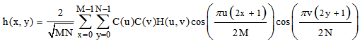
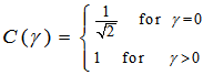
2.2. Discrete Cosine Transform
- The compression methods of EMG signals based on DCT admit an orthogonal transformation that is applied to the original signal. The redundancy of the signal in the new representation is reduced by means of existence of efficient algorithms for calculation. DCT has several advantages: it is real and can be calculated using a fast algorithm (hence its easy implementation). On the other hand, coefficients are decorrelated in the transformed domain. It has an excellent energy concentration for highly correlated data [20]. It tends to concentrate information, and thus making exploitable for image compression as used by the former JPEG standard [20]. In this paper, Discrete Cosine Transform in dimension 2 is applied to EMG transformed into 2D. The 2D DCT is given by the following formula:
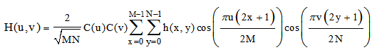 | (3) |
 | (4) |
 | (5) |
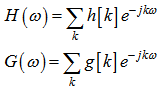
2.3. Discrete Wavelet Transform
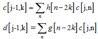
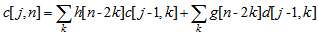
- Discrete Wavelet Transform is a multiresolution / multifrequency representation [23, 24, 25, 26]. It is a tool that decomposes image into several sub-bands in three different directions: horizontal, vertical and diagonal. The 2D DWT is built with separable orthogonal mother wavelets, having a given regularity. At every iteration of the DWT, the lines of the input image are low-pass filtered with a filter having the impulse response g and high-pass filtered with the filter h. Then the lines of the two images obtained at the output of the two filters are decimated with a factor of 2. Next, the columns of the two images obtained are low-pass filtered with g and high-pass filtered with h. The columns of those four images are also decimated with a factor of 2. Four new sub-images are generated [27, 28]. Finally, we have:
 | (6) |
 | (7) |
 | (8) |
 | (9) |
2.4. SPIHT Coding
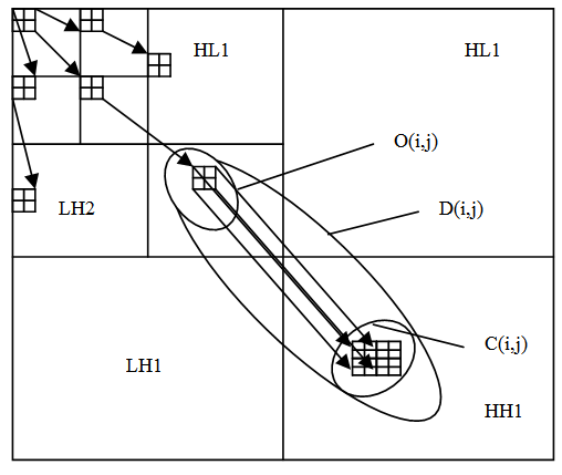
- Coding plays an important role in compression. SPIHT coding [22] is used. Its advantage depends on the threshold value, where each part of image can be seen or not as a detail. SPIHT coding was developed by Said and Pearlman in 1996 [29, 30]. This algorithm is an improvement of EZW proposed by Shapiro and is partitioned into three lists of coefficients.1. List of Significant Pixels (LSP)2. List of Insignificant Pixels (LIP)3. List of Insignificant Sets (LIS) The wavelet coefficients and trees are grouped into sets based on their significance information. Coefficients at the top of the pyramid have a strong spatial relationship with their children. The structure of coding SPIHT is represented by figure 2.A wavelet coefficient at location (i,j) in the pyramidal representation has four offspring at locations:
 | (10) |
 | Figure 2. Offspring dependencies in the pyramidal structure |
 ● If Sn(i,j) is 1, move (i,j) to the LSP and output the sign of wavelet coefficient Ci,j. For each entry (i,j) in LIS do and if the entry is of type A then ● Output Sn(D(i,j)). ●If Sn(D(i,j)) is 1 then for each instructions ● Output Sn(k,l) ● If Sn(k,l) is 1 then add (k,l) to the LSP and output the sign of Ck,l else add (k,l) to the end of LIS as entry of type B and go to step 2; else remove entry (i,j) from the LIS. If the entry is of type B then ● Output Sn(L(i,j)) ● If it is 1 then add each
  to the end of LIS as entry of type A. ● Remove (i,j) from LIS. 3. Refinement Pass: For all entries (i,j) in the LSP except those included in the last sorting pass, output the nth most significant bit of Ci,j. 4. Decrement n and go to step 2. O(i,j) - set of coordinates of offspring (i,j). D(i,j) - Set of coordinates of all descendants (i,j). H(i,j)- Set of all tree roots in highest level of pyramid. L(i,j)=D(i,j)-O(i,j)
3. Proposed Approach
3.1. EMG Signal into 2D
- The EMG signal in 2D is performed according the following algorithm:EMG signal is parted in Mi sequences multiple of 128, then align each after other and fulfill with zeros if necessary (zeros will be automatically removed during reconstruction) to give back sequence MN. Objective here is to achieve a 2D matrix of size M x N (M = N).If x is multiple of 128 (128*N = 2k, where k = 1 or 2), then sequence Mi is given by:
 | (11) |
 If X is not multiple of 128, then sequence Mi is given by:
 | (12) |
 And the last sequence is given by:
  | (13) |
 | (14) |
3.2. Compression Scheme
- A bloc diagram of compression process in shown in figure 3. EMG signal 1-D is processed by DPCM coding, reducing much redundancy of signal before switching to compression method. The method consists to segmenting EMG signal into 512 or 128 sample windows and arranging these segmenting as different columns of a 2D. Once EMG image is obtained, compression process can begin. Image is divided either into pixel blocks of size 32 x 32, in order to reduce noise and errors over a large portion of the signal. Processing that way also significantly reduces compression and decompression. Each block undergoes DCT and DWT and our algorithm only retains coefficients of the transform which yield best statistical parameters (entropy). It is advisable to choose parameters most sensitive and adapted to the program. All image blocks undergo the same technique. SPIHT coding is finally applied on selected coefficients. At the end, the reverse process is applied, to have an accurate reconstruction of original signal.
 | Figure 3. Block diagram of the proposed approach compression |
4. Results and Discussion
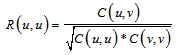
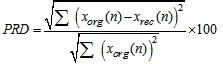
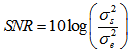
- Reconstruction quality is measured by signal to noise ratio (SNR) and percentage root mean square difference (PRD). These criteria are defined as follow:- The PRD is the most used in majority of scientific works and is defined as [32, 33, 34]:
 | (15) |
 | (16) |
 | (17) |
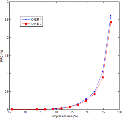 | Figure 4. Compression ratio as a function of the PRD |
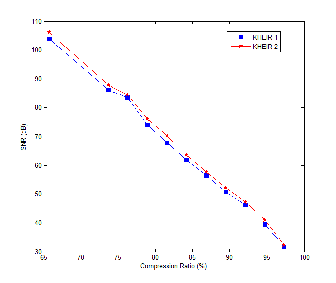 | Figure 5. Compression ratio as a function of the SNR |
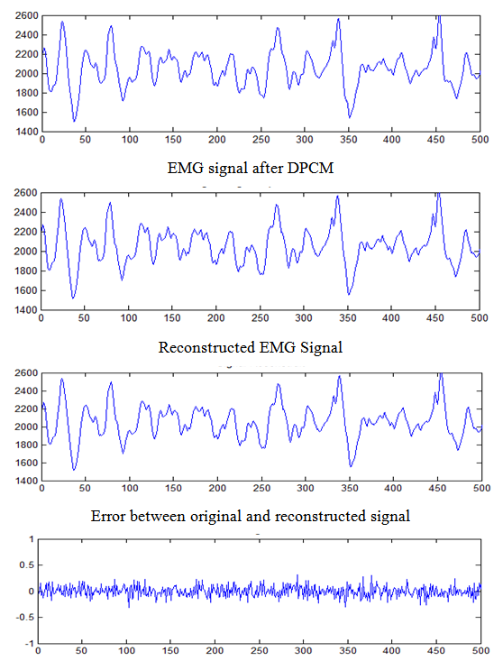 | Figure 6. Reconstruction of EMG signal called Kheir1: CR = 73.68 %; PRD = 0.0048 %; SNR = 86.24dB; Rate = 10 |
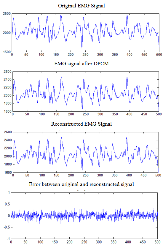 | Figure 7. Reconstruction of EMG signal called Kheir2: CR = 73.68 %; PRD = 0.0040 %; SNR = 87.94dB; Rate = 10 |
|
|
5. Conclusions
- In this paper, we have shown that it is possible to optimize and improve compression performance of an EMG signal by using the DPCM as preprocessing. The PRD can be improved. However, it is very difficult to find the optimal size of the DPCM dictionary for efficient signal processing, without damaging it. The signal to noise ratio continues to grow (for PRD = 0.0004%, SNR = 106.1 dB). For compression ratio 81.57, reconstructed signal begins to degrade. We intend to extend this approach to other electrophysiological signals and improve this method by a significant reduction in SNR. The research of optimal size of the codebook of DPCM will undoubtedly remain a prospect.
References
| [1] | Emre Z., "Compression multimodale du signal et de l’image en utilisant un seul codeur", Thèse de Doctorat, Université Paris-Est, Laboratoire Images Signaux et Systèmes Intelligents (LiSSi, E.A. 3956), Mars 2011 |
| [2] | Yvonnick B., "Projet de synthèse I2, Traitement de l’image Compression"; avril 2006.(http://seb.france.free.fr/eseo/I2-TST/traitement-d’image/). |
| [3] | Lee H., Buckley K.M., "Heart beat data compression using temporel beats alignment and 2-D transforms", Proceedings of the 1996 Conference Record of the Thirtieth Asilomar, Conference on Signals, and computers, Vol.2, pp.1224-1228, Nov.1996. |
| [4] | Lee H., Buckley K.M., "ECG data compression using cut and align beats approach and 2-D transforms", IEEE transactions on Biomedical Engineering, May 1999; 46(5): 556-564. |
| [5] | Uyar K., Ider Y.Z., "Development of a compression algorithm suitable for exercise ECG data", Proceedings of the 23rd Annual International Conferences of IEEE Engineering in Medicine and Biology Society, Oct.2001, Vol. 4: 3521-3524. |
| [6] | Moghaddam A.R.A., Nayebi K., "A two dimensional wavelet packet approach for ECG compression", Sixth International Symposium on Signal Processing and its Application Aug.2001, Vol.1: 226-229. |
| [7] | Bilgin A., Marcellin M.W., Siarry P., "An input-delay neural-network-based approach for JPEG 2000", IEEE Transactions on Consumer Electronics, Nov.2003; 49(4): 833-840. |
| [8] | Sahraeian S.M.E., Fatemizadeh E., "Wavelet-based 2D-ECG data compression method using SPIHT and VQ coding", International Conference on Computer as o Tool. EUROCON, Warsaw 9-12; Sep.2007 pp.133-137. ISBN 9781424408139. |
| [9] | Moazami G.M, Moradi M.H., Abbasabadi S., "High performance method for electrocardiogram compression using two dimensional multiwavelet", processing; Shanghai, Oct. 30-Nov.2, 2005, pp.1-5 ISBN 0780392884. |
| [10] | Rezazadeh I.M., Moradi M.H., Nasrabadi A.M., "Implementing of SPIHT and sub-band energy compression (SEC) method on two-diemensional ECG compression a novel approach", 27th Annual International Conference of the Engineering in Medicive and Biology Society, Shanghai, 17-18 Jan.2006, pp.3763-3766, ISBN 0780387414 |
| [11] | Marcus V.C.C., Joao L.A.C., Pedro A.B., Adson F.D.R., Francisco A.O.N., "Compression of Surface Electromyographic Signals Using Two-Dimensional, Techniques". Recent Advances in Biomedical Engineering, Ganesh R Naik (Ed), In Tech October 2009, ISBN: 978-953-307-004: 17-38. |
| [12] | Norris J.F, Lovely D.F., "Real-time compression of myoelectric data utilizing adaptive differential pulse code modulation.’, Medical and Biological Engineering and Computing, 1995; 33(5): 629-635. |
| [13] | Guerrero A.P, Mailhes C., "On the choice of an Electromyogram data compression method", Proceedings of the 19th Annual International Conference of the IEEE Engineering in Medicine and Biology Society, 1997, Oct.30-Nov.1; Chicago, USA, pp. 1558-1561. |
| [14] | Welling P., Zhenlan C., Semling M., Moschytz G.S., "Electromyogram data compression using single-tree and modified zero-tree wavelet encoding", Proceeding of the 20th Annual International Conference of the IEEE Engineering in Medicine and Biology Societ, 1998, Oct.29-Nov.1; Hong Kong, China, pp. 1330-1306. |
| [15] | Norris J.A., Englehart K., Lovely D., "Steady-state and dynamic myoelectric signal compression using embedded zero-tree wavelets", Proceedings of the 23rd Annual International Conference of the IEEE/EMBS, 2001, Oct. 25-28; Istanbul, Turkey, pp. 1879-1882. |
| [16] | Berger P.A., Nascimento F.A.O., Carmo J.C., Rocha A.F., "Compression of EMG signals with wavelet transform and artificial neural networks", Institute of physics Publishing: Physiological Measurement, 2006 March; 27(6): 457-465. |
| [17] | Paiva J.P.L.M., Kelencz C.A., Paiva H.M., Galvâo R.K.H., Magini M., "Adaptive wavelet EMG compression based on local optimization of filter banks", Physiological Measurement; 29(7): 843-856. |
| [18] | Filho E.B., Da S.E., Carvalho M.B., "On EMG signal compression with recurrent patterns", IEEE transactions on biomedical engineering, 2008; 55(7): 1920-1923. |
| [19] | Ntsama Eloundou P., Mbaidoun T.L., Ele P., Kabiena I. B.," EMG signal compression using 2D fractal", International Journal of Advanced Technology & Engineering Research, May 2013, 3(3): 58-68. |
| [20] | Ntsama Eloundou P., Ele P., Kabiena I.B., "Nouvel Algorithme Optimal de Compression des Signaux Biomédicaux par les Transformées en Ondelettes et Cosinus discrète associées au codage SPIHT", SETIT 2012, Sciences of Electronics Technologies of Information and Telecommunications. IEEE, Sousse 21-24 Mars 2012, Tunisie. |
| [21] | Xingyuan W., Juan M., "A 2-D ECG compression algorithm based on wavelet transform and vector quantization", School of Electronic and Information Engineering, Dalian University of Technology, Dalian 116024, PR China. 2008 |
| [22] | Gutzwiller J. L., Hariti M.,Barret M., Christophe E., Thiebaut C., Duhamel P., "Extension du codeur SPIHT au codage d’images hyperspectrales", Colloque CORESA, 6 pages, Toulouse (France), 2009. |
| [23] | Sana K., Kaïs O., "Comparaison des Méthodes de Compression Appliquées aux Electrocardiogrammes", Unité de Recherche Traitement du Signal, Traitement de l’Image et Reconnaissance de formes Ecole Nationale d’Ingénieurs de Tunis, ENIT, BP.37 Le Belvédère, 1002, Tunis, Tunisie. 2009. |
| [24] | Diab M.O., Marque C., khalil M., "Une approche de classification des contractions utérines basée sur la théorie des ondelettes et la statistique", Lebanese Science Journal, Vol 7, No. 1, pp. 91-103, 2006. |
| [25] | Pedro de A.B., Francisco A. de O.N., Adson F. da R., Joao L.A.C., "A New Wavelet Based Algorithm for Compression of Emg Signals", Proceedings of the 29th Annual International Conference of the IEEE EMBS Cité Internationale, Lyon, France August 23-26, pp. 1554-1557; 2007. |
| [26] | Baarir Z. E., Ouafi A., "Etude De La Transformée En Ondelettes dans La Compression D’images Fixes", Courrier du Savoir – N°05, Juin 2004, pp.69-74. |
| [27] | Rakotomalala M. A., Rakotondraina T. E., Rakotomiraho S., "Compression d’images dans des bases d’ondelettes", MADA-ETI, ISSN: 2220-0673; Vol.1,, 2010. www.madarevues.gov.mg |
| [28] | Daubechies I., "Ten Lectures on Wavelets", vol. 61 of Proc. CBMS-NSF Regional Conference Series in Applied Mathematics. Philadelphia, PA : SIAM, 1992. |
| [29] | Said A., Pearlman W. A., "A new fast and efficient image codec based on Set Partitioning In Hierarchical Trees", IEEE Trans. Circuits and Systems for Video Technology, vol. 6, no.3, 1996. |
| [30] | Sridevi S., Vijayakumar V.R., Sutha J. V., "Medical image compression based on set partitioning in hierarchical trees using quantized coefficients of self-organizing feature map for mages", Journal of Theoretical and Applied Information Technology, October 2013, Vol.,56; No.1: 46-51. |
| [31] | Ntsama E.P., Ele P., Kabiena I.B., "Compression Approach of EMG Signal Using 2D Discrete Wavelet and Cosine Transforms", American Journal of Signal Processing, 2013, 3(1): 10-16. |
| [32] | Kok-kiong P., Marziliano P., "Compressive sampling of EEG signals with finite rate of innovation", EURASIP journal on advances in signal processing, Volume 12, ID183105, February 2010. |
| [33] | Iulian B., Ciocoiu, "ECG Signal Compression Using 2D Wavelet Foveation", International Journal of Advanced Science and Technology Volume 13, December, 2009. |
| [34] | Mukhopadhyay S.K., Ghosh S., Chakraborty S., Das, M.M.S., Mitra S., "Lossless electrocardiogram compression technique and gsm based tele-cardiology application", international journal on smart sensing and intelligent systems vol. 6, no. 3, june 2013. |
| [35] | Nelson M., "Compression des données", Edition : Peter Burch, Dunod, Paris, 1993. |
| [36] | Ntsama P.E., Ele P., Tonye E., Marius B., ‘Compression des signaux EMG par la Transformée dite « impaire » : Lifting Scheme Modifié (LSM)", Journal des Sciences Pour l’Ingénieur. N° 7/2006, pages 1 à 9. |
| [37] | Hrubes J., Vitek M., Kozumplik J., "Multipoint Validation of Decompressed ECG Signal", Analysis of BioSignal 2010; 19(1): 1-4. |
| [38] | Pedro de A.B., Francisco A.D.O. N., Adson F.D.R., Joao L.A..C., "A New Wavelet-Based Algorithm for Compression of Emg Signals", Proceedings of the 29th Annual International Conference of the IEEE EMBS Cité Internationale, Lyon, France. August 23-26, 2007. |