-
Paper Information
- Previous Paper
- Paper Submission
-
Journal Information
- About This Journal
- Editorial Board
- Current Issue
- Archive
- Author Guidelines
- Contact Us
American Journal of Biomedical Engineering
p-ISSN: 2163-1050 e-ISSN: 2163-1077
2013; 3(5): 107-118
doi:10.5923/j.ajbe.20130305.02
Analysis of Oxy-Hb Signals to Determine Relationship between Jaw Imbalance and Arm Strength Using fNIRS
 Abstract
 Reference
 Full-Text PDF
 Full-text HTML
Hai Thanh Nguyen, Hoa Van Nguyen, Khoa Quang Dang Truong, Toi Van Vo
International University of Vietnam National Universities, Ho Chi Minh City, Vietnam
Correspondence to: Hai Thanh Nguyen, International University of Vietnam National Universities, Ho Chi Minh City, Vietnam.
| Email: |  |
Copyright © 2012 Scientific & Academic Publishing. All Rights Reserved.
This study is concerned with analyzing the relationship between jaw imbalance and arm strength regarding Oxy-Hb concentration changes using functional Near Infrared Spectroscopy (fNIRS). The jaw imbalance and arm activity, which possibly has a typical relation, was investigated based on Oxygen Hemoglobin (Oxy-Hb) concentration changes. From this relationship, one can find out the cause of the imbalance jaw for treatment. Six healthy volunteers, who participated in this study, were introduced to bite on a firm spacer at one side of the jaw (left), while holding different a typical pull-down force by the opposite arm (right) during Oxy-Hb measurements. In addition, the subjects performed another experiment to monitor the right arm strength by putting a 40N additional force on the same side of the jaw with bite of a spacer while holding a force of 70 N. In our method, a Savitzky-Golay filter was applied to remove spikes of the fNIRS Oxy-Hb data before analyzing them. From the smoothed data, a Polynomial Regression (PR) algorithm and the definite integral were employed to calculate the average volumes of Oxy-Hb concentration at brain motor zone for analyzing the relationship. Results indicated that there was the typical relationship between the jaw imbalance and arm strength through analyzing Oxy-Hb concentration changes.
Keywords: Polynomial Regression Model, Definite Integral, Oxy-Hb concentration volumes, Jaw Imbalance and Arm Strength
Cite this paper: Hai Thanh Nguyen, Hoa Van Nguyen, Khoa Quang Dang Truong, Toi Van Vo, Analysis of Oxy-Hb Signals to Determine Relationship between Jaw Imbalance and Arm Strength Using fNIRS, American Journal of Biomedical Engineering, Vol. 3 No. 5, 2013, pp. 107-118. doi: 10.5923/j.ajbe.20130305.02.
Article Outline
1. Introduction
- Human brain has a complex structure with a lot of different nerve cells which can communicate from one to another with or without external excitations to make typical decisions (pattern recognition, cognition, motion and others) [1, 2]. In recent decades, experimental techniques such as Electroencephalogram (EEG) and functional Magnetic Resonance Imaging fMRI have been investigated processing function of human brain for treatment or rehabilitation[3-7]. These technologies have also attracted researchers with many significant approaches to explore many problems related to human brain. An example of rehabilitation is that information obtained from human brain using EEG technique could be employed to perform shared control of motion wheelchairs[8, 9]. A brain simulator can lead to improve or to recover the cognitive/motor functions of tetraplegic patients with degenerative nerve diseases spinal cord injuriesIn many non-invasive techniques, fNIRS technique has been applied to measure cerebral hemodynamics as well as to detect localized blood area and oxygenation changes corresponding to human activities[10-13]. Moreover, there is no position obligation during experiment. The subject feels free for performing his or her brain activities. In practice, this technique has been successfully used to study human brain functions such as assessment of motor task from everyday living, athletic performance, and recovery from neurological illness[14] or assessment of verbal fluency[15] or quantification of brain function during finger tapping[16]. However, over recent years, there have not yet been applications using fNIRS technique employed to estimate the relationship between jaw and arm muscles. In this paper, we will analyse fNIRS data to find out the relationship between jaw and arm activities.It has been reported that there is a strong relationship between the temporomandibular joint, and the oral maxillofacial neck and arm muscles[17, 18]. Authors also found that the size of the jaw muscles was significantly related to the size of the limb muscles; however, maximal voluntary bite force moments were not significantly related to the moments of the arm flexion and leg extension forces. In addition, problems of jaw activities have been investigated in recent years, in which the quantification of the jaw healthy and pain muscle activities during different jaw movements (opening, chewing, closing, etc.) were performed[19-22]. In another study of finding a relationship between the height of bite plates and the strength of deltoid muscles, Chakfa et al.[23] carried out experiments with 20 female subjects using the bite plates of adjusted heights of 2, 4, 6 and 13 mm and a downward force on the wrist of the extended arm. The result was found that while increasing the height of the plate, the arm strength increased.An imbalance in the jaws is a big problem in human body. A few researchers have investigated to find its causes, in which one of causes is that loss of strength in the contra lateral arm can relate to an imbalance jaw. In this article[24] authors proposed a lever-based model to attempt to shed some light on this finding. A mechanical model was built and the mechanism can be represented by a weightless and rigid beam which rotates about a fulcrum placed at the center of the beam and 2 springs placed at each end of the beam. This model may constitute a basic element to build more elaborated models to include other reported data such EMG data and then suggest the ways to treat patients suffered from deformity. The relationship between dental occlusion and arm strength was investigated using EMG technique[25]. In particular, measurement data were employed to determine the loss of the arm strength related to the jaw and neck muscles. Khoa et al. investigated the relationship jaw imbalance and arm strength using EMG technique. In this research, a hypothesis is that an imbalance between the left and the right arm was shown in Figure 1. Authors in this paper collected EMG signals on muscles such as masseter, trapezoid, bicep and deltoid to analyse and the results showed that they found the loss of arm strength in the contralateral side during bite of a thick spacer. In our research, we will investigate the relationship between jaw imbalance and arm strength through fNIRS technique to find more understandings about this relationship related to Oxy-Hb concentration changes.
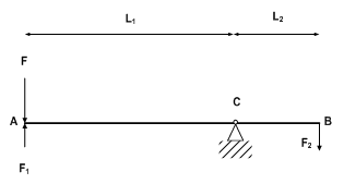 | Figure 1. The mechanical system with the jaw balances and imbalances |
2. Material and Methods
2.1. Subjects and the Experimental Setup
- Brain Oxy-Hb signals were acquired from a FOIRE 3000 fNIRS machine (SHIMAZU Co. LTD, Japan), located at Lab-104 of Biomedical Engineering Department, International University, VNU, Vietnam. The FOIRE 3000 system with the eight pairs of the probes, consisting of the illuminator and detector optodes, produces 24 channels. These probes were placed on the scalp to collect fNIRS data, in which the detectors were installed at a 4 cm distance from the illuminators. The optodes were arranged to install at the left hemisphere on the head of the subject as shown in Figure 2.
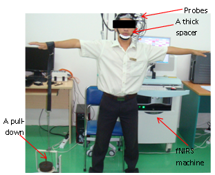 | Figure 2. FOIRE – 3000 system and probes were placed on the scalp at the left side of the human head while the subject is holding a pulldown force by the right arm |
 | (1) |
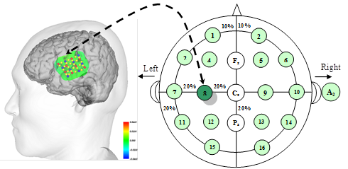 | Figure 3. Measured positions at the left brain based on international 10-20 system (T: transmitter (Red); R: Receiver (Blue); C: Channel (Yellow)) |
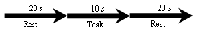 | Figure 4. Setting up Rest and Task time of measurements |
2.2. Savitzky – Golay Filter
- Oxy-Hb data often have noise (spikes) and artifacts. In particular, they can be generated from brain blood flow or the effect of measure environment. In addition, the fNIRS technology is also less sensitive to motion artifacts which are caused by head and body movement or around environment. In order to smooth the Oxy-Hb data, a filter as the Savitzky – Golay filter was applied in this research.A Savitzky – Golay filter[34, 35] was employed to smooth Oxy-Hb signal in the time domain. This filter uses a sliding window based on the samples of the Oxy-Hb signal. The simplest digital filter replaces each data value xi, in which xi is the value of Oxy-Hb data. The method is that the linear combination of a few nearby points was carried out to obtain point values gi at the filter output.
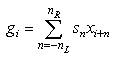 | (2) |
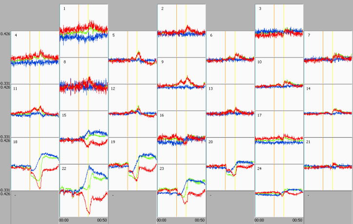 | Figure 5. A set of 24 fNIRS channels including Oxy-Hb (red), Deoxy-Hb (blue) and Total Oxy-Hb (green) |
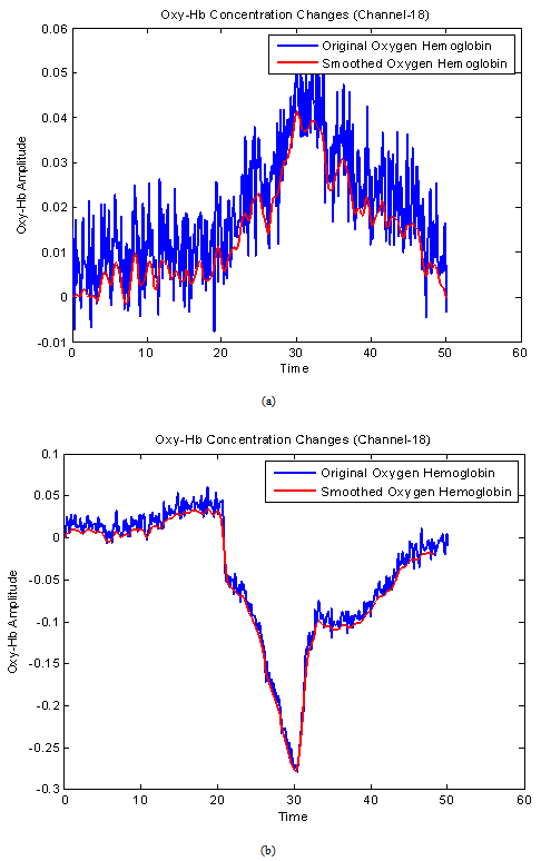 | Figure 6. Original Oxy-Hb signals (blue) and filtered Oxy-Hb signals (red) using the Savitzky-Golay filter, in which (a) the fNIRS signal in the case of the subject without bite of a spacer and (b) the fNIRS signal of the subject with a firm space |
2.3. Polynomial Regression Model for Filtered fNIRS Signals
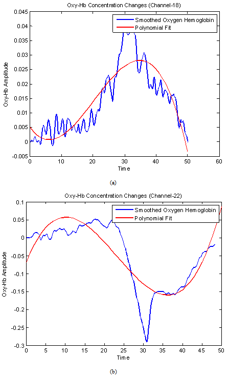 | Figure 7. Representation of the blue filtered signal and the red polynomial fit. in which (a) the fNIRS signal in the case of the subject without bite of a spacer and (b) the fNIRS signal of the subject with a firm space |
 were built based on the PR coefficients as:
 | (3) |
 are estimation values of
 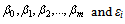 is a vector of an unobserved random error. From the PR coefficients obtained from Equation (3), one can build a polynomial curve corresponding to Oxy-Hb concentration changes during the activated tasks. In this research, the order-3 polynomial was chosen due to the same to the shape of the Oxy-Hb signal as shown in Figure 7a and Figure 7b. Moreover, we can calculate Oxy-Hb volumes by definite integral method.
2.4. Definite Integral Method to Determine Oxy-Hb Volume
- The relationship between the jaw imbalance and arm strength to Oxy-Hb in the brain can be found if the volumes of the Oxy-Hb concentration changes of the subject are determined during measurement tasks. In this case, from a polynomial curve of the Oxy-Hb determined, the volume of the Oxy-Hb concentration changes obtained from the channels was calculated.In this research, all channels (L channels) were introduced to determine the Oxy-Hb concentration changes related to pull-down forces of the subject with/without biting on a thick spacer at the left jaw of the same side of the brain motor cortex. The Oxy-Hb average
 of the channels was calculated as follows:
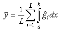 | (4) |
3. Results
3.1. Experiment 1: Subjects with Bite on a Spacer at the Left Jaw While Holding a Pull-down Force at the Right Arm
- In this experiment, six subjects were invited to do experiments. Each subject with/without bite of a firm spacer was asked to hold four types of different pull-down forces (40, 50, 60 and 70 in Newtons), respectively. The Oxy-Hb data filtered by the Savitzky-Golay filter were calculated to produce polynomial curves using the PR algorithm and then the polynomial curves were integrated to obtain the Oxy-Hb volumes.In the first case, six subjects without bite of the firm spacer, while holding four pull-down forces, respectively. Figure 8 showed the curves which represented the Oxy-Hb volumes synchronously changing with four types of the forces (40, 50, 60 and 70 in Newtons).
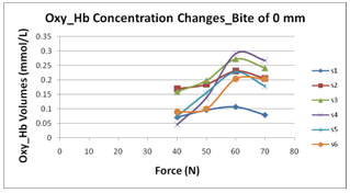 | Figure 8. Oxy-Hb volumes of six subjects without biting on a spacer corresponding to four different forces |
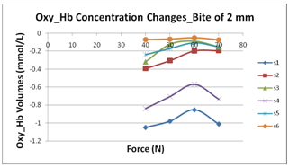 | Figure 9. Oxy-Hb volumes of six subjects with biting on a spacer of 2 mm |
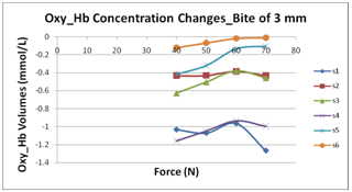 | Figure 10. Oxy-Hb volumes of six subjects with biting on a 3 mm spacer |
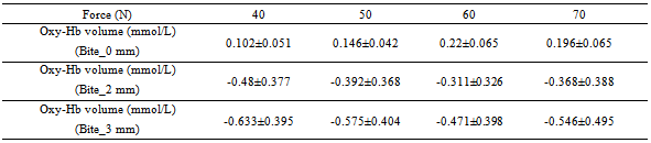
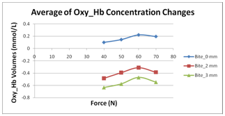 | Figure 11. The representation of Oxy-Hb mean volumes of six subjects with biting on the spacers of 0 mm, 2 mm and 3 mm, respectively |
|
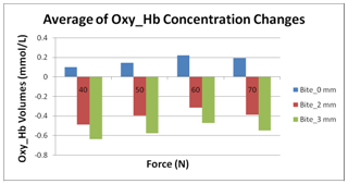 | Figure 12. The representation of the Oxy-Hb concentration changes in three cases (0 mm, 2 mm and 3 mm) |
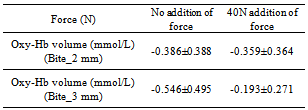
3.2. Experiment 2: Adding a Force of 40 N to the Opposite Arm
- This experiment is to observe the change of the Oxy-Hb concentration allowing us to get more evidences about the change of the Oxy-Hb concentration related to the right arm force. In order to obtain results, six subjects were introduced to bite on spacers (2 mm and 3 mm) at the left jaw, respectively, while holding a 70 N force and putting at the left shoulder a 40 N force. The result is that Oxy-Hb volume lightly increases back compared with the case of no force of 40 N at the left shoulder as shown in Table 2. The increase means that the force of the lifting arm (right) could lightly increase so that the subject is easier, (an increasing typical force) while holding a 70 N heavy force. The observation based on fNIRS technology allows us to predict that there is the Oxy-Hb concentration in relation to jaw and arm activities.
|
3.3. Experiment 3: Representing Oxy-Hb Concentration Changes Based on
- In relation to analysis of the relationship between jaw imbalance and arm strength, we did the third experiment. In particular, Oxy-Hb signals of 24 channels were processed to display brain images with different colors, which represent Oxy-Hb changes as shown in Figure 13. On these brain images, there are color dots corresponding to channels (yellow), the optical transmitters (red) and the optical receiver (blue).
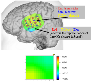 | Figure 13. Representation of the brain images based on Oxy-Hb channels, in which color changes correspond to Oxy-Hb changes |
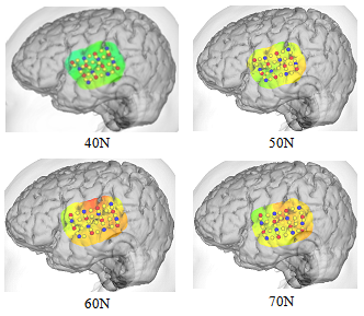 | Figure 14. The brain images of Subject-1 without bite of a thick spacer while asking to hold the pull-down force |
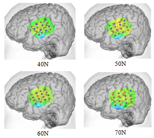 | Figure 15. The brain images of Subject-1 with bite of a 3 mm thick spacer while asking to hold the pull-down force |
4. Discussion
- In this study, we have shown that the changes of Oxy-Hb concentration in human brain blood flow depend on the pull-down forces and the bite of different spacers using fNIRS technique. In addition, potentials of arm strength loss relate to jaw imbalance, as well as the increasing of the arm strength can be performed by adding a force on the opposite arm. Finally, fNIRS images were shown to strongly see that the Oxy-Hb concentration always changes during biting on a spacer at the left jaw and holding a force by the right arm.Understanding of jaw and arm activities based on fNIRS signalsIn the normal occlusion, the Oxy-Hb concentration changes significantly increased according to the increasing of an applied-force. Therefore, it is clear that the levels of Oxy-Hb changes in motor cortex depend on the applied-force of the arm. It is possible that the neuron activities in motor cortex involved in the function of arm muscles. There are some studies also suggested that the neuronal activity in motor cortex is tightly coupled to muscle output. Neurons in motor cortex are active in correlation with a range of movement parameters including the movement direction of the hand through space, velocity, force, joint angle, and arm posture[36-40]. For every muscles activity, cells need a flow of oxygen in to support their activities. Oxygen related with Hemoglobin so when we do experiments, oxygen rushes into the place where we use the muscle so the arm muscles require more Oxygen supplies when it contracts strongly.Arm strength loss potential due to jaw imbalanceIn the imbalance occlusion, we considered both jaw and arm activities. In three above experiments, subjects without biting on a thick spacer (the normal jaw) and with biting on the thick spacers of 2 mm and 3 mm (called an imbalance at the jaw) at the left jaw held different pull-down forces for recording Oxy-Hb concentration changes, respectively. In these three cases, the variation of Oxy-Hb concentration is different. In particular, Oxy-Hb concentration lightly reduces following the increasing of the spacer thickness at the jaw. This can allow us to predict that the jaw imbalance can affect to arm strength. In other words, the jaw imbalance possibly causes the loss of the arm strength.Arm strength increasing related to added force of the opposite armIt is clear that when the subject holds the large pull-dowm force, Oxy-Hb concentration in brain blood partly flows to the arm to create enough force for lifting a large weight. In the case of the subject with bite of a spacer, the Oxy-Hb is decreased. This means that there is the relation to the activity of brain-jaw-arm. In another experiment, when the subject with biting on the 2 mm or 3 mm thick spacers at the left jaw is holding a 70 N force by the right arm side. At the same time, a 40 N force is added to the left shoulder, the Oxy-Hb concentration in human brain increases back compared with the case of not adding the force of 40 N. This means that the right arm strength is lightly increased. Therefore, we possibly found that Oxy-Hb concentration always changes between the jaw and arm in the brain blood flows under controlling the brain system. Thus, one could predict that the jaw imbalance can affect to the loss of the arm strength or inversely the arm strength loss can be affected by the imbalance of jaw.Understanding jaw imbalance and arm based on fNIRS imagesThe activities of the jaw imbalance and arm muscle related to the Oxy-Hb in brain blood as well as electrodes were also represented through fNIRS images. In particular, Oxy-Hb concentration changes containing the activities of jaw and arm muscles were described by different colors. Based on the colors, one can know the Oxy-Hb changes in the body motions and then give estimates and predictions. Therefore in this study, we determined the case of the imbalance jaw or normal jaw based on the changes of the Oxy-Hb flowing in brain blood. Similarly, we could recognize the change of the arm strength through the change of the Oxy-Hb concentration.In previous researches, authors investigated jaw muscles relative to other muscles such as arms, legs, neck etc.[15, 18-21]. In one of these investigations, Wang et. Al. measured jaw imbalance, neck and arm muscles using the EMG technique on subjects to determine their relationship between them. In addition, authors showed jaw muscles related to alternative parts of human body. Brinkworth et al. investigated the response of the masseter and digastric muscles by the stimulation of the upper left central incisor [41]. This study found that there are problems of jaw reflexes when stimulating the tooth in either the orthogonal or axial directions. In the study about jaw muscles related to human motions such as walking and running, Miles et al. did experiments by measuring mandibular movements and masseter muscles in humans to determine the problem of jaw position during locomotion and then considered reflex responses in the masseter EMG[42]. The result was found that the mandible had faster and larger displacements in the jaw-closing muscles during running. In this study with fNIRS technique used to measure Oxy-Hb changes in brain blood, when the subject without bite of a spacer is holding a pull-down force, immediately a typical Oxy-Hb volume increases. We did experiments with different forces many times. The result is that the obtained Oxy-Hb concentration strongly changed corresponding to the change of the pull-down force.In the previous research, Khoa et al.[25] also analyzed the relationship between the jaw imbalance and arm strength using EMG signals, in which a hypothesis related to a balance between the left and the right arm was shown in Figure 1. When there could be an imbalance of the jaw, the arm strength is possibly lost through investigating EMG signals. In order to understand more about this relationship, fNIRS technique, which is non-invasive, was used in this study. Our hypothesis is that whenever the jaw imbalance occurs, the Oxy-Hb signal of the control cortex area will be reduced. The fNIRS technique was applied to measure Oxy-Hb concentration changes in human brain when the subject is introduced to bit on a thick space while holding a typical pull-down force. Obtained results in this research allow us to understand more about losing the arms strength due to the biomechanical effects of the jaw imbalance.
5. Conclusions
- In this paper, a protocol, which plays an important role in data acquisition, was built for a pool of six subjects. For analyzing Oxy-Hb signals, the Savitzky Golay was applied to smooth Oxy-Hb signals obtained from fNIRS channels. The smoothed signals were calculated to produce Oxy-Hb volumes using polynomial regression and integral algorithms. After analyzing Oxy-Hb volumes, we found that there is the sharing of Oxy-Hb concentration from brain into some place, particularly the Oxy-Hb in the brain blood flows into the jaws and arms. This also indicates that fNIRS tools allow us understand more about the relationships between jaw imbalance and arm strength. Moreover, the results showed that whether jaw imbalance is the main cause of the arm strength loss or not for diagnosis and treatment of jaw imbalance patients.
ACKNOWLEDGEMENTS
- We would like to thank Vietnam National Foundation for Science and Technology Development-NAFOSTED for supporting research grant No. 106.99-2010.11. Furthermore research was partly supported by a research fund from the Vietnam National University in Ho Chi Minh City. Finally, an honorable mention goes to our volunteers, families and friends for their supports on us in completing this project.