O. Mokhlessi 1, H. M. Rad 1, N. Mehrshad 1, A. Mokhlessi 2
1Eng. Faculty, University of Birjand, Iran
2Eng. Faculty, University of Mashhad-Ghasem Abad, Iran
Correspondence to: H. M. Rad , Eng. Faculty, University of Birjand, Iran.
| Email: |  |
Copyright © 2012 Scientific & Academic Publishing. All Rights Reserved.
Abstract
Classification of the sound heart into different valve-physiological heart disease categories is a complex pattern recognition task. In this paper application of various types of neural networks are introduced for diagnosing heart disease). At first a method is described for extracting useful features from the sound hearts and then a simple algorithm is introduced for heart sounds recognition. In fact, feature vectors are formed based on a wavelet decomposition of the sounds. The heart sound diseases are classified into normal heart sound and the other six valve physiological heart categories. Different types of artificial neural networks (ANNs) are used for this purpose. Those are Multilayer perceptron (MLP) with back propagation training algorithm, Elman Neural Network (ENN) and Radial Basis Function (RBF) Network. Expensive experimental results show an average recognition score of 81.25% to 96.42%.
Keywords:
Artificial Neural Network Classifier, Back Propagation Algorithm, Elman Neural Network, Heart Valve Diseases, Heart Sound Recognition, RBF Neural Network
Cite this paper:
O. Mokhlessi , H. M. Rad , N. Mehrshad , A. Mokhlessi , "Application of Neural Networks in Diagnosis of Valve Physiological Heart Disease from Heart Sounds", American Journal of Biomedical Engineering, Vol. 1 No. 1, 2011, pp. 26-34. doi: 10.5923/j.ajbe.20110101.05.
1. Introduction
Heart auscultation, defined as listening and interpretation of the sound produced by the heart, has been a very important method for diagnosing heart diseases from the early stages of medicine, since most heart diseases are reflected to the sound that the heart produces[1]. The timing and pitch of a murmur are of significant importance in the diagnosis of a heart condition, for example, murmurs during diastole are signs of malfunctioning of heart valves but murmurs during systole may correspond to either a pathological or healthy heart, depending on the acoustic characteristics of the murmurs.At present, the wavelet transform is popular in extracting feature vectors from heart sound signal because of its ability to characterize time-frequency information which is important in this context. Each 7 class of heart sounds contains characteristic and distinctive information that exists in time and frequency domains. In the other hand totally Artificial Neural Networks (ANNs) are used as classifiers to increase the classification performance. The most prominent advantages of using an ANN as a classifier are:(i) Weights representing the solution are found by iteratively training, (ii) ANN has a simple structure for physical implementation, (iii) ANN can easily map complex class distributions, and (iv) generalization property of the ANN produces appropriate results for the input vectors that are not present in the training set[2]. A variety of methods have been reported for the classification of heart diseases. Sharif et al. have reported a classification technique based on the instantaneous energy and frequency estimations of heart sound signal. They have reported an accuracy of 70% for classification of normal heart sound, mitral stenosis and mitral regurgitation. Time-frequency analysis techniques like wavelet[3]. Olmez and Dokur have given a classification technique that utilizes Daubechies-2 wavelet detail coefficients at the second decomposition level for classification of 7 different heart sounds collected from 28 subjects using ANN[4].Omran and Tayel presented a wavelet based feature extraction technique using ANN as a model for classifying 10 different heart sounds with accuracy of 98%[5].This paper is organized as follows: In Section 2, we review some basic properties of the pattern recognition, the heart sound diseases class's description, wavelet packet decomposition and Feature extraction. A comparison between the 4 type of Artificial Neural Network inclusive Back Propagation Algorithm (BPA), Elman Neural Network (ENN) and Radial Basis Function Network (RBF) is described in Section 3. In Section 4 presents results and conclusion.
2. Preliminaries
2.1. Pattern Recognition
Pattern recognition can be divided into a sequence of stages, starting with feature extraction from the occurring patterns, which is the conversion of patterns to features that are regarded as a condensed representation, ideally containing all the necessary information. In the next stage, the feature selection step, a smaller number of meaningful features that best represent the given pattern without redundancy is identified. Finally, classification is carried out: a specific pattern is assigned to a specific class according to its characteristic features, selected for it. This general abstract model, which is demonstrated in Figure 1, allows a broad variety of different realizations and implementations. Applying this terminology to the medical diagnostic process, the patterns can be identified, for example, as particular, formalized symptoms, recorded signals, or a set of images of a patient[6,7].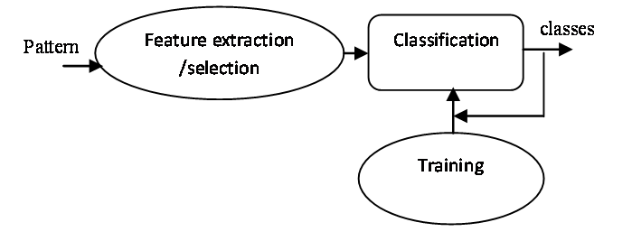 | Figure 1. The pattern recognition approach. |
2.2. Heart Sound Diseases Classes
The heart sound diseases classes considered for the purpose of this study were classified into normal heart sound and the other six valve physiological heart categories, as below:
2.2.1. Aortic Stenosis
The most common cause of Aortic Stenosis (AS) in adults is calcification of a congenital bicuspid valve. This calcific disease progresses from the base of the cusps to the leaflets, eventually causing a reduction in leaflet motion and effective valve area without commissural fusion.
2.2.2. Aortic Regurgitation
There are a number of common causes of Aortic Regurgitation (AR). Some of these include idiopathic dilatation of the aorta, congenital abnormalities of the aortic valve (most notably bicuspid valves), rheumatic disease, infective endocarditis, Systemic hypertension, myxomatous degeneration, dissection of the ascending aorta, and Marfan syndrome.
2.2.3. Mitral Stenosis
In patients with Mitral Stenosis (MS) due to rheumatic fever, the pathological process causes leaflet thickening and calcification, commissural fusion, chordal fusion, or a combination of these processes .The result is a funnel-shaped mitral apparatus in which the orifice of the mitral opening is decreased in size.
2.2.4. Mitral Regurgitation
The common causes of organic Mitral Regurgitation (MR) include MVP syndrome, rheumatic heart disease, infective endocarditis, certain drugs, and collagen vascular disease. MR may also occur secondary to a dilated annulus from dilatation of the left ventricle. In some cases, such as ruptured chordae tendineae, ruptured papillary muscle, or infective endocarditis, MR may be acute and severe. Alternatively, MR may worsen gradually over a prolonged period of time. These 2 ends of the spectrum have quite different clinical presentations.
2.2.5. Pulmonary Stenosis
Because the pulmonary valve is the least likely valve to be affected by acquired heart disease, virtually all cases of pulmonary valve stenosis are congenital in origin. Most patients with stenosis have a conical or dome-shaped pulmonary valve formed by fusion of the valve leaflets. Occasionally, the valve may be thickened and dysplastic, with the stenosis caused by inability of the valve leaflets to separate sufficiently during ventricular systole.
2.2.6. Tricuspid Regurgitation
Tricuspid Regurgitation (TR) should be used to evaluate the results of a tricuspid valve repair immediately after cardiopulmonary bypass to assess for residual regurgitation and restriction of the tricuspid valve opening with stenosis. If a prosthetic tricuspid valve is implanted, transesophageal echocardiography can detect technical problems such as paravalvular regurgitation or abnormal leaflet motion [8].
2.3. Noise Reduction
Filters are classified according to the functions that they are to perform, in terms of ranges of frequencies. In this paper, 5th order butter worth low pass filter with 0.2 Hz cut off frequency, which has the property that low-frequency excitation signal components down to and including PCG signals, are transmitted, while high-frequency components, up to and including noises are blocked.Results from this noise reduction are shown for 6 heart cycles in Figure. 2. The difference of these insufficiency are shown in Figure.3.
2.4. Wavelet packet decomposition and Feature Extraction
2.4.1. Wavelet Transform
In intelligent systems, feature extraction is of paramount importance. The process of converting a signal from the time domain to the frequency domain is achieved conventionally With the Fourier transform (FT). Fourier transform does not provide enough information when used on non-stationary signals. FT determines only the frequency components of a signal, but not their location in time. 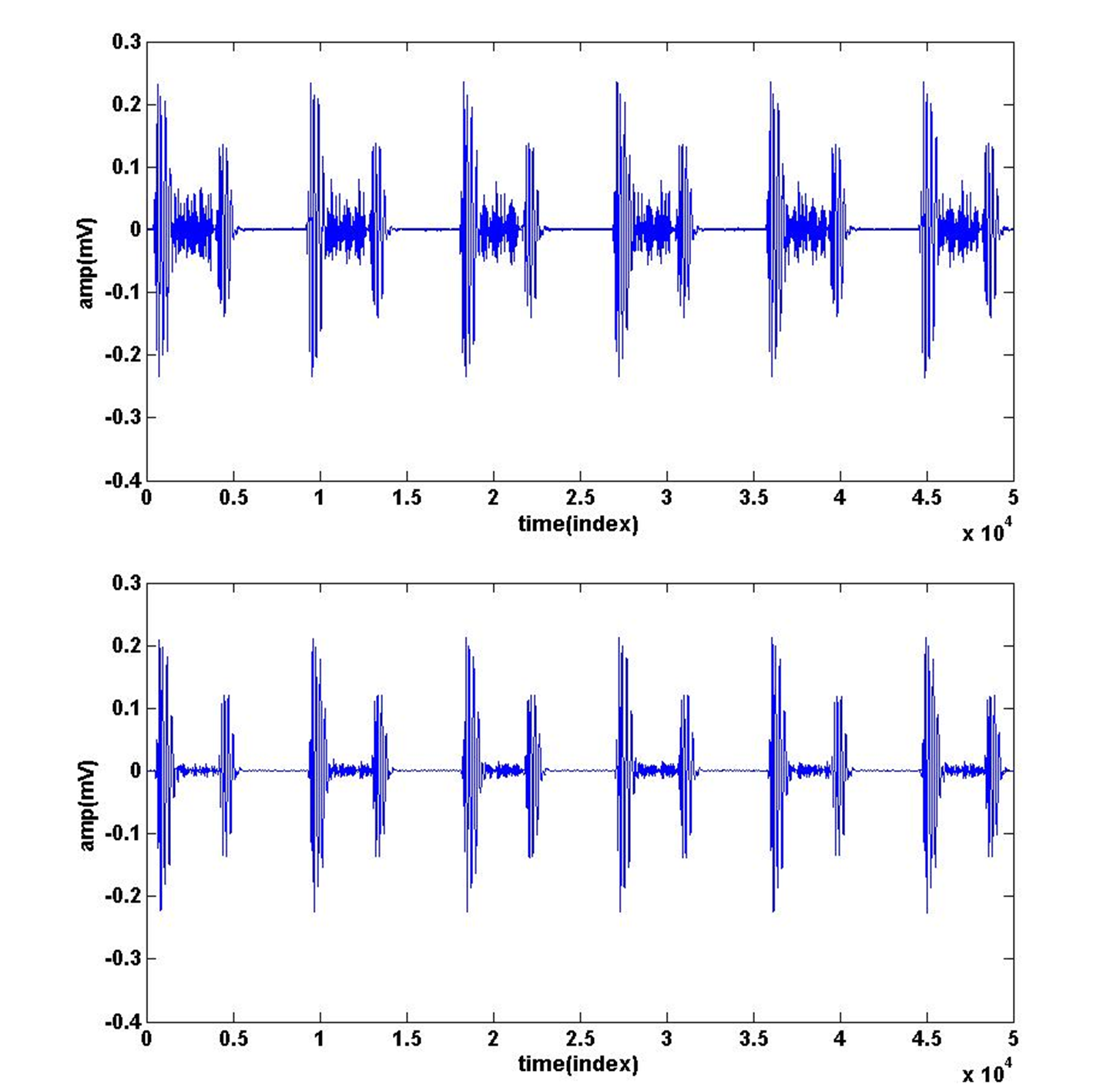 | Figure 2. An example of noise reduction for heart sounds. |
In order to overcome this drawback, short time Fourier Transform (STFT), using a technique called windowing, was proposed. STFT maps the signal into a two-dimensional space of time and frequency using a single fixed window.Wavelet transform enables analysis with multiple window durations that allow for a coarse to fine multi-resolution perspective of the signal. Being able to dilate or compress the variable sized window region (wavelet), different features of the signal will be extracted in WT[9].Mathematical equation describing the continuous wavelet transform (CWT) of the signal x(t) is: | (1) |
Continuous wavelet coefficients (CWC) show how well a wavelet function correlates with the signal, supposing signal energy and wavelet function energy are equal to one and the signal x(t) procuring from the Continuous wavelet coefficients (CWC): | (2) |
2.4.2. Method for Feature ExtractionAccording to Theorem Naykvyst, coefficients of WT are gotten if the PCG signals are passed through lowpass filter. These filters will Eliminate all frequencies higher than π (rad/s). thereupon According to Nyquist sampling theorem, High frequency signals will be π (rad/s). So after these signal passing, half the samples can be eliminated because the signal after applying these filters, the highest frequency will be π/2 (rad/s). In this work both of Details and Approximations from CA15 to CD1 in 15 levels wavelet decomposition has been calculated. Then for each of these 15 levels, we are calculated each signal Energies for each samples. | (3) |
These decompositions and extraction is shown in Figure.4.The wavelet based feature extraction technique is used, mentioned in where Daubechies-4 wavelet is employed for determining the wavelet coefficients. This forms 15 element initial feature vectors for the each cases under study: Aortic Stenosis, Aortic Regurgitation, Mitral Stenosis, Mitral Regurgitation, Pulmonary Stenosis, Tricuspid Regurgitation, Normal heart sound respectively. These vectors are shown in Figure.5 For example.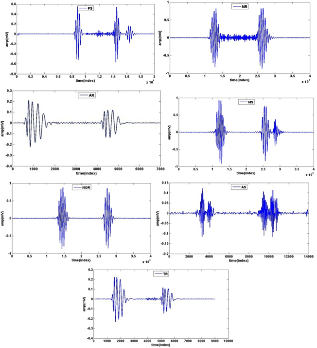 | Figure 3. An example of PCG signals contain each insufficiency. |
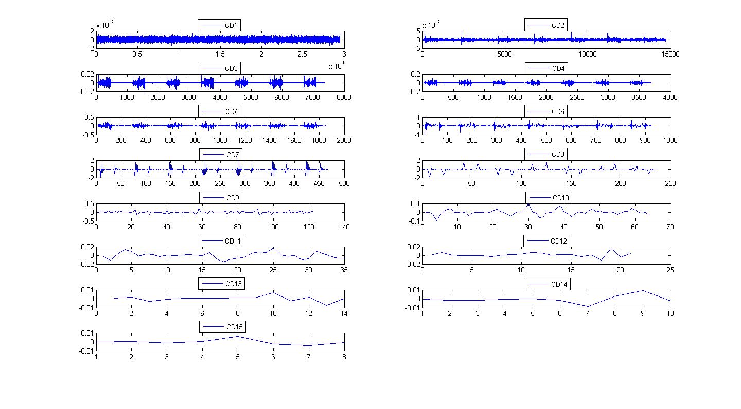 | Figure 4. Details of PCG signal with 15 levels. |
 | Figure 5. Difference energy calculation between 15 Decomposition levels. |
3. Artificial Neural Networks
The pattern classification theory has been a key factor in fault diagnosis methods development. Some classification methods for process monitoring use the relationship between a set of patterns and fault types without modeling the internal processes or structure of an explicit way. Nowadays, the ANN’s constitute the most popular method. An artificial neural network (ANN) is an information processing paradigm inspired by biological nervous systems .The human learning process may be partially automated with ANN’s, which can be configured for a specific application, such as pattern recognition or data classification, through a learning process. An artificial neuron is composed for some connections, which receive and transfer information, also there is a net function designed for collect all information (weights * inputs + bias) and send it to the transfer function, which process it and produces an output[11,12]. Generally, the number of nodes in the output layer of ANN is equal to the number of classes. Here, we are used 4 type Artificial neural networks and totally are compared ability of them with same wavelet coefficients for input. The number of nodes in output layer is generally equal to the number of heart sounds classes to be classified. The number of nodes in the hidden layer is usually chosen empirically to find a superior performance of the system. The experiment starts with a relatively large number and let the proposed algorithm select the final number of hidden node.
3.1. Back Propagation Algorithm (BPA)
Once the architecture of MLP has been determined, the connection weights of the network have to be computed through a training procedure based on the training patterns and the desired output. BP is one of the simplest and most general methods for the supervised training of MLP[13].The basic BPA works as follows:Initialize all the connection weights W with small random values from a pseudorandom sequence generator. Repeat until convergence (either when the error E is below a preset value or until the gradient  is smaller than a preset value).2.1 Compute the update using:
  2.2 Update the weights with W(t+1) =W(t) +
 2.3 Compute the error E(t+1)Where “t” is the iteration number, W is the connection weight, and η is the learning rate. The error E can be chosen as the mean square error (MSE) function between the actual output yj and the desired output dj:
 | (4) |
The BPA described above has some shortcomings. If the learning rate is set small enough to minimize the total error, the learning process will be slowed down. On the other hand, a larger learning rate may speed up learning process at the risk of potential oscillation. Another problem is that, partial minimal points or stable stages on error surface are often encountered during the learning process. Using a momentum term is the simplest method to avoid oscillation problems during the search for the minimum value on the error surface [14].
3.1.1. BPA Architecture
The BPA is used in this study consists of four layers including an input layer, two hidden layers and an output layer. The number of neurons in the input layer, 1th hidden layer, 2th hidden layer and the output layer are used respectively 4, 4, 5 and 7. In this work we are used Sigmoid transfer function in hidden and output layer as Transfer function. Training tolerance is considered 0.01.
3.2. Elman Neural Network (ENN)
Elman recurrent network is a well-known recurrent topology, developed by Jeffrey Elman. An Elman network has a set of context nodes. Each context node receives input from a single hidden node and sends its output to each node in the layer of its corresponding hidden node. Since the context nodes depend only on the activations of the hidden nodes from the previous input, the context nodes retain state information among inputs. The context layer carries the memory. The hidden layer activates the output layer and refreshes the context layer with the current state of the hidden layer, see Figure. 6[15,16]. | Figure 6. BPA architecture. |
3.2.1. ENN Architecture
The Elman network commonly is a two-layer network with feedback from the first-layer output to the first-layer input. This recurrent connection allows the Elman network to both detect and generate time-varying patterns. The Elman network has tansig neurons in its hidden (recurrent) layer, and purelin neurons in its output layer. This combination is special in that two-layer networks with these transfer functions can approximate any function (with a finite number of discontinuities) with arbitrary accuracy. The only requirement is that the hidden layer must have enough neurons. More hidden neurons are needed as the function being fitted increases in complexity. The number of neurons in the hidden layer is 37 neurons. | Figure 7. ENN architecture. |
 | Figure 8. RBF architecture. |
3.3. Radial Basis Function Neural Network (RBF)
The radial basis function (RBF) neural network is a kind of feed forward neural network. RBF neural network is embedded in a three layers neural network, where each hidden unit implements a radial activated function. The input units implement the data input to the network. The output units implement a weighted sum of hidden unit outputs. The input into a RBF network is non-linear while the output is linear. Due to their non-linear approximation properties, RBF networks are able to model the complex mappings, which perception neural network can only be modeled by means of multiple intermediary layers.An RBF neural network bases on the Cover Theorem. Radial basis functions are embedded into a three-layer feed-forward neural network. Such a network is characterized by a set of inputs and a set of outputs. Between the inputs and outputs there is a layer of processing units which is called hidden units. Each of them implements a radial basis function. The network is used as a pattern classification, as shown in Figure. 6[16].
3.3.1. RBF Architecture
RBF network has to learn three parameters: the center of radial basis function, the variance of radial basis of function and the weight. Here the self-organizing learning algorithm of selecting the radial basis function center with 25 neurons is used in this paper.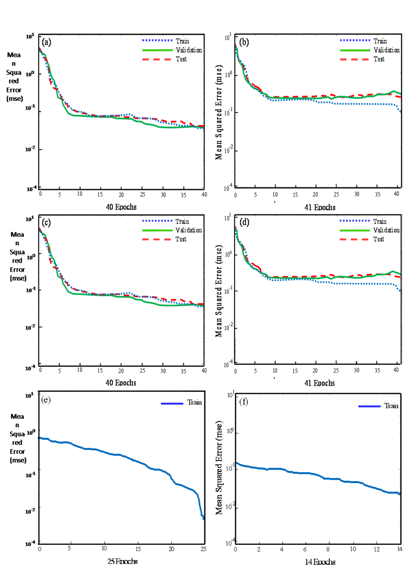 | Figure 9. Networks Performance are: a,b) Mlp Performance with 100% and 80% features, c,d) ENN Performance with 100% and 80% features, and e, f) RBF Performance with 100% and 80% features. |
4. Results
To compare the performances of different networks, we kept the same condition in initializing the networks, and also we adopted the same training parameters and learning function during the training process. The output classes were based on the final diagnosis of physicians according to the patient's record.The exclusive properties of the networks are shown in Table.1 in below.The experimental results of ensemble classification with different Network classifiers for valvular heart disease data set are given in Table 2. MSE (Mean Square Error) shows the ability of the network performance to recognize different possible diagnoses or the other pattern recognition approaches. We performed , ten runs per parameter set on our proposed networks and chose the best training results and acquire the mean of ten run for 100% and 80% of features .We believe that the training process can be repeated a few times for selecting the best network feature extraction. Finally Utilization of four types of Artificial Neural Network is aimed in diagnosing accuracy on Table below (see Table. 3).
4.1. Training Performance
The performances of the four proposed networks with the overall architecture as shown in Figure 9 were evaluated respectively. However, it should be noted that the Training performance may vary significantly between consecutive runs due to the nature of training algorithms.| Table 1. Properties of the networks |
| | Networks | Neurons | Transfer Functions | Training tolerance | | MLP | [5 36] | tansig | 5*10-2 | | BPA | [4 5] | tansig | 1*10-2 | | ENN | [37] | tansig | 7*10-6 | | RBF | [25] | radbas | 1*10-5 |
|
|
| | Networks | 100% | 80% | | MSE | MEAN | MSE | MEAN | | MLP | 0.491 | 0.493 | 0.521 | 0.526 | | BPA | 0.210 | 0.215 | 0.218 | 0.224 | | ENN | 0.482 | 0.485 | 0.519 | 0.523 | | RBF | 0.466 | 0.470 | 0.537 | 0.594 |
|
|
| Table 3. Diagnosing accuracy |
| | Networks | 100% | 80% | | MLP | 81.25 | 77.53 | | BPA | 87.17 | 83.71 | | ENN | 91.59 | 86.56 | | RBF | 96.42 | 81.30 |
|
|
5. Conclusions
The Define Researchers showed that the most of human deaths in the world are due to heart diseases. The heart valve disorders are of importance among the heart diseases. Among them, mitral and aortic valve disorders are the most common ones. For this reason, early detection of heart valve disorders is one of the most important medical research areas. Currently sound recognition techniques for diagnosis of heart valve disorders are the most preferred because they are completely non-invasive and without risk in the serial studies.In this paper, we have demonstrated that four types Artificial Neural Network for detection and identification of valve-physiological heart disease from heart sounds can result in accurate classifiers. The outcomes of the all classification approaches assert that the extracted features using the db4 wavelet transform in fifteen resolution levels create acceptable distinctions among every pairs of the AS, AR, MS, MR, PS, TR disorders and normal heart sounds. This can, furthermore, facilitate infinitely unique reconstruction of the modified wavelet coefficients without any group interference. In this work, we have used RBF and Elman networks as classifiers which has two characteristics.1. RBF classifiers have memory of recent events and may use in the real-time classification, whereas ENN is the best in the memorial functionality uses.2. The train of the both of them can be easily configured, which allows end users to tune them for acceptable tolerances.What causes the ENN homogeneity and its performance precision improvement against RBF by reducing the instructional data to 80% may be due to two factors:1. The reduction influence of instructional data quantity on RBF instruction2. The existence of some dynamic data which, in regard to the dynamic nature of ENN, can exhibit better response by reducing the instructional data reduction.The results demonstrate that the RBF can detect heart diseases with higher Diagnosing accuracy and have best Training performance than other ANNs. While, ENN extracts temporal contents from the whole signal epoch after epoch and may support for productive behavior or support inference. Besides, BPA can announce heart diseases with lower Neurons numbers; have best MSE quantity than others.The present work also furnishes a feasible approach for non-stationary generation of signals for machine aided diagnosis of the heart disorders. The same methodology can be applied to simulate other heart diseases dynamically for example ECG and the other non-stationary signal diseases.
References
| [1] | B. Ericson, Heart Sounds, and Murmurs: A Practical Guide, Mosby-Year Book Inc., 1997 |
| [2] | T. olmez, Z. Dokur, “Classification of heart sounds using an artificial neural network”, Pattern Recognition Letters 24, pp. 617–629, 2003 |
| [3] | Z. Sharif, M. S. Zainal, A. Z. Sha’ameri, and S. H. S. Salleh, “Analysis and classification of heart sounds and murmurs based on the instantaneous energy and frequency estimations”, Proceedings of the TENCON, vol. 2, pp.130–134, Sep. 2002 |
| [4] | T. Olmez, and Z. Dokur, “Classification of heart sounds using an artificial neural network”, Pattern Recognition Letters 24, pp. 617–629, 2003 |
| [5] | S. Omran, M. Tayel, “A heart sound segmentation and feature extraction algorithm using wavelets”, Proceedings of the First International Symposium on Control, Communication and Signal Processing, pp. 235–238, 2004 |
| [6] | H. Dickhous, and H. Heinrich, “Classifying biosignals with wavelet networks”, IEEE Eng. Med. pp. 103–111, 1996 |
| [7] | C. M. Bishop, “Neural Networks for Pattern Recognition, Clarendon Press”, Oxford, 1996 |
| [8] | R. O. Bonow & other thirteen Writing Committee Members, 2008 Focused Update Incorporated Into the ACC/AHA 2006 Guidelines for the Management of Patients With Valvular Heart Diseases, Journal of the Amwerican College Cardiology, Elsevier (2008) |
| [9] | S. G. Mallat, “A theory for multi-resolution signal decomposition: the wavelet representation”, IEEE Trans. Pattern Anal. Mach. Intel. pp. 674–693, 1989 |
| [10] | A. Kandaswamy, C. Sathish Kumar, Rm. Pl. Ramanathan, S. Jayaraman, and N. Malmurugan, “Neural classification of lung sounds using wavelet coefficients”, Computer Physics Communications , pp. 523-53, sep. 2004 |
| [11] | S. Haykin, a Comprehensive Foundation, Neural Networks, Macmillan College Publishing Co., New York, 1996 |
| [12] | C. Navin Gupta, R. Palaniappan, and S. Swaminathan, S. Krishnan, “Neural network classification of homomorphic segmented heart sounds”, Appl. Soft Computing. 7 (1) pp. 286–297, 2005 |
| [13] | R. O. Duda, P. E. Hart, D. G.Strok, Pattern classification. New York: Wiley, 2001 |
| [14] | H. Yan. Y. Jiang, J. Zheng, C. Peng, Q. Li, H. Yan. Y. Jiang, J. Zheng, C. Peng, and Q. Li, “A multilayer perceptron-based medical decision support system for heart disease diagnosis”, Expert Systems with Applications 30, pp. 272–281, 2006 |
| [15] | C. Y. Liou, J. C. Huang, and W. C. Yang, “Modeling word perception using the Elman network”, Neuro computing 71, pp. 3150– 3157, 2008 |
| [16] | X. Tong, and Z. Wang, H. Yu, “A research using hybrid RBF/Elman neural networks for intrusion detection system secure model”, Computer Physics Communications 180, pp. 1795–1801, 2009 |
 Abstract
 Reference
 Full-Text PDF
 Full-Text HTML