
-
Paper Information
- Paper Submission
-
Journal Information
- About This Journal
- Editorial Board
- Current Issue
- Archive
- Author Guidelines
- Contact Us
American Journal of Biochemistry
p-ISSN: 2163-3010 e-ISSN: 2163-3029
2026; 16(2): 21-28
doi:10.5923/j.ajb.20261602.02
Received: Oct. 16; Accepted: Nov. 3, 2026; Published: Nov. 7, 2026

Association between HBV Viral Load and Serum Aminotransferase Activity in HBsAg-Positive Young Recruits in Dakar, Senegal: A Cross-Sectional Study
 Abstract
 Reference
 Full-Text PDF
 Full-text HTML
Moustapha Djite1, 2, 3, Aïssatou Sow3, Ndèye Marème Thioune4, Niokhor Ndane Diouf5, Pape Matar Kandji1, 2, Néné Oumou Kesso Barry1, 2, Mame Ndoumbé Mbacke2, Najah Fatou Coly-Gueye6, El Hadji Malick Ndour1, Fatou Gueye-Tall1, Dominique Doupa4, Papa Madieye Gueye1, 2, Babacar Ndao3
1Pharmaceutical Biochemistry Laboratory, Faculty of Medicine, Pharmacy, Cheikh Anta Diop University, Dakar, Senegal
2Biochemistry Laboratory, Fann National University Hospital, Dakar, Senegal
3Medical Analysis Laboratory of the National Gendarmerie Health Service
4Gaston Berger University, Saint-Louis, Senegal
5Department of Medical Biochemistry, Assane Seck University, Ziguinchor, Senegal
6Diamniadio Children's Hospital, Dakar, Senegal
Correspondence to: Moustapha Djite, Pharmaceutical Biochemistry Laboratory, Faculty of Medicine, Pharmacy, Cheikh Anta Diop University, Dakar, Senegal.
| Email: |  |
Copyright © 2026 The Author(s). Published by Scientific & Academic Publishing.
This work is licensed under the Creative Commons Attribution International License (CC BY).
http://creativecommons.org/licenses/by/4.0/

Background: Hepatitis B is a major public health problem in sub-Saharan Africa, where serum aminotransferases are often the only affordable laboratory test available to follow HBV-infected patients. The aims of this study were to estimate the prevalence of HBsAg among young recruits in Dakar and to assess the association between HBV viral load and serum alanine (ALT) and aspartate (AST) aminotransferase activity in HBsAg-positive recruits. Methods: This cross-sectional study was conducted from November 20, 2024 to December 31, 2025 at the laboratory of the Health Service of the National Gendarmerie (SSGN) in Dakar. HBsAg was screened with a rapid test and positive results were confirmed by CLIA; in HBsAg-positive subjects, HBV DNA was quantified by real-time PCR (IU/mL) and serum ALT and AST were measured once. Viral load was classified as undetectable, < 2,000, 2,000–20,000 and > 20,000 IU/mL. Associations were assessed with Spearman correlation, Kruskal–Wallis, Mann–Whitney and chi-square tests, and multivariable linear regression on log-transformed values. Results: Among 7,600 recruits screened, 773 were HBsAg-positive (10.17%; 95% CI 9.51–10.87). Median age was 25 years (IQR 24–26) and 94.30% were men. HBV DNA was undetectable in 32.08% of subjects, < 2,000 IU/mL in 55.76%, 2,000–20,000 IU/mL in 5.05% and > 20,000 IU/mL in 7.12%. ALT was > 45 IU/L in 12.16% and AST in 13.71%; with the sex-specific WHO 2024 ALT limits, 40.88% had elevated ALT. Median ALT and AST increased across viral load categories (p < 0.001) and correlated weakly with log10 viral load (Spearman ρ = 0.18 for ALT and 0.22 for AST; p < 0.001). After adjustment for age and sex, each 1-log10 increase in viral load was associated with an 11.9% higher ALT (95% CI 8.0–15.9). ALT > 45 IU/L was present in only 41.5% (39/94) of subjects with HBV DNA > 2,000 IU/mL. Conclusion: In this single-time-point study, aminotransferase activity was weakly to moderately associated with HBV viral load and, with the conventional limit, missed most subjects with HBV DNA > 2,000 IU/mL. These data do not allow aminotransferases to be evaluated as a monitoring tool; longitudinal studies including HBeAg and fibrosis assessment are needed.
Keywords: Hepatitis B, HBV, Viral load, Aminotransferases, ALT, AST, Senegal
Cite this paper: Moustapha Djite, Aïssatou Sow, Ndèye Marème Thioune, Niokhor Ndane Diouf, Pape Matar Kandji, Néné Oumou Kesso Barry, Mame Ndoumbé Mbacke, Najah Fatou Coly-Gueye, El Hadji Malick Ndour, Fatou Gueye-Tall, Dominique Doupa, Papa Madieye Gueye, Babacar Ndao, Association between HBV Viral Load and Serum Aminotransferase Activity in HBsAg-Positive Young Recruits in Dakar, Senegal: A Cross-Sectional Study, American Journal of Biochemistry, Vol. 16 No. 2, 2026, pp. 21-28. doi: 10.5923/j.ajb.20261602.02.
Article Outline
1. Introduction
- Viral hepatitis B is a systemic infection that preferentially affects the liver and causes hepatocellular alterations and inflammatory lesions [1]. It remains a major public health problem worldwide: according to the World Health Organization (WHO), 254 million people were living with chronic hepatitis B in 2022, with approximately 1.2 million new infections that year [2]. Viral hepatitis caused 1.3 million deaths in 2022, 83% of which were attributable to hepatitis B virus (HBV) [2].Africa is particularly affected: the WHO African Region accounts for 63% of new HBV infections worldwide [2], and the prevalence exceeds 8% in many sub-Saharan countries [3]. In Senegal, the prevalence of chronic viral hepatitis B is estimated at 11% according to the National Hepatitis Control Program (PNLH) [4].HBV infection is characterized by acute hepatitis, which is very rarely symptomatic before the age of 5 years and resolves spontaneously in most cases. Two complications may nevertheless occur: fulminant hepatitis (less than 1% of symptomatic cases) and progression to chronicity, with a risk of evolution to cirrhosis and hepatocellular carcinoma (HCC) [5].Among the laboratory parameters available for the surveillance of chronic hepatitis B, serum aminotransferases (alanine aminotransferase [ALT] and aspartate aminotransferase [AST]) play a central role in the assessment of liver damage. ALT, which is more specific to liver tissue, is the reference biochemical marker of hepatic inflammatory activity, and its measurement is widely used in routine practice [6,7]. In sub-Saharan Africa, where HBV DNA quantification, HBeAg testing and fibrosis assessment are often unavailable or unaffordable, aminotransferases are frequently the only test that can be offered to patients.However, the actual value of aminotransferases as a marker of viral activity remains debated. Normal levels may be observed in patients with active viral replication or significant fibrosis [6,8], whereas fluctuating elevations do not always reflect worsening liver damage. This potential discordance between aminotransferase activity, virological activity and histological damage raises questions about the reliability of this marker when used alone.The aim of this cross-sectional study was therefore (i) to estimate the prevalence of HBsAg among young recruits of the national gendarmerie screened in Dakar, and (ii) to assess, in HBsAg-positive recruits, the association between HBV viral load and serum ALT and AST activity measured at a single time point. Because each subject was tested once, the study does not evaluate aminotransferases as a longitudinal monitoring tool.
2. Materials and Methods
2.1. Study Design and Setting
- This was a descriptive and analytical cross-sectional study conducted from November 20, 2024 to December 31, 2025 at the medical biology laboratory of the Health Service of the National Gendarmerie (SSGN) in Dakar, where samples were collected and analyzed.
2.2. Study Population and Ethical Considerations
- The study involved prospective recruits of the national gendarmerie. All young subjects who attended the laboratory for the routine pre-enlistment examination during the study period were screened for HBsAg (n = 7,600); those who tested positive were included in the analysis of viral load and aminotransferases. Subjects who did not give their consent, pregnant women, and subjects with a condition likely to interfere with the study were not included. Age, sex and region of origin were recorded for HBsAg-positive subjects; HBV DNA, ALT and AST were measured in all of them.This study was approved by the Research Ethics Committee of Cheikh Anta Diop University (UCAD), in accordance with the rules laid down by Senegal's National Health Research Ethics Committee, under number 0227/2017/CER/UCAD. The recruits' free and informed consent was obtained, and anonymity was maintained throughout the study. Refusal to participate had no impact on care. Subjects who tested positive for HBsAg were referred to the gendarmerie physician for further care.
2.3. Sample Collection
- Blood samples were collected in the morning, in a fasting state, into dry tubes (serum, used for HBsAg and aminotransferase assays) and EDTA tubes (used for HBV DNA extraction), promptly transported to the laboratory and centrifuged at 2,054 × g for 5 minutes.
2.4. Analytical Methods
- Hepatitis B surface antigen (HBsAg) testing was performed on serum using Cypress Diagnostics immunochromatographic cassettes (qualitative one-step detection on 50 µL of serum), the simultaneous appearance of the control and test bands indicating a positive result. The manufacturer reports an average sensitivity of 100% and an average specificity of 99%. All positive results, and any faint bands, were verified by chemiluminescent immunoassay (CLIA).Serum AST and ALT activities were determined by the IFCC kinetic enzymatic method at 37 °C with pyridoxal-5′-phosphate (P5P), on the XL 200 biochemistry analyzer (Erba Lachema), by measuring at 340 nm the decrease in absorbance proportional to enzyme activity. Two levels of controls (normal and pathological) were used, and validation was based on the Westgard rules. Two upper limits of normal (ULN) were applied. The conventional laboratory limit of 45 IU/L was used for AST and ALT in both sexes. For ALT, the sex-specific limits recommended by WHO in 2024 (> 30 IU/L in men and > 19 IU/L in women) were applied in a sensitivity analysis [9].Nucleic acids were extracted with the automated MagPurix 12 EVO system (Zinexts), based on magnetic bead separation. HBV DNA was amplified and quantified by real-time PCR on the Amplix NG instrument using the GenProof HBV kit. Results were obtained in copies/mL and in IU/mL (conversion factor of 4.2 copies per IU). The linear measurement range of the assay is from 13.9 IU/mL to 10¹⁰ IU/mL, and viral load is expressed in IU/mL throughout this article. Viral load was classified as undetectable (target not detected), < 2,000, 2,000–20,000 and > 20,000 IU/mL, in line with the thresholds used in current and previous WHO and EASL guidelines [6,9].
2.5. Statistical Analysis
- Data were entered into Microsoft Excel 2016 (double entry, anonymous coding of patients) and analyzed with Python 3 (SciPy and statsmodels libraries). Because viral load and aminotransferase activity are strongly right-skewed, continuous variables are presented as median [interquartile range, IQR] together with mean ± standard deviation (SD) where relevant. For correlation and regression analyses, viral load was log10-transformed; undetectable results were assigned a value of 6.95 IU/mL (half of the 13.9 IU/mL lower limit of the linear range). Prevalence is given with its 95% Wilson confidence interval (CI).The association between viral load and aminotransferases was assessed with Spearman's rank correlation coefficient (primary analysis; 95% CI by Fisher's z transformation) and, secondarily, with Pearson's coefficient computed on log10 viral load; both were also computed after exclusion of subjects with undetectable HBV DNA. The Mann–Whitney U test was used to compare two groups (men and women), the Kruskal–Wallis test to compare the four viral load categories (followed by pairwise Mann–Whitney tests with Bonferroni correction), and the chi-square test to compare the proportions of subjects with elevated ALT or AST. Multivariable linear regression models (heteroscedasticity-robust standard errors), with log10 ALT or log10 AST as the dependent variable and log10 viral load, age and sex as independent variables, were used to adjust the association for age and sex. A two-sided p value < 0.05 was considered statistically significant.
3. Results
3.1. General Characteristics of the HBsAg-Positive Subjects
- The characteristics of the 773 HBsAg-positive subjects are summarized in Table 1. Age ranged from 19 to 30 years (mean 24.9 ± 1.73; median 25 [IQR 24–26]). There were 729 men (94.30%) and 44 women (5.70%), i.e., a male-to-female sex ratio of 16.6. Women were slightly younger than men (median 24 vs 25 years; p < 0.001).
|
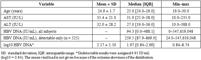
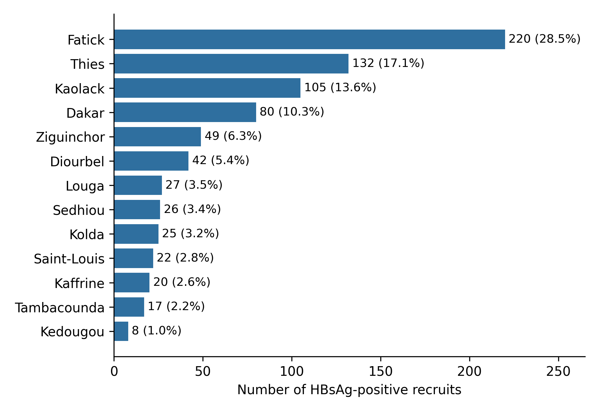 | Figure 1. Distribution of HBsAg-positive recruits by region of origin (n = 773) |
3.2. Prevalence of HBsAg
- Among the 7,600 recruits screened, 773 were HBsAg-positive, i.e., a prevalence of 10.17% (95% CI 9.51–10.87).
3.3. Assessment of Viral Load
- HBV DNA was detectable in 525 of the 773 HBsAg-positive subjects (67.92%). The median viral load was 94.3 [0.0–488.1] IU/mL in all subjects and 259.5 [87.9–869.0] IU/mL in those with detectable HBV DNA; the maximum value was 5.5 × 10⁸ IU/mL (2.3 × 10⁹ copies/mL). Median viral load did not differ significantly between men (101.7 [0.0–497.6] IU/mL) and women (40.5 [0.0–182.2] IU/mL; Mann–Whitney p = 0.126). According to the thresholds used, HBV DNA was undetectable in 248 subjects (32.08%), < 2,000 IU/mL in 431 (55.76%), between 2,000 and 20,000 IU/mL in 39 (5.05%) and > 20,000 IU/mL in 55 (7.12%) (Figure 2). In total, 94 subjects (12.16%) had an HBV DNA level above 2,000 IU/mL.
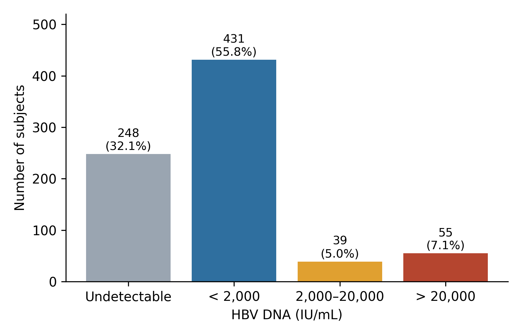 | Figure 2. Distribution of HBsAg-positive subjects by HBV DNA level (IU/mL), n = 773 |
3.4. Assessment of Aminotransferase Activity
- The median ALT and AST activities were 27 [19–36] IU/L and 31 [25–38] IU/L, respectively (Table 1). Both were higher in men than in women (ALT 28.0 [20.0–37.0] vs 18.5 [15.0–25.2] IU/L; AST 31.0 [26.0–39.0] vs 25.5 [23.0–32.0] IU/L; Mann–Whitney p < 0.001 for both). With the conventional upper limit of 45 IU/L, ALT was elevated in 94 (12.16%; 95% CI 10.04–14.65) and AST in 106 (13.71%; 95% CI 11.47–16.32). When the sex-specific WHO 2024 limits were applied to ALT (> 30 IU/L in men, > 19 IU/L in women), 316 (40.88%; 95% CI 37.47–44.38) of the subjects had elevated ALT (men: 295/729, 40.47%; women: 21/44, 47.73%).Aminotransferase activity differed across the four viral load categories (Kruskal–Wallis p < 0.001 for ALT and p < 0.001 for AST; Table 2 and Figure 3). Median ALT and AST did not differ between undetectable HBV DNA and HBV DNA < 2,000 IU/mL (adjusted p = 1.00 for both), whereas subjects with 2,000–20,000 IU/mL and > 20,000 IU/mL had higher values than the two lower categories (all adjusted p < 0.01) and differed from each other (AST: adjusted p = 0.005; ALT: adjusted p = 0.018). The proportion of subjects with ALT > 45 IU/L rose from 6.5% (undetectable) and 9.0% (< 2,000 IU/mL) to 23.1% (2,000–20,000 IU/mL) and 54.5% (> 20,000 IU/mL) (chi-square p < 0.001); with the sex-specific limits, the corresponding proportions were 39.5%, 35.7%, 56.4% and 76.4% (p < 0.001) (Figure 4).
|
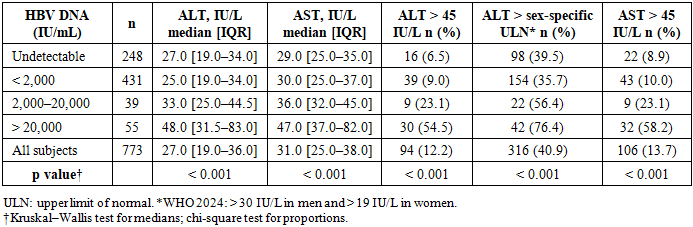
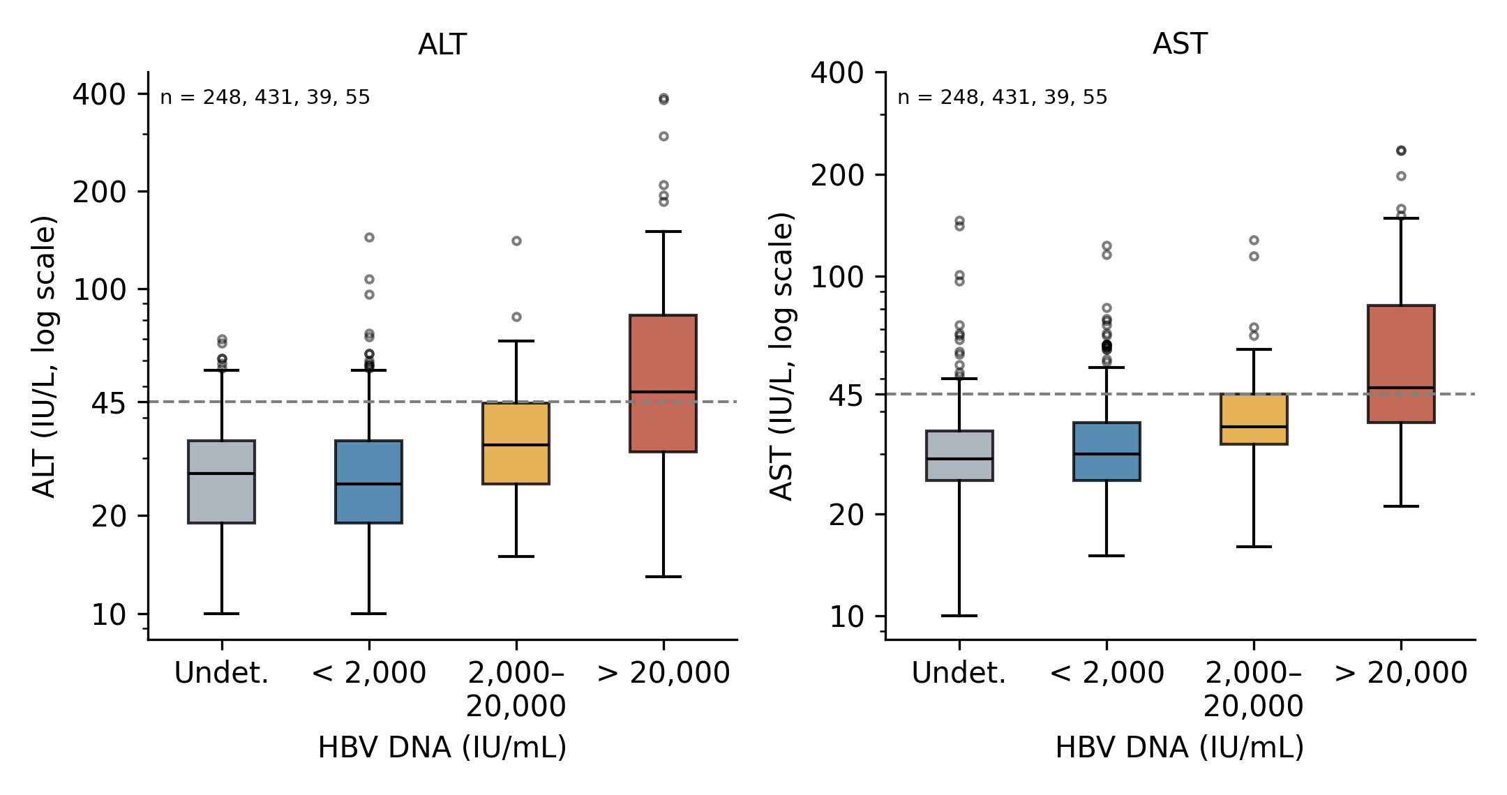 | Figure 3. ALT and AST activity according to HBV viral load category. Boxes show the median and interquartile range; the dashed line indicates 45 IU/L |
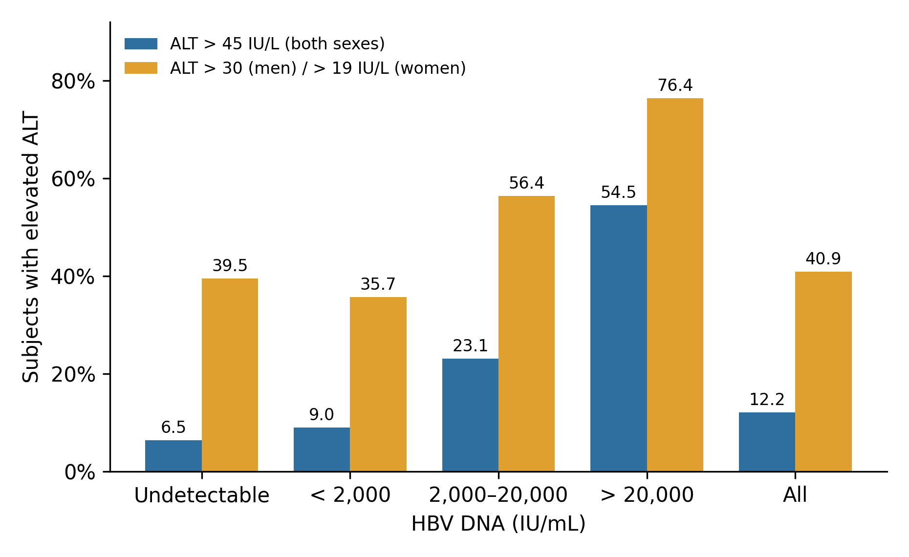 | Figure 4. Proportion of subjects with elevated ALT according to HBV viral load category, using the 45 IU/L limit for both sexes or the sex-specific WHO 2024 limits |
3.5. Association Between Viral Load and Aminotransferase Activity
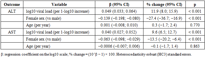
- ALT and AST were positively but weakly correlated with log10 viral load (Spearman ρ = 0.18 for ALT and 0.22 for AST; both p < 0.001; Table 3). The Pearson coefficients computed on log10 viral load were higher (r = 0.38 for ALT and 0.37 for AST), and the rank correlations were stronger when the analysis was restricted to the 525 subjects with detectable HBV DNA (ρ = 0.29 and 0.28). Viral load therefore explains only a limited part of the variability of aminotransferase activity.
|

|
3.6. Aminotransferase Activity in Subjects with HBV DNA Above 2,000 IU/mL
- Of the 94 subjects with HBV DNA > 2,000 IU/mL, 39 (41.5%) had ALT > 45 IU/L and 64 (68.1%) had ALT above the sex-specific WHO 2024 limits. Overall, 39 (5.05%) and 64 (8.28%; 95% CI 6.54–10.43) of the 773 HBsAg-positive subjects combined HBV DNA > 2,000 IU/mL with elevated ALT according to the 45 IU/L limit and to the sex-specific limits, respectively. Fibrosis markers (APRI, FIB-4) were not available, and ALT was measured only once.
4. Discussion
- In this cross-sectional study of 7,600 young recruits screened in Dakar, the prevalence of HBsAg was 10.17%, and, among the 773 HBsAg-positive subjects, serum ALT and AST activities were weakly to moderately associated with HBV viral load. Because aminotransferases were measured once, the study describes an association at a single time point; it cannot assess their value for longitudinal monitoring or for treatment decisions.The prevalence found (10.17%; 95% CI 9.51–10.87) is consistent with the 8% to 12% reported in sub-Saharan Africa [3] and with the 11% estimated for Senegal [4], and places the population in a highly endemic area. Similar results have been reported in Burkina Faso, with an HBsAg prevalence of 12.1% among health professionals in the Nanoro district [10] and 14.3% among blood donors in Nouna [11]. Conversely, the prevalence remains below 2% in Western Europe [12]. Our estimate concerns young adults attending a pre-enlistment examination, was obtained with a rapid test whose positive results were confirmed by CLIA, and should not be extrapolated to the general population.The age of the subjects (19 to 30 years, median 25 years) reflects a population of young adults, because recruitment into the defense and security forces targets only this age group. Male predominance among HBsAg-positive individuals has been described [13], and a female predominance has been observed among patients followed for chronic hepatitis B in Madagascar [14] and during a screening campaign in Saint-Louis, Senegal (sex ratio 0.19) [15]. In our study, however, the marked male predominance (94.30%; sex ratio 16.6) mainly reflects the composition of the recruits: sex was available only for HBsAg-positive subjects, so prevalence by sex could not be estimated, and comparisons with community-based studies are of limited value. The same reasoning applies to the regional distribution: Fatick, Thiès and Kaolack together accounted for 59.12% of the HBsAg-positive subjects, but without the regional distribution of all 7,600 recruits this cannot be interpreted as a difference in prevalence. HBV genotypes and prevalence do vary across regions of Africa [16], and limited access to screening and vaccination may contribute, but our data cannot test these hypotheses.The median ALT (27 IU/L) and AST (31 IU/L) and the low proportion of abnormal values with the 45 IU/L limit (12.16% for ALT and 13.71% for AST) are consistent with West African data showing that most HBsAg-positive young adults have normal or only moderately elevated aminotransferases [8]. In the natural history of chronic infection, aminotransferases are frequently normal during immune tolerance and inactive carriage [17]; HBeAg status and disease phase could not be determined here. The choice of the upper limit of normal is nevertheless critical. With the sex-specific WHO 2024 limits (30 IU/L in men and 19 IU/L in women) [9], 40.88% of the subjects had elevated ALT, more than three times the proportion obtained with 45 IU/L; the AASLD 2018 limits (35 and 25 IU/L) [7] are intermediate. A single limit of 45 IU/L, particularly in women, probably underestimates hepatic inflammatory activity, although only 44 women were included and estimates in women are imprecise. Since AST is less specific to the liver than ALT, an isolated elevation must be interpreted with caution in the presence of extrahepatic factors (muscle damage, alcohol, comorbidities).ALT and AST increased with the level of viral replication, but only above 2,000 IU/mL: median values did not differ between undetectable HBV DNA and HBV DNA < 2,000 IU/mL. The correlation was weak on a rank basis (ρ = 0.18–0.22) and moderate on the log scale (r ≈ 0.38), and the association persisted after adjustment for age and sex (about 12% higher ALT per 1-log10 increase in viral load). Viral load therefore explains only part of the variation in aminotransferases, which also depends on the host immune response, the stage of the disease, fibrosis and cofactors (alcohol, metabolic syndrome, co-infections) [6], none of which could be measured here. Sex was an independent determinant of aminotransferase activity, which supports the use of sex-specific limits.According to the WHO 2024 guidelines, antiviral treatment is considered in adults without cirrhosis when HBV DNA exceeds 2,000 IU/mL and ALT is elevated, in addition to the assessment of fibrosis [9]. In our population, 94 subjects (12.16%) had HBV DNA > 2,000 IU/mL, but only 41.5% of them had ALT > 45 IU/L, against 68.1% with the sex-specific limits. The number of subjects combining high viral load and elevated ALT thus ranged from 39 to 64 depending on the limit used. These figures are descriptive only: APRI or FIB-4 and HBeAg were not available, and persistence of ALT elevation could not be verified, so no subject can be classified as eligible for treatment on these data. They do show that, with the conventional limit, ALT alone failed to identify more than half of the subjects with HBV DNA > 2,000 IU/mL. Shimakawa et al. likewise reported in West Africa that significant liver damage may exist despite normal aminotransferases [8], and an association between high viral load and cytolysis has been reported in other African cohorts [18].This study has several limitations. First, each subject was tested once: neither the persistence of HBsAg for more than 6 months nor the variation of ALT over time could be assessed, chronic infection was therefore not confirmed, and the treatment-naïve status of the subjects was not verified. Second, HBeAg, anti-HDV, anti-HCV and HIV status, alcohol consumption, body mass index and markers of fibrosis were not available, so confounding by these factors cannot be excluded. Third, the population consisted of young, mostly male recruits attending an enlistment examination, which limits generalizability. Fourth, HBsAg was screened with a rapid immunochromatographic test and only positive results were confirmed by CLIA, so false-negative results cannot be excluded and prevalence may be slightly underestimated. Fifth, sex and region of origin were known only for HBsAg-positive subjects, and only 44 women were included. Longitudinal studies with repeated measurements, HBeAg testing and fibrosis assessment are needed to define the role of aminotransferases in the follow-up of HBV-infected patients in sub-Saharan Africa.
5. Conclusions
- In this cross-sectional study of young recruits in Dakar, the prevalence of HBsAg was 10.17%. ALT and AST were weakly to moderately associated with HBV viral load and, with the conventional limit of 45 IU/L, identified fewer than half of the subjects with HBV DNA > 2,000 IU/mL. These findings do not allow aminotransferases to be evaluated as a monitoring tool; longitudinal studies including HBeAg and fibrosis assessment are needed.
ACKNOWLEDGEMENTS
- The authors thank the staff of the medical biology laboratory of the Health Service of the National Gendarmerie (SSGN), Dakar, for their technical assistance and collaboration, and all the young recruits who agreed to take part in this study.
CONFLICTS OF INTEREST
- The authors declare that they have no conflict of interest in relation to this article.