American Journal of Biochemistry

p-ISSN: 2163-3010    e-ISSN: 2163-3029

2016;  6(5): 130-135

doi:10.5923/j.ajb.20160605.03


Effects of Aqueous Stem Bark Extract of Jatropha curcas on Some Biochemical Indices of Mice Infected with Plasmodium berghei

S. Sarkiyayi, H. A. Zailani, Simon J. G.

Department of Biochemistry School of Pure and Applied Sciences, Modibbo Adama University of Technology, Yola, Nigeria

Correspondence to: S. Sarkiyayi, Department of Biochemistry School of Pure and Applied Sciences, Modibbo Adama University of Technology, Yola, Nigeria.

Email:

Copyright © 2016 Scientific & Academic Publishing. All Rights Reserved.

This work is licensed under the Creative Commons Attribution International License (CC BY).
http://creativecommons.org/licenses/by/4.0/

Abstract

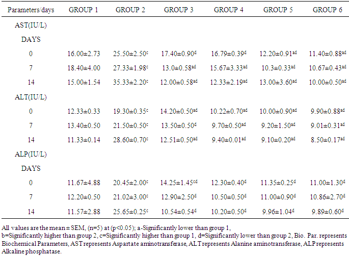
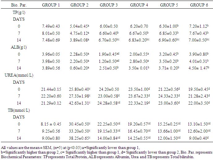
The aim of this research work was to investigate the effect of Jatrophacurcasplant in Mice infected with Plasmodium berghei in order to ascertain the efficacy of the plant stem bark extract in the treatment of malaria as claimed by traditional practitioners in Nigeria. The LD50 of the plant extract was determined. Six groups of fifteen mice each were used for the research. Group 1 was uninfected and untreated, group 2 was infected, but untreated, Groups 3, 4 and 5 were infected and treated with 250mg/kg, 500mg/kg, and 750mg/kg of aqueous stem bark extract of Jatrophacurcas respectively. While Group 6 was infected and treated with Arthemether (20mg/kg). Treatment commenced on the third day and lasted for a period of 12 days. Then, the animals were sacrificed, blood was collected and used for biochemical investigations. Our findings shows that infection caused a significant (p< 0.05) increase in the activities of AST, ALT, ALP and the concentration of total bilirubin and urea when compared to control. However, treatment with the plant extract and arthemether significantly decreased (p<0.05) the activities of liver enzymes and the concentration of total bilirubin and urea compared untreated control. The total protein in infected extract-treated and Arthemether treated animals were found to increase (p<0.05) significantly when compared with the infected untreated mice. Our findings revealed that the decrease in values for AST, ALT, and ALP activity by the plant extract may suggest that the extract has some inhibitory effect on malaria parasite, since the there were no physiological disturbances in the biochemical parameters investigated. Our research had revealed the LD50 was greater than 5000mg/kg, suggesting that the plant extract was relatively safe for oral medication. The percentage parasitemia has decreased from 9.25 to 7.80 for the treated group, where as the infected and untreated has the parasitemia increased from 9.30% to 18.70%. Suggesting that the plant extract possess some anti-malarial properties. Our findings support the possibility of using Jatrophacurcas plant leaf as anti bacterial agent.

Keywords: Plasmodium berghei, Jatrophacurcas, Biochemical indices, Parasitemia Malaria parasite

Cite this paper: S. Sarkiyayi, H. A. Zailani, Simon J. G., Effects of Aqueous Stem Bark Extract of Jatropha curcas on Some Biochemical Indices of Mice Infected with Plasmodium berghei, American Journal of Biochemistry, Vol. 6 No. 5, 2016, pp. 130-135. doi: 10.5923/j.ajb.20160605.03.

1. Introduction

Malaria remains one of the major killers of humans worldwide, threatening the lives of more than one-third of the world’s population. It thrives in the tropical areas of Asia, Africa, and Central and South America, where it affects millions of people. Each year 350 to 500 million cases of malaria occur worldwide. Sadly, more than 1 million of its victims, mostly young children, die yearly [1]. Although malaria has been virtually eradicated in the United States and other regions with temperate climates, it continues to affect hundreds of people in Nigeria every year. The Centers for Disease Control and Prevention (CDC) estimated 1,200 cases of malaria are diagnosed each year in the United States.
Plants have been recognized for their several life saving and therapeutic properties. Several herbs are parts of socio-cultural and socio-economic heritage. Even in the present times rural populations turn to herbal medicine as the most preferred therapeutic source. Jatropha curcas is a drought resistant large shrub or small tree, belonging to the genus Euphorbiaceae, producing oil containing seeds [2]. Jatropha curcas is the commonest specie found in Nigeria, but many species exist in different parts of the world. Traditionally, Jatropha curcas is used for the treatment of fever, mouth infections, jaundice, guinea worm, sores and joint rheumatism [3, 4]. Previous studies also reveals the presence of antibacterial agents in different parts of Jatropha curcas [5]. For developing herbal treatment for malaria from various parts of Jatropha curcas which part will be more effective against Plasmodium berghei. The present study aims to investigate the antiplasmodial potential of the stem bark of Jatropha curcas using the aqueous extract on Plasmodium berghei. The people of Jada local government area and its inhabitants widely use Jatropha curcas stem bark for the treatment of many ailments including malaria. However, there are no scientific bases for this treatment. Therefore, this research is designed to provide the scientific evidence for the use of the aqueous stem bark extract of Jatropha curcas in the treatment of malaria and its safety in the medication.

2. Materials and Methods

Collection of Plant Material
Fresh Jatropha curcas Linn plantwas collected from the Staff quarters of Government Secondary School, Jada Local Government Area of Adamawa State, Nigeria and was taxonomically identified and authenticated in the Plant Science Department of Modibbo Adama University of Technology, Yola.
Chemicals and Reagents
Assay kits for enzymes (ALT, AST and ALP) and liver function indices (Albumin, Total and direct Bilirubin and total protein) were obtained from Randox Laboratories Ltd, UK while those of creatinine and urea were obtained from Agappe Diagnostics, Switzerland. All other reagents and solvents used were of analytical grade.
Experimental Animals
Ninety mice weighing between18-22g were obtained from Animal Facility Centre, National Veterinary Research Institute Vom, Jos, Nigeria. The mice were housed in polypropylene cages, and given standard laboratory diet and water and maintained under standard laboratory conditions.
Malaria Parasite
The malaria parasite (Plasmodium berghei NK-65) was obtained from National Institute for Medical Research (NIMR), Lagos, Nigeria.
Extract Preparation
The stem bark was carefully removed from the Jatropha curcas plant and washed thoroughly with tap water then rinsed twice with distilled water and allowed to sun dry. The dried stem bark was crushed to powder with pestle and mortar and two hundred grams of the powder was cold macerated in 1000ml of distilled water for 24 hours with constant shaking and filtered using Whitman’s filter paper No. 1. It was then concentrated to dryness on water bath at 40°C and the crude extract was kept in desiccator and dried.
Acute Toxicity Test
The LD50 of the aqueous extract of the stem bark was tested using [6] method in order to determine its safety. The aqueous extract of the stem bark of Jatropha curcas was administered in doses of 1000, 3000 and 5000 mg/kg body weight to four groups of mice (n = 3). The mice were kept under same conditions and observed for toxic signs and mortality for 24hs.
Parasite Inoculation
The inoculation of the parasite was carried out by determining both the percentage parasitemia and erythrocytes count of the donor mouse using a haemocytometer and appropriate dilutions of the infected blood with isotonic saline were made. Each mouse was inoculated intraperitoneally with 0.2 ml of infected blood containing about 1 x 10⁷ Plasmodium berghei parasitized red blood cells. After 72 hours the mice were treated for malaria conditions for twelve days.
Experimental Design
Ninety Swiss albino mice were divided into six groups of fifteen [15] mice each. Groups 2-6 were inoculated with the rodent malaria parasite Plasmodium berghei from the same donor mouse. Groups 3, 4 and 5 were treated with 250, 500 and 750 mg/Kg body weight of the aqueous leaf stem bark extract of Jatrophacurcas respectively. Group 6 was treated with 20 mg/Kg body weight of arthemether. Groups 1 and 2 served as normal and untreated controls respectively.
Determination of Biochemical Parameters
Test for liver function indices
Bilirubin was determined by the method of Walter and Gerard [7]. Total protein was determined by the Method of Tietz [8] while albumin was determined as described by Doumas et al [9].
Test for kidney function indices
Serum urea concentration was determined according to the method described by Fawcett and Scott [10] while Creatinine was determined as described by Bartels et al [11].
Enzyme assays
Alkaline phosphatase activity of serum was determined as described by Wright et al [12]. Alanine and aspartate aminotransferase activities were assayed by the method of Reitman and Frankel [13].
Statistical Analysis
Results were expressed as mean ± SEM. Student t-test was used to analyse the data between groups at p values(p<0.05) was considered significant in all cases (SPSS version 21 was used).

3. Results and Discussion

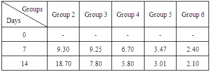
Acute Toxicity Tests
Our findings had shown that there was no mortality recorded in the mice at a dose of 5000 mg/kgbody weight of the aqueous extract of the stem bark of Jatropha curcas, suggesting that the plant extract was not toxic and relatively safe for oral consumption.
The antimalarial properties observed in this study could be attributed to bioactive constituents in the extract. Guido et al [14] reported that antimalarial activity could be attributed to alkaloids in plants extract. Our findings in table 1 had shown that the aqueous stem bark extract of Jatropha curcas produced some antiplasmodial effects on the parasitemic levels when compared to the standard anti malarial drug. in a related development Abiodun et al [15] reported that ethyl acetate extract of leaves of Ocimum gratissimum Linn. (Labiatae) and hexane extract of stem bark of Trema orientalis (L.) Blume (Ulmaceae) showed the highest antiplasmodial activity against P. falciparum K1 strain but elicited low cytotoxicity. Thus the percentage of parastitemia has decreased from 9.25 to 7.80 for the treated group, where as the infected and untreated has the parisetemia increased from 9.30 to 18.70. However, it was observed that at higher dose of the extract was able to exhibit a considerable effect comparable to the standard drug. Hence use of standard drug has percentage parastitemia decreased from 9.30 to 2.10, suggesting that aqueous extract of the stem bark of Jatropha curcas posessess some anti malaria activities. A review conducted by Sabandar et al [16] had shown that, the genus Jatropha (Euphorbiaceae) comprises of about 170 species of woody trees, shrubs, sub shrubs or herbs are used in medicinal folklore to cure various diseases of 80% of the human population in Africa, Asia and Latin America. Species from this genus have been popular to cure stomachache, toothache, swelling and inflammation.
Table 1. Percentage (%) Parasitemia
     
Biochemical tests related to the hepatocyte integrity can be observed to follow hepatocellular integrity and liver injury. Serum activities of AST and ALT were employed to access liver status. Within an hour after being bitten by aninfected anopheles mosquito, the sporozoites enter the liver and multiply into exo-erythrocytic merozoites. The changes in the activity of the hepatocyte due to the infection of plasmodium may change the activities of the liver enzymes. Alanine aminotransferase (ALT), aspartate aminotransferase (AST), Alkaline phosphatase (ALP) are considered indices for hepatocellular health [17, 18]. Infection caused asignificant (p<0.05) increase in the activities of AST, ALT, and ALP in the blood ofparasitized mice compared to the normal control group. The increased activities of the enzymes may be as a result of liver injury and altered hepatocyte integrity caused by the Plasmodium infection and consequent release of the enzymes into the blood stream. However, the administration of the plant extract improved the situation. This finding is in agreement with previous reports by Cotran et al [19]. Although the liver enzymes normalization were effective at higher doses of 500mg and 750mg/kg body weight of the extract. It could be suggesting that plant extract possess some anti-malarial activities. Treatment with the plant extract had some protective effects the hepatocyte integrity of the parasitized mice.
Our findings also show an increase in serum total bilirubin concentration in the infected untreated mice. Bilirubin is the major bile pigment produced in heme catabolism. The increase in the concentration of serum bilirubin in severe malaria are due to numerous factors which include intravascular haemolysis of parasitized red blood cells, hepatic dysfunction and possibly an element of microangiopathic haemolysis associated with disseminated intravascular coagulation [20]. Consequently, jaundice in malaria has been reported to range from 20-31.7% and it is predominantly haemolytic rather than hepatic, as shown by pre-eminence of unconjugated bilirubinemia [21]. Therefore, the increased serum bilirubin observed in the infected untreated mice in this study signifies the impairment of hepatic bilirubin excretion or haemolysis which is in agreement with the report by Kochar et al [22]. But, the administration of the extract to the infected mice was able to ameliorate these anomalies to a considerable extent, suggesting that the extract may be hepatoprotective [23, 24].
Furthermore, this study showed significant (p<0.05) decrease in the concentration of total protein of infected-animal groups as shown in table 3. This agrees with many other studies that reported significant decrease of plasma total proteins in malaria infections compared with normal control [24]. The total protein in infected extract-treated and Arthemether treated animals was found to increase (p<0.05) significantly when compared with the infected untreated mice. This study agreed to the report by Balaji, et al [25] that the values of total protein can be influenced by the degree of infection. Amah et al (26) reported significant reduction in serum levels of albumin in malaria patients in endemic regions of Calabar, Nigeria. Many researchers have proposed the use of serum albumin levels as a reliable biochemical marker for establishing severe pathologic conditions such as malnutrition and infectiousdiseases [27, 28]. Malaria infections are accompanied with significant decrease in plasma albumin concentrations as well as in malnutrition and pregnancy [28]. However, the prevailing plasma albumin concentration in malaria infection is dependent on the nutritional status of the affected individual and hepatic functionality [29, 30]. Possibly, based on the nutritional and hepatic status of the infected animals in this study groups, showed significant (p<0.05) reduction in serum albumin levels in the infected animals compared to normal control. However, in another studies, Oguche et al [31] reported that there was initially significant (p<0.05) increase in serum levels of albumin in low and moderate malaria infections, but decreased as the malaria density increased. This study agrees with that the previous report by Oguche et al [31] observed a significant (p<0.05) decrease in the levels of albumin on day 14. But the administration of the extract appeared to increase the levels of albumin with increase in concentration of the dose of the extract in all the treated mice.
Table 2. Effects of Aqueous Stem Bark Extract of Jatropha curcas on some Biochemical indices in Mice
     
Table 3. Effects of Aqueous Stem Bark Extract of Jatropha curcas on TP, ALB, Urea and Creatinine in Mice
     
The observed increased serum urea levels in the infected untreated from days 7 to 14 is supported by a study reported by [32] in malaria patients in Minna. Studies also reported that increased Urea: Creatinine ratio in malaria patients also indicate that the causes of Ureamia in these patients are largely prerenal and may be due to reduced renal blood flow, rather than organic renal involvement [32]. The reduced blood flow to the glomeruli due to malaria-associated hypotension may be responsible for the reduced glomerular filtration rate and hence decreased renal excretion of the analytes [32]. However, the extract reduced the levels of urea in the treated groups showed an improvement malaria attenuation or parasite clearance. Increased urea levels in children with typhoid fever and also in malaria in children, adults and also in sickle celled patients [32].
The result presented in this study indicated that serum creatinine was significantly higher (p<0.05) in the infected untreated mice. In a study reported by Idonije et al [32] showed that increase in serum creatinine level is mostly likely as a result of impaired glomerular filtration of urea and creatinine; and that is also an indicator for malaria severity. Creatinine level can also increase temporarily as a result of muscle injury and are generally slightly lower during pregnancy. High levels of creatinine in children and adults with malaria infection, and also in disease and infections such as hepatitis, typhoid, urinary infections, kidney infections, diabetes which indicates acute renal failure [32]. However, the administration of the extract and the Artemether significantly (p<0.05) reduced the values of the creatinine at the various doses on days 7 to 14. This suggests that the plant extract has not affected the excretory function of the kidney in the infected treated mice as revealed in table 3.

4. Conclusions

The aqueous stem bark extract of Jatropha curcas possess some antimalarial properties. The extract showed protective effect on liver tissues and kidney indices. This suggest the possibility of using Jatropha curcas extract in the treatment of malaria and perhaps a promising future for this new class of anti bacterial agent could be actualized.

5. Recommendations

Further studies should be carried out other parts of Jatropha curcas as a source of antimalarial remedy and isolation and characterization of the active components responsible for the antimalarial property should be investigated.

References

[1]  Centers for Disease Control and Prevention (2013). 1600 Clifton Rd. Atlanta, GA 30333, USA 800-CDC-INFO (800-232-4636) TTY: (888) 232-6348.
[2]  Jongschaap, R.E., Corre, W.J., Bindraban, P.S. and Brandenburg, W.A. (2007). Claims and Facts on Jatropha curcas L: Global Jatropha curcas Evaluation, Breeding and Propagation Programme. Plant Research International, B.V. Wageningen, the Netherlands. P1-10.
[3]  Vaughan, A.M., Aly, A.S., and Kappe, S.H. (2008). Malaria Parasite Pre-Erythrocytic Stage Infection: Gliding and Hiding”. Cell Host and Microbe 4(3): 209-18.
[4]  Villegas, L.F., Fernandez, I.D., Maldonado, H., Torres, R., Zavaleta, A., Vaisberg, A.J. and Hammond, G.B. (1997). Evaluation of the Wound-Healing Activity of Selected Traditional Medicinal Plants from Peru. Journal of Ethnopharmacology.55: 193-200.
[5]  Mishra, S.B. Mukerjee, A. and Vijya Kumar, M. (2010). Pharmacognostic and Phytochemical Evaluation of Leaves Extract of Jatrophacurcas L. Pharmacognosy Journal. 2(15); 9-14.
[6]  Lorke D. (1983). A new Approach for Acute Toxicity Testing. Architectural Toxicology, 54: 275-287.
[7]  Walter, M and Gerard, H. (1970). Ultramicromethod for the determination ofconjugated and total bilirubin in serum or plasma.Microchem.J.15:231–236.
[8]  Tietz, N.W. (1976). Fundamentals of Clinical Chemistry. Philadelpia, W.B. Saunders, pp. 299.
[9]  Doumas B.T., Watson W.A. and Biggs H.G. (1971). Albumin Standard and Measurement of Serum Albumin with Bromocresol Green. Clinica Chimica Acta. 31:87-96.
[10]  Fawcett, J.K. and Scott, J.E. (1960).A Rapid and Precise Method for the Determination of Urea. Journal of Clinical Pathology. 13, 156-159.
[11]  Bartels H., Bohmer M. and Heierli C. (1972). Serum Creatinine Determination without Protein Precipitation. Clinica Chimica Acta, 37:193-197.
[12]  Wright, P.T., Leathwood, P.D., and Plummer, (1972). Enzymes in Rats Urine: Alkaline Phosphatase. Enzymology. 42: 317– 327.
[13]  Reitman, S. and Frankel, S.A. (1957). A Colorimetric Method for the Determination of Serum Glutamic Oxalacetic and Glutamic Pyruvic Transaminases. American Journal of Clinical Pathology. 28(1): 56-63.
[14]  Guido, R., Menavanza, N. and Kurt, H. (1995). Antifungal Anthaquinones from Morindalucida. International Journal of Pharmacology 33(2): 107-114.
[15]  Abiodun O, Gbotosho G, Ajaiyeoba E, Happi T, Falade M, Wittlin S, Sowunmi A, Brun R, Oduola A. (2011) In vitro antiplasmodial activity and toxicity assessment of some plants from Nigerian ethnomedicine. Pharm Biol.; 49(1): 9-14.
[16]  Sabandar CW, Ahmat N, Jaafar FM, Sahidin I. (2013) Medicinal property, phytochemistry and pharmacology of several Jatropha species (Euphorbiaceae): a review. Phytochemistry.; 85: 7-29.
[17]  Vozarova, B., Stefan, N., Lindsay, R. S., Saremi, A., Pratley, R. E., Bogardus, C. and Tataranni, P. A. (2002). High Alanine Aminotransferase is Associated with Decreased Hepatic insulin Sensitivity and Predicts the Development of type 2 Diabetes. Diabetes. 51. 1889-1895.
[18]  Yang, J.L., Chen, H.C. (2003). Serum Metabolic Enzyme Activities and Hepatocyte Ultra Structure of Common carp after Gallium Exposure. Zoological Studies, 42: 455-461.
[19]  Cotran, R. S., Kumar, V., Fausto, N., Nelso, F., Robbins, S. L. and Abbas, A. K. (2005). Robbin and Cotran Pathologic Basic of Disease. St. Louis, Mo.: Elsevier Saunders. Pp. 637.
[20]  Adebayo, J.O., Zailani, A.H. and Balogun, E.A. (2009). Effects of Clerodendrum violaceum Leaf Extract on Some Liver Function Indices in P. berghei-Infected Mice. Research Journal of Parasitology, 4:12-21.
[21]  Mishra, S. K., Mohanty, S., Das, B. S., Patnail, J. K., Satpathy, S. K., Mohanty, D. and Bose, T. K. (1992). Hepatic Changes in P. falciparum Malaria. Indian journal of Malariology. 29, 167-171.
[22]  Kochar, D.K., Agarwal, P., Kochar, S.K., Jain, R., Rawat, N., Pokharna, R.K., Kachhaawa, S., and Srivastava, T. (2003). Hepatocyte Dysfunction and Hepatic Encephalopathy in Plasmodium falciparum. Qualitative Journal of Medicine. 96: 505-512.
[23]  Naengchomnong, W., Thebtaranonth, Y., Wiriyachitra, P., Okamoto, T.K., and Clardy, J. (1986). Isolation and Structure Determination of Two Novel Lathyrene Tetrahedron Letters. 47(27: 2439-2442.
[24]  Adeosun, O.G., Oduola, T., Akanji, B.O., Sunday, A.M., Udoh, S.J. and Bello, I.S. (2007). Biochemical Alteration in Nigerian Children with Acute falciparum Malaria. African Journal of Biotechnology. 6(7):881-885.
[25]  Balaji, R., Rekha, N., Deecaraman, M and Manikandan, L. (2009). Antimetastatic and Antiproliferative Activity of Methanolic Fraction of Jatrophacurcas against B16F10 Melanoma Induced Lung Metastasis in C57BL/6 Mice. African Journal of Pharmacy and Pharmacology. Vol. 3(11), pp. 547-555.
[26]  Amah U.K., Ahaneku J.E., Usoro CAO, Ezeoke ACJ, Okwara JE, Amah AK, Etukudo MH, Okwara E.C. and Amah B.C. (2011). Comparative Study of C-reactive Protein and other Biochemical Parameters in Patients with Hepatitis B and Malaria in Calabar, Nigeria. Nigerian Journal of Physiology and Science. 26:109-112.
[27]  Das B.S., Thurnham D.J. and Das D.B. (1997). Influence of Malaria on Markers of Iron Status in Children: Implications for Interpreting Iron Status in Malaria–Endemic Communities. British Journal of Nutrition. 78:751-760.
[28]  Kwena, A., Wakhisi, J. and Mambo, F. (2012). Possible Biochemical Markers in Plasmodium falciparum Malaria Infected Children with or without Malnutrition at Webuye and Eldoret, Western Kenya. Advance Bioresources. 3(2): 49-54.
[29]  Crawly J. (2004). Reducing the Burden of Malaria in Infants and Young Children in Malaria Endemic Countries of Africa: from Evidence to Action. American Journal of Tropical and Medical Hygiene. 71(2): 25-34.
[30]  Ogbodo, S.O., Okeke, A.C., Obu, H.A., Shu, E.N. and Chukwurah, E.F. (2010). Nutritional Status of Parasitemic Children from Malaria Endemic Rural Communities in Eastern Nigeria. Current Pediatric Research. 14(2):131-135.
[31]  Oguche, S., Watila, I.M., and Ikpa, T.F. (2011). In vitro Antimalarial Activity of the Extracts of Vernonia amygdalina Commonly Used in Traditional Medicine in Nigeria. Science World Journal Vol. 6, No. 2.
[32]  Idonije, O.B., Festus, O., Okhiai, O. and Akpamu, U. (2011). Comparative Study of the Status of aBiomarker of Lipid Peroxidation (Malondialdehyde) inPatients with Plasmodium falciparum and Plasmodium vivax Malaria Infection. Asian Journal of Biological Sciences.4, 506-513.