American Journal of Biochemistry

p-ISSN: 2163-3010    e-ISSN: 2163-3029

2016;  6(3): 59-71

doi:10.5923/j.ajb.20160603.01


Toxicological Evaluation of Ficus thonningii Blume (Moraceae) Stem Bark Extract on the Liver, Kidney and Testes of Adult Wistar Rats

Badiora A. S.1, Agbi A. J.1, Ogunleye S. G.1, Areola J. O.2, Babalola O. O.1

1Department of Biochemistry, Obafemi Awolowo University, Ile-Ife, Nigeria

2Department of Medical Biochemistry, Obafemi Awolowo University, Ile-Ife, Nigeria

Correspondence to: Babalola O. O., Department of Biochemistry, Obafemi Awolowo University, Ile-Ife, Nigeria.

Email:

Copyright © 2016 Scientific & Academic Publishing. All Rights Reserved.

This work is licensed under the Creative Commons Attribution International License (CC BY).
http://creativecommons.org/licenses/by/4.0/

Abstract

This study investigated the effect of the ethanol extract of the stem bark of Ficus thonningii Blume on the liver, kidney, and testes of adult Wistar rats. Eighteen mice (19 - 30 g bwt) were used for acute oral toxicity study of Ficus thonningii ethanol extract (EE) while twenty five male Wistar rats (145 - 265 g bwt) were used for sub-chronic toxicity study of EE. The rats were divided randomly into five groups of five rats per group and treated orally for 42 days. On the 43rd day, the rats were sacrificed and blood, liver, kidneys, testes were collected aseptically for biochemical study. Organ, sperm and biochemical parameters were determined. The data obtained were analyzed using descriptive and inferential statistics. The acute toxicity test showed no signs of morbidity or mortality and the median lethal dose (LD50) of the extract was higher than 5000 mg/kg body weight. Compared to the control, extract administration for 42 days at all the doses resulted in a dose-dependent significant (p < 0.05) increase and decrease in testicular aspartate aminotransferase and γ-glutamyl transferase respectively. A dose-dependent significant (p < 0.05) decrease and increase was also observed in sperm count and motility, and in percentage total sperm abnormalities respectively. There was no significant (p > 0.05) difference in the plasma alanine aminotransferase, aspartate aminotransferase, γ-glutamyl transferase, alkaline phosphatase, total protein, bilirubin, urea, and creatinine of the treated groups when compared to the control. Histological studies on the liver, kidney and testes revealed degenerative changes only in the testes. The study concluded that the ethanol extract of F. thonningii had deleterious effects on the testes but not on the liver and kidney, at all administered doses (250, 500, 750, 1000 mg/kg body weight) of the ethanol extract.

Keywords: Ficus thonningii, Acute toxicity, Sub-chronic toxicity, Histology, Testes

Cite this paper: Badiora A. S., Agbi A. J., Ogunleye S. G., Areola J. O., Babalola O. O., Toxicological Evaluation of Ficus thonningii Blume (Moraceae) Stem Bark Extract on the Liver, Kidney and Testes of Adult Wistar Rats, American Journal of Biochemistry, Vol. 6 No. 3, 2016, pp. 59-71. doi: 10.5923/j.ajb.20160603.01.

1. Introduction

The use of traditional and herbal medicine is widely practiced in Nigeria and Ficus thonningii is one of such plant used in Nigeria folkloric medicine. Ficus thonningii is a multistemmed tree of the Moraceae family. It is a fruit bearing tree that has been used traditionally for treating diseases. The tree is mainly distributed in the upland forests of tropical and subtropical Africa and it grows best in light, deep and well-drained soil [1].
Ficus thonningii leaves has been used traditionally to treat diarrhoea, gonorrhoea, diabetes mellitus [2], bronchitis, urinary tract infections [3], mental illness [4], bone movement disorders, ringworm, thrush, scabies, and athlete’s foot rot [5, 6]. Ficus thonningii exudes white, sticky latex that turns pinkish with time [7]. The latex has been traditionally used for treating fever, tooth decay and ringworm [5, 7], cataract in the eye [5], and as a vermifuge [8]. The roots of the plant have been reportedly used for preventing miscarriages and for stopping nose-bleeding [9]. Additionally, the roots are also used for relieving stomach pains, diarrhoea, pneumonia and chest pains [2, 10]. Stem bark extract of Ficus thonningii is commonly used for the treatment of sore throat, arthritis, diarrhoea, ulcers [10] and to enhance fertility [11]. Furthermore, modern pharmacological studies have demonstrated that F. thonningii extract or its active constituents have antimicrobial [12], antiprotozoal [13], antifungal [14], antihelmintic [15], antioxidant [16, 17] anti-inflammatory [18] and antipsychotic [19] effects.
There were no histopathological changes in the liver, kidney, spleen, ovary, uteri and lungs of adult female Wistar rats treated with methanol extracts (200, 400 and 1000 mg/kg body weight) of F. thonningii leaves for 21 days [20]. Oral administration of aqueous leaf extracts (250 and 500 mg/kg body weight) for 15 days in adult Wistar rats resulted in no mortality, no haematological derangements and no clinical signs of toxicity observed within 72 hours after administration [21]. The cytotoxicity of stem bark ethanol extract of Ficus thonningii on kidney cell lines was investigated and there was no observed toxicity in both distal and proximal tubule cell lines after treatment with plant extracts (600-100 μg/ml). Distal tubule cell lines showed a dose dependent increase in metabolism and viability [22].
Preparations of stem bark extract of Ficus thonningii are administered in most disease conditions over a long period of time without proper dosage monitoring and consideration of toxic effects that might result from prolonged usage. The study is therefore designed to investigate the toxic effects of ethanol extract of the stem bark of Ficus thonningii on the liver, kidney and testicular functions of male Wistar rats.

2. Materials and Methods

2.1. Collection and Identification of Plant Material

Fresh stem barks of Ficus thonningii Blume (Moraceae) were collected from Moore Street, Ile-Ife, Osun State. The plant was identified and authenticated at IFE Herbarium, Department of Botany, Obafemi Awolowo University, Ile-Ife, Nigeria. The voucher specimen was deposited in the Herbarium and the specimen identification number is IFE 17426.

2.2. Experimental Animals

The male albino rats used in this study were obtained from the animal house, Faculty of Pharmacy, Obafemi Awolowo University, Ile-Ife, Nigeria. The animals were acclimatized for six weeks in colony cages (5 rats or mice per cage) under standard laboratory conditions (12h light/dark cycle), fed with standard commercial pellet diet obtained from Ogooluwa feeds, Ile-Ife and given access to water ad libitum.

2.3. Preparation of Ethanolic Extract of Ficus thonningii

The dried barks of the Ficus thonningii (2.0 kg) was ground into fine smooth powder using impact mill. The powdered stem bark (500 g) was soaked in 70% ethanol (3.0 L) for 72 hours, and filtered afterwards with a double layered cheese cloth. This was then filtered using filter paper (Whatman No. 1) and concentrated to dryness using a rotary evaporator Model ED-100.

2.4. Phytochemical Screening

The phytochemical screening of the extract and fractions was carried out using standard procedures as described by the earlier reports of Trease and Evans, (2002) [23] and Sofowora (2006) [24].

2.5. Acute Toxicity Study (LD50 Determination)

Acute toxicity study was carried out in two phases, according to the procedure of Lorke (1983) [25]. The LD50 was estimated from the plot of percentage mortality versus logarithm of concentrations.

2.6. Sub-chronic Toxicity Study

The study was carried out as described by Tchamadeu et al. (2010) [26] with slight modifications. A total of forty five albino rats of average weight between 145 - 200 g were randomly distributed into five groups of five rats per group. Rats in group 1 (control group) were administered normal saline (2 ml), while groups 2 to 5 were orally administered 250 mg/kg, 500 mg/kg, 750 mg/kg, and 1000 mg/kg body weight of the ethanolic extract in normal saline every day respectively. The rats were weighed before the commencement of treatment and thereafter weighed weekly throughout the duration (42 days) of the study. At the end of the 42-day experiment, the animals, fasted for 24 hours, were sacrificed. The blood samples were collected by cardiac puncture into heparinized bottles for estimation of biochemical parameters and the organs such as the testes, seminal vesicle, prostate, epididymis, liver, and kidney were excised, weighed, rinsed with normal saline and stored for further biochemical and histomorphological studies.
2.6.1. Preparation of Blood Plasma
The blood collected in heparinized bottle was centrifuged using Bench centrifuge (Model 90-2) at 4000 rpm for 15 min to separate supernatant and residue. The supernatant (plasma) was collected using dry Pasteur pipette, stored in sterile vial and kept in freezer for biochemical analyses.
2.6.2. Preparation of Tissue Homogenate
Testes, liver and kidney were surgically removed and a 10% (w/v) tissue homogenates were prepared by homogenizing the testes, liver and kidney separately in phosphate buffer solution, pH 7.4. The homogenates were centrifuged at 4000 rpm for 15 min and the supernatant was collected as a source for the assessment of testicular and hepatic marker enzymes, and kidney function parameters.
2.6.3. Biochemical Assays
2.6.3.1. Estimation of Creatinine Concentration
The creatinine concentration was estimated using Jaffe’s-alkaline picrate method as described by Chawla (1999) [27] using Randox kit.
2.6.3.2. Estimation of Urea Concentration
Plasma urea was estimated as described by Weatherburn (1967) [28] using Randox kits.
2.6.3.3. Estimation of Protein Concentration
Protein concentrations were determined according to Lowry et al., (1951) [29].
2.6.3.4. Estimation of Albumin Concentration
The bromocresol purple (BCP albumin) procedure as modified (Pinnell and Northam, 1978) [30] was employed to estimate the concentration of albumin in the plasma using commercially available Randox kit.
2.6.3.5. Estimation of Total Bilirubin Concentration
Bilirubin concentration in the plasma was determined according to the method of Jendrassisk and Grof (1938) [31].
2.6.3.6. Estimation of Alkaline Phosphatase Activity
The activity of alkaline phosphatase in plasma and testicular homogenate was assayed according to the recommendations of the Deutsche Gesellschaft fur Klinische Chemie (Rec. GSCC DGKC) (1972) [32], using Randox Diagnostic Kit.
2.6.3.7. Assay for Total Acid Phosphatase (ACP) Activity
The activity of ACP in the testes was assayed according to the method of Seiler, (1983) [33], using Randox Diagnostic Kit.
2.6.3.8. Assay for Alanine Aminotransferase (ALT) Activity
The activity of alanine aminotransferase in the plasma and testes was assayed based on the colorimetric method of Reitman and Frankel (1957) [34].
2.6.3.9. Assay for Aspartate Aminotransferase (AST) Activity
The activity of aspartate aminotransferase was assayed based on the colorimetric method of Reitman and Frankel (1957) [34] using a commercially available Randox kit.
2.6.3.10. Assay for γ-Glutamyl Transferase (GGT) Activity
GGT activity was estimated using commercially available Randox kit according to the method of Szasz, (1969) [35].
2.6.4. Histopathological Analysis
Portions of the tissue from testis, liver and kidney were used for histopathological examination using standard procedure. The stained tissues were observed under the microscope (LEICA DM750) interfaced with a LEICA (ICC50) camera.
2.6.5. Sperm Evaluation Studies
2.6.5.1. Collection of Semen and Analysis
The methods of collection used were as described by Akusu et al. (1985) [36] and Oyeyemi and Ubiogoro (2005) [37].
2.6.5.2. Sperm Volume
The volume was determined by reading out the volume in a calibrated cylinder.
2.6.5.3. Sperm Count and Motility Analysis
The spermatozoa were counted by hemocytometer using improved Neubauer chamber as described by Pant and Srivastava (2003) [38].
2.6.5.4. Percentage Viability Assay
Live/dead ratio was determined according to the method described by Wells and Awa (1970) [39].
2.6.5.5. Morphological Abnormalities Assay
These were determined from a total count of 400 spermatozoa in smears obtained with Wells and Awa stains (0.2 g of Eosin and 0.6 g of Fast green dissolved in distilled water and ethanol in ratio 2:1) [39]. A drop of the stain was added to the sperm suspension and kept for 5 min at 37°C. Following this, a drop of sperm suspension was placed on a clean slide and spread gently to make a thin film. The film was air dried and then observed under a microscope for changes in sperm morphology. The criteria chosen for abnormality were tailless head, headless tail, bent tail, curve tail, bent mid-piece, coiled tail, looped tail, rudimentary tail and curved mid-piece. The results are presented as the percentage mean sperm abnormalities.
2.6.6. Statistical Analysis
Data are expressed as mean ± SEM. Comparisons between different groups were done using one way analysis of variance (ANOVA) followed by Tukey–Kramer multiple comparisons test using the software GraphPad InStat. A probability level of less than 0.05 was accepted as statistically significant.

3. Results

3.1. Percentage Yield

The percentage yield of the ethanolic extract obtained from the starting plant material is given in Table 3.1.
Table 3.1. Percentage Yield of the Ethanol Extract of F. thonningii
     

3.2. Phytochemical Screening

The ethanolic extract revealed the presence of flavonoids, tannins, alkaloids, saponins, cardiac glycosides and triterpenes.

3.3. Acute Toxicity Test

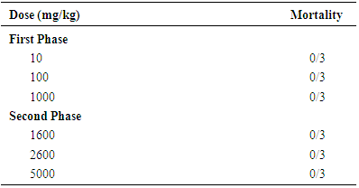
Table 3.2 showed the acute toxicity effect of ethanolic stem bark extract of Ficus thonningii in mice. There was no mortality recorded in animals treated with a single dose of 10 - 5000 mg/kg body weight. The median lethal dose (LD50) of ethanolic extract in the experimental mice was estimated to be greater than 5000 mg/kg body weight.
Table 3.2. Acute Toxicological Effect of Ethanolic Extract of Ficus thonningii in Mice
     

3.4. Sub-chronic Toxicity Studies

3.4.1. Clinical Observation
Throughout the duration of the experiment, the rats did not show any observable signs of toxicity or morbidity as they looked bright, were feeding well and their feces looked normal. Also no mortality was recorded.
3.4.2. Body Weights
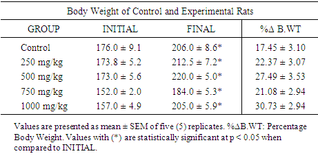
Table 3.3 shows the changes in the body weights of rats administered different doses (250, 500, 750 and 1000 mg/kg) of Ficus thonningii. Within each group there was a significant (p < 0.05) increase in the body weights from day 1 to day 42 including the control group but it was more pronounced in the extract treated rats. Rats in the control and 1000 mg/kg body weight treatment group showed the highest and the least percentage body weight change respectively.
Table 3.3. Effect of Ethanolic Extract of F. thonningii on the Body Weight of Control and Experimental Rats
     
3.4.3. Relative Organ Weights
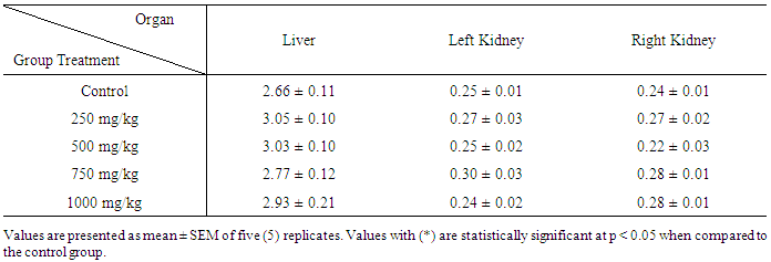
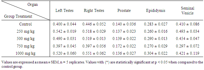
There was no significant (p < 0.05) difference in the percentage relative organ weight (ROW) of the liver, left and right kidney of the treated groups when compared to the control ROW (2.66 ± 0.11, 0.25 ± 0.01, 0.24 ± 0.01for liver, left and right kidney respectively). However, a marginal increase was observed in the percentage ROW of the liver of the treated group when compared to the control group of rats (Table 3.4). Also, there was no significant (p < 0.05) difference in the percentage ROW of the testes and accessory organs of the treated groups when compared to the control ROW (Table 3.5).
Table 3.4. Effect of Ethanolic Extract of F. thonningii on Relative Liver and Kidney Weights (%) of Control and Experimental Wistar Rats
     
Table 3.5. Effect of Ethanolic Extract of F. thonningii on Relative Testes and Accessory Organs Weight (%)
     
3.4.4. Liver Function Parameters
3.4.4.1. Activity of Aspartate Aminotransferase (AST)
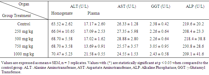
The result shows that there is no significant (p < 0.05) difference in the activity of plasma aspartate aminotransferase of the treated groups when compared to the control group (Table 3.6). However, a partial increase was observed in some of the treated groups (250 mg/kg and 500 mg/kg) while a partial reduction was observed in other treated groups (750 mg/kg and 1000 mg/kg) when compared to the control group (Table 3.6).
Table 3.6. Effect of Ethanolic Extract of F. thonningii on Liver Marker Enzymes
     
3.4.4.2. Activity of Alanine Aminotransferase (ALT)
There is no significant (p < 0.05) difference in the activity of alanine aminotransferase (both in the plasma and liver homogenate) of the treated groups when compared to the control group (Table 3.6). Rats in the 1000 mg/kg body weight treatment group showed the highest activity of alanine aminotransferase (both in the plasma and liver homogenate).
3.4.4.3. Activity of Alkaline Phosphatase (ALP)
The activity of alkaline phosphatase in Table (3.6) did not reveal significant (p < 0.05) difference between the control and treated groups. ALP activity in the control, 250, 500, 750 and 1000 mg/kg body weight treated groups are: 219.6 ± 20.2, 208.4 ± 23.3, 218.4 ± 38.8, 230.8 ± 28.6, and 209.1 ± 41.6 U/L respectively.
3.4.4.4. Activity of γ-Glutamyl Transferase (GGT)
The differences observed in the activity of γ-glutamyl transferase between the control and the treated groups were statistically significant at p < 0.05. The GGT activity in the control and treated groups (250, 500, 750, 1000 mg/kg body weight) are 2.38 ± 0.42, 2.26 ± 0.64, 2.26 ± 0.64, 3.05 ± 0.95, 2.43 ± 0.58 U/L respectively.
3.4.4.5. Total Protein Concentration
Total protein concentration (both in the plasma and liver homogenate) estimated by Lowry method showed that the difference between the control group and the extract treated groups (250, 500, 750, and 1000 mg/kg body weight) are not statistically significant at p < 0.05 (Table 3.7). However, the total protein concentrations (both in the plasma and liver homogenate) in the control group were lower than those of the treated groups.
Table 3.7. Effect of Ethanolic Extract of Ficus thonningii on other Liver Function Parameters
     
3.4.4.6. Total Bilirubin Concentration
The concentration of total bilirubin in the control group was higher than the treated groups. The concentrations are 0.82 ± 0.13, 0.73 ± 0.11, 0.73 ± 0.66, 0.58 ± 0.21, and 0.57 ± 0.14 mg/dl for the control, 250 mg/kg, 500 mg/kg, 750 mg/kg and 1000 mg/kg body weight treated groups respectively. The differences between the control and treated groups are not statistically different at p < 0.05 (Table 3.7).
3.4.4.7. Albumin Concentration
Dose-dependent marginal increase was observed in the plasma albumin concentration of rats in the treatment groups compared with the control group. There is no significant (p < 0.05) difference between the control and treated groups (Table 3.7). Rats in the control and 1000 mg/kg body weight treatment group showed the highest and the least albumin concentration respectively.
3.4.5. Kidney Function Parameters
3.4.5.1. Total Protein Concentration in the Kidney
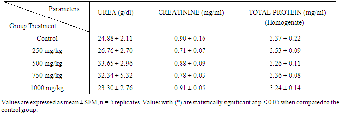
There is no significant (p < 0.05) difference in the total protein concentration (in kidney homogenate) between the control and the treated groups as estimated by the method of Lowry (Table 3.8). The highest total protein concentration was observed at 250 mg/kg body weight treated group.
Table 3.8. Effect of Ethanolic Extract of Ficus thonningii on Kidney Function Parameters
     
3.4.5.2. Creatinine Concentration
Creatinine concentrations presented in table (3.8) showed that there were no significant differences between the control and treated groups. 1000 mg/kg treated group has the highest creatinine concentration of 0.91 ± 0.05 mg/dl.
3.4.5.3. Urea Concentration
The results presented in Table 3.8 showed that urea concentration in the control and treated groups are not significantly different at p < 0.05. The highest and the least urea concentration was observed in the 500 mg/kg and 1000 mg/kg body weight treated group respectively.
3.4.6. Testicular Function Parameters
3.4.6.1. Total Protein Concentration in the Testis
Total protein concentration (testicular homogenate) estimated by Lowry method showed that the difference between the control group and the extract treated groups (250, 500, 750, and 1000 mg/kg body weight) are not statistically significant at p < 0.05 (Table 3.9).
Table 3.9. Effect of Ethanolic Extract of Ficus thonningii on Testicular Function Parameters
3.4.6.2. Activity of Testicular Acid Phosphatase (ACP)
The activity of testicular acid phosphatase in Table (3.9) did not reveal significant (p < 0.05) difference between the control and treated groups. Rats in the 750 mg/kg and 500 mg/kg body weight have the highest and the least testicular ACP activities respectively.
3.4.6.3. Activity of Testicular Alkaline Phosphatase (ALP)
Marginal increase was observed in the testicular ALP activities of treated groups when compared to the control group. There is no significant (p < 0.05) difference between the control and treated groups (Table 3.9). The highest testicular ALP activity of 120.40 ± 58.75 U/g protein was observed in the 750 mg/kg body weight treated group.
3.4.6.4. Activity of Testicular Alanine Aminotransferase (ALT)
The activity of testicular acid phosphatase as shown in Table (3.9) did not reveal significant (p < 0.05) difference between the control and treated groups. The highest testicular ALT activity of 33.44 ± 4.80 U/L was observed in the 750 mg/kg body weight treated group.
3.4.6.5. Activity of Testicular Aspartate Aminotransferase (AST)
There is no significant (p < 0.05) difference in 250 mg/kg body weight treated group when compared with the control group. A dose dependent significant (p < 0.05) increase was observed in 500 mg/kg, 750 mg/kg and 1000 mg/kg body weight treated group when compared with the control group. The highest activity of testicular aspartate aminotransferase was observed in 1000 mg/kg body weight treated group.
3.4.6.6. Activity of Testicular γ-Glutamyl Transferase (GGT)
The activity of testicular GGT as presented in Table (3.9) did not reveal significant (p < 0.05) difference between the control and treated (250 mg/kg and 500 mg/kg body weight) groups. A significant (p < 0.05) reduction in testicular GGT activity was observed in the treated (750 mg/kg and 1000 mg/kg body weight) groups when compared to the control group.
3.4.7. Sperm Parameters
3.4.7.1. Sperm Motility
The degree of motility of spermatozoa of rats in the control group was significantly (p < 0.05) higher than what was observed in rats in each of the treatment groups (Table 3.10).
Table 3.10. Effect of Ethanolic Extract of Ficus thonningii on sperm morphology, count, motility, viability, and sperm volume
3.4.7.2. Sperm Volume
The mean sperm volume was almost constant for the rats in the control group and each of the test groups (Table 3.10).
3.4.7.3. Sperm Count
The mean value of sperm count (millions/ml) of rats in 250 mg/kg body weight treated group (102.50 ± 5.70), 500 mg/kg body weight treated group (92.20 ± 4.36), 750 mg/kg body weight treated group (92.20 ± 4.36), and 1000 mg/kg body weight treated group (94.40 ± 3.44) were significantly (p < 0.05) lower compared with that of the control rats (126.40 ± 5.87) (Table 3.10).
3.4.7.4. Sperm Viability
The mean value of percentage liveability or live: dead ratio of sperm cells was not significant (p < 0.05) for rats in each of the treatment groups compared with that of rats in the control group (Table 3.10).
3.4.7.5. Total Sperm Abnormality
The total mean sperm abnormality (%) was significantly (p < 0.05) lower than the values obtained for the rats in the test groups (Table 3.10).
3.4.8. Histomorphological Changes
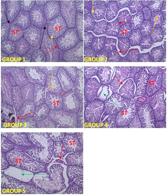
Plate 3.1 exhibits photomicrographs of the testes; scale enlargement: ×400. Normal seminiferous tubules were noted in the control group. Interstitial spaces were widened in rats treated sub-chronically with 250, 500, 750 and 1000 mg/kg dose of Ficus thonningii stem bark extract. Degeneration of cells in the seminiferous tubules of rats treated sub-chronically with 750 and 1000 mg/kg dose of F. thonningii were also noted.
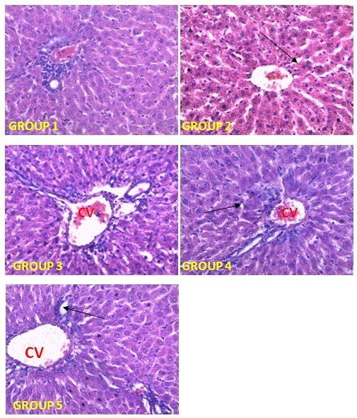
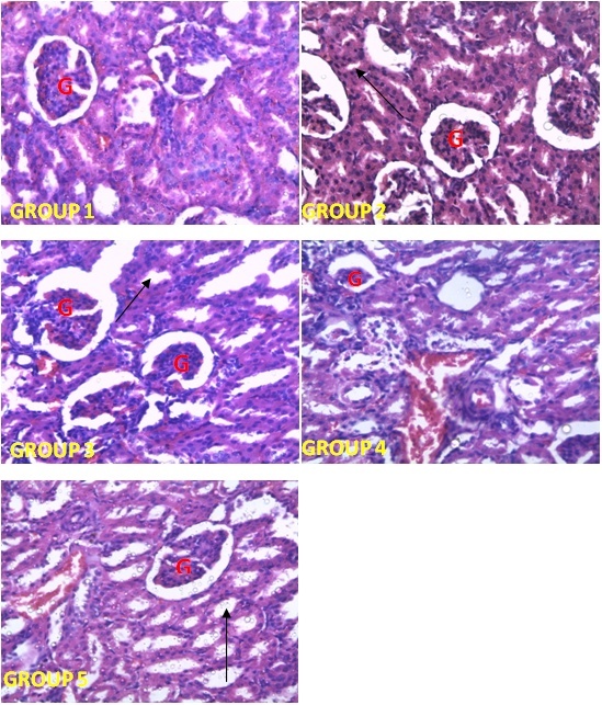
Plate 3.2 and 3.3 exhibits photomicrographs of the liver and the kidney respectively; scale enlargement: ×400. Histological features of the liver of control rats showed normal structures. Rats treated sub-chronically with 250, 500, 750 and 1000 mg/kg dose of Ficus thonningii stem bark extract did not cause any adverse effect on the histoarchitecture of hepatocytes. There was no effect on the kidneys of rats treated sub-chronically with 250, 500, 750 and 1000 mg/kg dose of Ficus thonningii stem bark extract, as the glomeruli, and the distal and proximal tubules appeared normal, as shown in Plate 3.3.
Plate 3.1. Photomicrographs of the testes after 42 days of treatment with Ficus thonningii stem bark extract showing the seminiferous tubules (ST) of the different groups. Stain H & E ×100. Normal Seminiferous tubules were noted in Group 1 with full complement of spermatogenic (Spermatogonia-Yellow arrow; Spermatocyte- Orange arrow) and Leydig cells (Black arrow). Interstitial spaces were widened in Group 2, 3, 4 and 5. Degeneraton of the cells in the seminiferous tubules of Groups 4 and 5 were noted (represented with Green arrow)
Plate 3.2. Photomicrographs of liver sections from control and experimental rats (group 2, 3, 4, and 5) after 42 days of treatment with Ficus thonningii stem bark extract showing normal central vein (CV). Hepatic plates appear normal in all groups. Slightly widened sinusoid is noted in group 2 (Black arrow)
Plate 3.3. Photomicrographs of kidney section from the control and experimental rats (group 2, 3, 4 and 5) after 42 days of treatment with Ficus thonningii stem bark extract showing apparently normal glomerulus (G) and renal tubules (arrow). H & E (×400)

4. Discussion and Conclusions

4.1. Discussion

The use of herbs is very common in developing countries, particularly in rural settings. However, during the last decade an increase in the use of plants has been observed in metropolitan areas of developed countries [40]. Plants and plant extracts, in traditional herbal medicine, play important roles in the treatment of various illnesses [41]. The common wild fig, Ficus thonningii, is one of the many fruit-bearing trees that have traditionally been used for treating diseases in Africa and beyond [1]. Although, stem bark extract of Ficus thonningii is commonly used in ethnomedicine for the treatment of sore throat, arthritis, diarrhoea, ulcers [10] and to enhance fertility [11], experimental studies referring to the risks of the oral use of the ethanol stem bark extract and pharmacological studies are lacking. The present investigation was carried out to estimate safety limits of its oral administration through toxicological assessment in adult Wistar rats.
Currently, studies involving animals are used in the estimation of safety of drugs and plant products. A good correlation has been reported between toxicological insults in rats and humans; correlation is weaker between humans and mice [42]. Therefore numerous studies investigate the acute effects of high doses in mice and the chronic effects of lower doses in rats including the doses potentially usable in humans [43, 44].
The effect of ethanolic extract of F. thonningii when administered to mice showed no mortality within 48 hours after treatment with extract. There were no signs of weakness and/or loss of appetite even up to doses of 5000 mg/kg body weight. Thus, the lethal dose (LD50) was estimated to be higher than 5000 mg/kg body weight, therefore, the LD50 being greater than 5000 mg/kg body weight is thought to be safe as suggested by Lorke (1983) [25]. This correlates with results of earlier works carried out on the ethyl acetate leaf extract of the plant [45].
In the sub-chronic toxicity study using all male adult wistar rats, ethanolic Ficus thonningii stem bark extract was administered at doses ranging from 0 to 1000 mg/kg body weight. There were no significant changes in animal behaviour and body weight. Consistent increase in weight was observed in all the groups of animals treated with ethanolic extract of Ficus thonningii which could mean that the extract may not be toxic at a concentration up to 1000 mg/kg body weight investigated in this study. A decrease in body weight would be an indicator of adverse effects [46, 47]. No difference was observed in the relative organ body weight of the liver, kidney, testes, prostate, epididymis and seminal vesicle between the control and the treated groups. An increase or decrease in weight could indicate a potential toxic effect [48].
Liver is the major organ involved in drug biotransformation. The liver is prone to xenobiotic-induced injury because of its central role in xenobiotic metabolism [49]. Levels of serum liver biomarker enzymes are biochemical parameters usually performed in order to evaluate any toxic effects on the liver [50]. Increases in the levels of AST, ALT, ALP and GGT in the serum are associated with liver toxicity by drugs or any other hepatotoxin [51]. However, ALT is more specific to liver and thus a better parameter for detecting liver injury as AST is also associated with diseases of other organs such as heart and muscle [52]. ALP is present mostly in cells lining the biliary duct of the liver and is used to diagnose obstruction to the biliary system. Therefore, its elevation in the blood indicates cholestatic diseases such as gallstone or tumor blocking the bile duct [53]. GGT is an enzyme which is found in liver, kidney and pancreatic tissues, the enzyme concentration being low in liver as compared to kidney [52]. GGT is a specific biomarker of hepatobiliary injury, especially cholestasis and biliary effects [54]. In this study, sub-chronic exposure of rats to Ficus thonningii stem bark extract at different doses (250, 500, 750 and 1000 mg/kg) caused no significant increase in AST, ALT, ALP, and GGT. These results, associated to the observed normal levels of liver marker enzymes suggest that the stem bark extract of Ficus thonningii did not alter liver function at the doses studied.
Bilirubin is a breakdown product of hemoglobin and is associated with hepatic diseases like jaundice and ineffective erythropoiesis and increased bilirubin levels reflect the depth of jaundice [55]. Albumin is the most abundant of the plasma proteins with the physiological role of maintenance of osmotic pressure and transportation of both endogenous and exogenous substances. The ability of the liver to synthesize albumin is diminished if the organ is damaged [56]. In this study there was no significant change in the levels of plasma bilirubin, total protein, and albumin of both the treated and control rats. This suggests that the extract may have no toxic effect on the hepatic and erythropoietic system.
Urea and creatinine are considered as important markers of kidney dysfunction [57]. Higher than normal levels of serum urea and creatinine and total protein concentration in the kidney are indications of deficiency in renal function [58]. The differences in the plasma concentration of creatinine and urea, and total protein in the kidney observed in the treated groups were not statistically significant when compared to the control. The result suggests that the glomerular filtration rate (GFR) was not affected by the oral administration of ethanolic extract at a maximum dose of 1000 mg/kg body weight.
In testis, alkaline phosphatase is involved in mobilizing carbohydrates and lipid metabolites to be utilized either within the cells of the accessory sex structure or by the spermatozoa in the seminal fluid [59]. Acid phosphatase is widely distributed in the testes and is important in the physiology of sperm [60]. Acid phosphatase is one of the markers of dyszoospermia associated with the denaturation of seminiferous epithelium and phagocytosis of Sertoli cells [61]. Elevated acid phosphatase activities result in indiscriminate hydrolysis of phosphate esters, which are potential energy source for the cell [62]. γ-Glutamyl transferase is a membrane bound enzyme which catalysis the transfer of γ-glutamyl group between peptides and amino acids [63] and is considered a ‘marker’ enzyme of Sertoli function of testes [64].
No significant difference was observed in the testicular ALP and ACP activity of the treated groups when compared with the control group. This probably imply that the mobilization of carbohydrates and lipid metabolites for use by accessory sex structure were unperturbed by the administration of Ficus thonningii stem bark extract but the significant decrease in the activity of testicular γ-glutamyl transferase of the 750mg/kg and 1000 mg/kg body weight treated groups probably indicates impairment of the function of Sertoli cells [59] by the plant extract.
Similarly, the activities of AST and ALT increase when the membrane of spermatozoa is damaged, and the rate of intact acrosome spermatozoa decreases [65]. The present result showed that there was no significant difference in testicular ALT activity but a dose-dependent elevated testicular AST activity of the 500, 750, and 1000 mg/kg body weight treated groups when compared to the control group was observed. This significant increase in AST activity could probably indicate a possible threat on the functions of spermatozoa and the testis.
The sperm cell count, motility, live/dead sperm cell ratio, morphology, and the seminal volume were used in this study to evaluate the effect of sub-chronic administration of Ficus thonningii stem bark ethanolic extract on male reproductive system. These andrological parameters are usually evaluated to determine the fertility of a male subject [66]. A significant increase of total sperm abnormality in the treated groups was observed when compared with the control group. This could mean that the administration of the extract caused aberrations in the process of spermatogenesis [67, 68] or maturation of abnormal sperm cells from damaged seminiferous tubules [69].
The sperm motility of rats in the treatment groups were significantly lower, but the live/dead sperm cells ratio were within the same range as those of the control rats, which shows that the extract did not affect the viability of spermatozoa but it caused deformation of the cells and rendered them less motile or immotile. These observations are contrary to what was reported for Kolaviron (a biflavonoid from seeds of Garcinia kola) which was discovered to prevent peroxidative changes in the sperm and testicular membrane, thus enhancing sperm motility and decreasing spermatozoa abnormalities [70, 71]. Tribulus terrestris with protodioscin as its active component is another plant proven to improve spermatogenesis, sperm motility and morphology [72].
In this study, the sperm count was observed to have reduced significantly (p<0.05) which is an indication that the stem bark extract of Ficus thonningii reduced or inhibited spermatogenesis. This is similar to what was observed in some medicinal plants with detrimental effects on male fertility such as Carica papaya and Quassia amara. C. papaya was reported by Chinoy and Padman (1996) [73] to have anti-fertility effect by reduction of testicular mass, sperm count and sperm motility when the benzene extract of the seeds was administered to male albino rats. The chloroform extract of the bark of Q. amara has been shown to decrease sperm count, motility and viability in albino rats [74], while on the contrary, aqueous extracts of root, leaf, or whole plant of Withania somnifera is known to increase sperm count [75].
The varying degrees of distortion in the testicular seminiferous tubules following the administration of the plant extract at the doses of 250, 500, 750, and 1000 mg/kg bwt when compared with the control further point to the toxicity risk of the plant extract as revealed by the testicular function parameters investigated in this study. Gametogenesis occurs in the seminiferous tubules while the interstitial cells secrete the testicular hormone, mainly testosterone. Therefore, any alteration in the seminiferous tubules as observed in the histopathological studies will have its consequential effect on gametogenesis [76].
Photomicrographs of the sections of liver and kidney showed that the administrations of the ethanolic plant extract at the doses of 250, 500, 750, and 1000 mg/kg bwt did not have adverse effect on the histoarchitecture of hepatocytes and glomerulus as revealed by the liver and kidney functions parameters respectively.

4.2. Conclusions

The study concluded that the ethanolic stem bark extract of F. thonningii had deleterious effects on the testes, but not on the liver and kidney, at all administered doses (250, 500, 750, 1000 mg/kg body weight) of the extract. Thus, at the doses empirically used in traditional medicine, the ethanolic stem bark extract of the plant could not be considered as safe for the purpose for which it is traditionally employed, particularly, as fertility enhancer.

References

[1]  Schmidt, E., Lutter, M. and McCleland, W. (2002). Trees and shrubs of Mpumalanga and Kruger National Park, Jacana, South Africa, pp. 80.
[2]  Njoronge, G. N. and Kibunga J. W. (2007). Herbal medicine acceptance, sources and utilisation for diarrhoea management in a cosmopolitan urban area (Thika, Kenya). African Journal of Ecology. 45, 65-70.
[3]  Cousins, D. and Huffman, M. A. (2002). Medicinal properties in the diets of gorillas: an ethno-pharmacological evaluation. African Study Monographs. 23, 65-89.
[4]  Igoli, J. O, Ogaji, O.G, Tor-Anyiin T. A, Igoli N. P. (2005) Traditional Medical Practices Amongst the Igede People of Nigeria. African Journal of Traditional, Complementary and Alternative Medicine. 2(2):134–152.
[5]  Alawa, J. P., Jokthan, G. E. and Akut, K. (2002). Ethnoveterinary medical practice for ruminants in the sub-humid zone of the northern Nigeria. Preventive Veterinary Medicine. 54, 79-90.
[6]  Moshi, M. J., Otieno, D. F., Mbabazi P. K. and Weisheit. A. (2009). The ethnomedicine of the Haya people of Bugabo ward, Kagera Region, North-western Tanzania. Journal of Ethnobiology and Ethnomedicine. 5, 24.
[7]  Arbonier, M. A. (2004). Trees, Shrubs and Lianas of West African Dry Zones. 2nd edition, Margraf publishers, Netherlands, pp. 412.
[8]  Mali, R. G. and Mehta, A. A. (2007). A review on anthemintic plants. Natural Product Radiance. 7, 466-475.
[9]  Gelfand, M., Mavi, S., Drummond, R. B. and Ndemera, B. (1985). The Traditional Medicinal Practitioner. His principles of practice and pharmacopoeia. Mambo Press, Gweru, Zimbabwe, pp. 411.
[10]  Teklehaymanot, T. and Gidday, M. (2007). Ethnobotanical study of medicinal plants used by people in Zegie Peninsula, North Western Ethiopia. Journal of Ethnobiology and Ethnomedicine. 3, 12.
[11]  Obata, O. O. and Aighokhan, E. I. (2012). Ethnobotanical practices among the people of Oka-Akoko, Nigeria. Plant Archive.12 (2), 627-638.
[12]  Cowan, M. M. (1999). Plant products as antimicrobial agents. Clinical Microbiology Reviews. 12, 564-582.
[13]  Jansen, O., Angenot, L., Tits, M., Nicolas, J. P., De Mol, P., Nikiema, J. B. and Frederich, M. (2010). Evaluation of 13 selected medicinal plants from Burkina Faso for their antiplasmodial properties. Journal of Ethnophamacology. 13, 143-150.
[14]  Oyelana, O. A., Durugbo, E. U., Olukanni, O. D., Ayodele, E. A., Aikulola, Z. O. and Adewole, A. I. (2011). Antimicrobial activity of Ficus leaf Extracts on some fungal and bacterial pathogens of Dioscorea rotundata from Southwest Nigeria. Journal of Biological Sciences. 11, 359-366.
[15]  Krief, S., Hladik, C. M. and Haxaire, C. (2005). Ethnomedicinal and bioactive properties of plants ingested by wild chimpanzees in Uganda. Journal of Ethnopharmacology. 101, 1-15.
[16]  Olas, B., Wachowicz, B., Nowak, P., Stochmal, A., Oleszek, W., Glowacki, A. and Bald, E. (2008). Comparative studies of the antioxidant effects of a naturally occurring resveratrol analogue trans 3, 3’, 5, 5-tetrahydroxy-4-methoxystilbene and resveratrol- against oxidation and nitration of biomolecules in blood platelets. Cell Biology and Toxicology. 24, 331-340.
[17]  Grassi, D., Desideri, D. and Ferri, C. (2010). Flavonoids antioxidants against atherosclerosis. Nutrients. 2, 889-902.
[18]  Otimenyin, S. O., Uguru, M. O. and Atang, B. L. (2004). Anti-inflammatory and analgesic activities of Ficus thonningii and Pseudocedrela kotschyi extracts. Nigerian Journal of Pharmaceutical Research. 3, 82-85.
[19]  Maiha, B. B., Mohammed, B. and Magaji, M. G. (2013). Psychopharmacological potential of methanol leaf extract of Ficus thonningii (Blume) in mice. Nigerian Journal of Pharmaceutical Science. 12 (2), 30-34.
[20]  Coker, M. E., Emikpe, B. O., Adeniyi, B. A. and Budale, B. A. (2009). The inflammatory potential, haematological and histological changes induced in rats due to the administration of methanol extracts of Ficus thonningii leaves. African Journal of Pharmacology. 3, 273-276.
[21]  Aniagu, S. O., Nwinyi, F. C., Akumka, D. D., Agbain, E. O., Dzarma, S., Ajoku, P., Adelusola, K..A., Ibe, J., Inyang, U. S. and Gamaniel, K. S. (2008). Short term toxicity studies of Ficus thonningii Blume Moraceae leaf extracts in rats. International Journal of Food Science and Technology. 43, 456-464.
[22]  Musabayane, C. T., Gondwe, M., Kadyamaapa, D. R., Chuturgoon, A. A. and Ojewole, J. A. O. (2007). Effects of F. thonningii (Blume) Moraceae stem bark ethanol extract on blood glucose, cardiovascular and kidney cell lines of the proximal (LLC-PK1) and distal tubules (MDBK). Renal Failure. 29, 389-397.
[23]  Trease, G. E. and Evans, W. C. (1978). A Textbook of Pharmacognosy. 11th Edition, Bailliere Tindall, London. Pp. 530.
[24]  Sofowora, E. A. (2006). Phytochemical screening: In “Medicinal plants and Traditional Medicine in Africa”, 2nd edition, Spectrum Books Limited Ibadan, Nigeria. Pp. 150-153.
[25]  Lorke, D. A. (1983). A new approach to practical acute toxicity testing. Archives of Toxicology. 54, 275-287.
[26]  Tchamadeu, M. C., Dzeufiet, P. D. D., Nana, P., Kouambou Nouga, F., Allard, J., Blaes, N., Siagat, R., Zapfack, L., Girolami, J. P., Tack, I., Kamtchouing, T. and Dimo, T. (2011). Acute and sub-chronic oral toxicity studies of an aqueous stem bark extract of Pterocarpus soyauxii Taub (Papilionaceae) in rodents. Journal of Ethnopharmacology. 133, 329-335.
[27]  Chawla, R. (1999). Biochemical tests for total proteins, albumin and pyruvate. In: Practical Clinical Biochemistry Methods and Interpretations, 2nd Edition, JAYPEE. pp 107-134.
[28]  Weatherburn, M. W. (1967) Phenolhypochlorite reaction for the determination of ammonia. Analytical Chemistry. 39, 971.
[29]  Lowry, O. H., Rosebrough, N. J., Farr, A. L. and Randall, K. J. (1951). Protein measurement with the Folin phenol reagent. Journal of Biologic Chemisry. 193, 265-275.
[30]  Pinnell, A. E. and Northam, B. E. (1978). New automated dye-binding method for serum albumin determination with bromocresol purple. Clinical Chemistry. 24, 80-86.
[31]  Jendrassisk, L. and Grof, P. (1938). Simplified photometric methods for the determination of blood bilirubin. Biochemistry Zeitschrift. 297, 81-89.
[32]  Deutsche Gesellschaft fur klinische Chemie, (1972). Empfehlungen der deutschen Gesellschaft fur Klinische Chemie (DGKC). Standardisierung von Methoden zur Bestimmung von Enzymaktivit ten in biologischen Flussigkeiten. (Recommendation of the German Society of Clinical Chemistry. Standardization of methods for measurement of enzymatic activities in biological fluids). Journal of Clinical Chemistry and Clinical Biochemistry. 10, 182-192.
[33]  Seiler, D. (1983). Saure Phosphatase in Serum (Substrate: α-Naphthylphosphat) Referenczwerte und Diagnostische Aussage. Journal of Clinical Chemistry and Clinical Biochemistry. 21, 519-525.
[34]  Reitman, S. and Frankel, S. (1957). A colorimetric method for the determination of serum glutamic oxaloacetic and glutamic pyruvic transaminases. American Journal of Clinical Pathology. 28, 58-63.
[35]  Szasz, G. A. (1969). Kinetic photometric method for serum γ-glutamyl transpeptidase. Clinical Chemistry. 15(2), 124-137.
[36]  Akusu, M. O., Akpokodje, J. U., Ogwnegbu, S. O. and Oke, B. O. (1985). Differences in morphology of Bull spermatozoa from normal and pathological testis during epididymal transit. Nigerian Veterinary Journal. 14(2), 30-33.
[37]  Oyeyemi, M. O. and Ubiogoro, O. (2005). Spermiogram and morphological chacteristics in testicular and epididymal spermatozoa of large white boar in boar in Nigeria. International. Journal of. Morphology. 23(3), 235-239.
[38]  Pant, N. and Srivastava, S. P. (2003). Testicular and spermatotoxic effect of quinaphos in rats. Journal of Applied Toxicology. 23, 271-274.
[39]  Wells, M. E. and Awa, O. A. (1970). New technique for assessing acrosomal characteristics of spermatozoa. Journal of Diary Science. 53, 227.
[40]  Harnack, L. J., Rydell, S. A. and Stang, J. (2001). Prevalence of use of herbal products by adults in the Minneapolis/St Paul, Minn, metropolitan area. Mayo Clinic Proceedings. 76, 688–694.
[41]  Ogle, B. M., Tuyet, H. T., Duyet, H. N. and Dung, N. N. (2003). Food, Feed or Medicine: The Multiple Functions of Edible Wild Plants in Vietnam. Economic Botany. 57(1), 103-117.
[42]  Olson, H., Betton, G., Robinson, D., Thomas, K., Monro, A., Kolaja, G., Lilly, P., Sanders, J., Sipes, G., Bracken, W., Dorato, M., Van Deun, K., Smith, P., Berger, B. and Heller, A. (2000). Concordance of the toxicity of pharmaceuticals in humans and in animals regulatory. Toxicology and Pharmacology. 32, 56-57.
[43]  Rhiouani, M., Al-Shabanah, O. A. and Al-Majed, A. A. (2002). Effect of prolonged vigabatrin treatment on haemaological and biochemical parameters in plasma, liver and kidney of Swiss albino mice. Scientia Pharmaceutica. 70, 135-145.
[44]  Lahlou, S., Zafar, H. I. and Badiaa, L. (2008). Acute and chronic toxicity of a lyophilized aqueous extract of Tanacetum vulgare leaves in rodents. Journal of Ethnopharmacology. 117, 221-227.
[45]  Ahur, V. M., Madubunyi, I., Adenkola, A. Y. and Udem, S. C. (2010). The effect of acetyl acetate extract of Ficus thonningii (Blume) leaves on erythrocyte osmotic fragility and haematological parameters in acetaminophen treated rats. Comparative Clinical Pathology. 10, 1107-1111.
[46]  Raza, M., Al-Shabanah, O. A., El-Hadiya, T. M. and Al-Majed, A. A. (2002). Effect of prolonged vigabatrin treatment on haematological and biochemical parameters in plasma, liver and kidney of Swiss albino mice. Science Pharmaceutica. 70, 135-145.
[47]  Teo, S., Stirling, D., Thomas, S., Hoberman, A., Kiorpes, A. and Khetani, V. (2002). A 90-day oral gavage toxicity study of D-methylphenidate and D, L methyphenidate in Sprague Dawley rats. Toxicology. 179, 183-196.
[48]  Ezeja, M. I., Anaga, A. O. and Asuzu I. U. (2014). Acute and sub-chronic toxicity profile of methanol leaf extract of Gouania longipetala in rats. Journal of Ethnopharmacology. 151, 1155–1164.
[49]  Bass, N. M. and Ockner, B. A. (1996). Drug-induced liver disease. In: Zakin, D. and Boyer, T. D. (Eds.), Hepatology, a Text Book of Liver Diseases, 3rd edition W. B. Saunders, Philadephia, pp.962–1017.
[50]  Mukinda, J. and Syce, J. A. (2007). Acute and chronic toxicity of aqueous extract of Artemisia afra in rodents. Journal of Ethnopharmacology. 112, 138–144.
[51]  Ramaiah, S. K. (2011). Preclinical safety assessment. Current gaps, challenges and approaches in identifying translatable biomarkers of drug-induced liver damage. Clinics in Laboratory Medicine. 31, 161–172.
[52]  Ozer, J., Ratweb, M., Shawc, M., Baileya, W. and Schomaker, S. (2008). The current state of serum biomarker of hepatoxicity. Toxicity. 245, 194–205.
[53]  Burtis, C. A. and Ashwood, E. R. (2001). Enzymes: In Tietz Fundamentals of Clinical Chemistry, 5th edition W. B. Saunders Company, New York, USA, pp.352–369.
[54]  Mauro, P., Renze, B. and Wouter, W. (2006). In Tietz text book of clinical chemistry and molecular diagnostics. 5th edition, editors: Carl A.B., Edward, R, David E.B, Elsevier; Enzymes. Pp. 604-616.
[55]  Thapa, B. R. and Walia, A. (2007). Liver function tests and their interpretation. Indian Journal of Pediatrics. 74, 663-671.
[56]  Whitby, L. G., Smith, A. F. and Becket, B. J. (1989). Lecture notes on clinical chemistry, 4th edition. Blackwell Scientific Publications, London. pp 38-178.
[57]  Mukinda, J. T. and Eagles, F. K. (2010). Acute and sub-chronic oral toxicity profile of the aqueous extract of Pohygala fruticosa in female mice and rats. Journal of Ethnopharmacology. 128, 236–240.
[58]  Mukinda, J. and Syce, J. A. (2007). Acute and chronic toxicity of aqueous extract of Artemisia afra in rodents. Journal of Ethnopharmacology. 112, 138–144.
[59]  Ramalingam, V. and Vimaladevi, V. (2002). Effect of mercuric chloride on membrane-bound enzymes in rat testis. Asian Journal of Andrology. 4, 309–311.
[60]  Breton, S., Smith, P. T. S., Lui, B. and Brown, D. (1996). Acidification of the male reproductive tract by a proton pumping (H+)-ATPase. Natural Medicine. 2, 470–472.
[61]  Samarth, R. M. and Samarth, M. (2009). Protection against radiation induced testicular damage in Swiss albino mice by Mentha piperita (Linn.). Basic Clinical Pharmacology and. Toxicology. 104, 329–334.
[62]  Butterworth, P. J. and Moss, D. W. (1966). The effect of urea on human kidney alkaline phosphatase. Biochemical Journal. 99, 9–10.
[63]  Tate, S. S. and Rose, M. E. (1977). Human kidney γ-glutamyl transpeptidase: catalytic properties, subunit, structure and localization of the γ-glutamyl binding site on the large subunit. Journal of Biological Chemistry. 252, 6042–6045.
[64]  Sherin, R. J. and Hodgen, G. D. (1967). Testicular γ-glutamyl transpeptidase: an index of Sertoli cell function in man. Journal of Reproduction and Fertility. 48, 191–194.
[65]  Yang, J., Wu, G., Feng, Y., Lu, Q., Lin, S. and Hu, J. (2010). Effects of taurine on male reproduction in rats of different ages. Journal of Biomedical Science. 17 (Suppl 1), S9.
[66]  Garner, D. L. and Hafez, E. S. E. (1993). Spermatozoa and seminal plasma. In Hafez E.S.E (eds.). Reproduction in Farm animals. (6th ed.). Lea and Febiger, Philadelphia, USA. Pp. 165-187.
[67]  Moss, J. A., Melrose, D. R., Reed, H. C. B. and Vanderplassche, M. (1979). Spermatozoa, semen and artificial insemination. Baillire Tindal, London. pp. 59-91.
[68]  Hafez, E. S. E. (1987). Advances in reproductive biology and semen evaluations. In: Reproduction in farm animals. 5th edition. Lea and Febiger, Philadelphia, USA. pp. 649.
[69]  Thomas, M. J. and Thomas, J. A. (2001). Toxic responses of the reproductive system. In: Casarett and Doull’s Toxicology. The basic science of poisons (6th edition), McGraww-Hill Medical Publishing Division, New York, pp. 677- 681.
[70]  Farombi, E. O., Adedara, I. A., Akinrinde, S. A., Ojo, O. O. and Eboh, A. S. (2012). Protective effects of kolaviron and quercetin on cadmium-induced testicular damage and endocrine pathology in rats. Andrologia. 44(4), 273-284.
[71]  Ishihara, M., Itoh, M., Miyamoto, K., Takeuchi, Y., Takenaka, I. and Jitsunari, F. (2000). Spermatogenic disturbance induced by di-(2-ethylhexyl) phthalate is significantly prevented by treatment with antioxidant vitamins in rat. International Journal of Andrology. 23, 85-94.
[72]  Adimoelja, A., Setiawan, L. and Djojotananjo, T. (1995). Tribulus terrestris (protodioscin) in the treatment of male infertility with idiopathic oligoasthenoterato-zoospermia. In: First International Conference of Medical Plants for Reproductive Medicine in Taipei, China.
[73]  Chinoy, N. J. and Padman, P. (1996). Anti-fertility investigation on the benzene extract of Carica papaya seeds in male albino rats. Journal of Medicinal and Aromatic Plant Science. 19(2), 422-426.
[74]  Parveen, S., Das, S., Kundra, C. P. and Pereira B. M. (2003). A comprehensive evaluation of the reproductive toxicity of Quassia amara in male rats. Reproductive Toxicology. 17(1), 45-50.
[75]  Abdel-Magnied, E. M, Abdel-Rahman, H. A. and Hanaz, F. M. (2001). The effect of Aqueous extracts of Cynomorium coccineum and Withania somnifera on testicular development in immature Wistar rats. Journal of Ethnopharmacology. 75, 1-4.
[76]  Yakubu, M. T., Akanji, M. A. and Oladiji, A. T. (2008). Effects of oral administration of aqueous extract of Fadogia agrestis (Schweinf. Ex Hiern) stem on some testicular function indices of male rats. Journal of Ethnopharmacology. 115, 288–292.