-
Paper Information
- Paper Submission
-
Journal Information
- About This Journal
- Editorial Board
- Current Issue
- Archive
- Author Guidelines
- Contact Us
American Journal of Biochemistry
p-ISSN: 2163-3010 e-ISSN: 2163-3029
2015; 5(2): 35-41
doi:10.5923/j.ajb.20150502.03
Anti-Inflammatory and Analgesic Effect of Leptadenia hastata on Albino Rats
 Abstract
 Reference
 Full-Text PDF
 Full-text HTML
Sarkiyayi S., Umaru H. A, Onche H. O.
Department of Biochemistry, Modibbo Adama University of Technology, Yola, Nigeria
Correspondence to: Sarkiyayi S., Department of Biochemistry, Modibbo Adama University of Technology, Yola, Nigeria.
| Email: |  |
Copyright © 2015 Scientific & Academic Publishing. All Rights Reserved.
The anti-inflammatory and analgesic effects of the leaf-stem extract of leptadeniahastatawere investigated in albino rats. The phytochemical screening of the extract was also carried out and at the end of the experiment; all the albino rats were tested to check the any abnormality in the liver enzymes which could have been caused by the administration of the plant extract. The anti-inflammatory and analgesic activities were determined by formalin induced inflammation of the rat paw. The phytochemical screening revealed the presence of alkaloids, saponin, tannins, flavonoids, steroidal ring, resins and terpenoids. The leaf-stem extract of leptadeniahastata (200 to 800mg/kg) caused significant inhibition on the formalin induced inflammation in albino rats (p ≤ 0.05). I fact, 800mg/kg test group showed maximum inhibition of inflammation (96.2%) when compared with the control and the reference groups. This effect was comparable to the observed effect with paraceutamol (2mg/kg) which was used as a standard. Furthermore, the leaf-stem extract of leptadeniahastata caused a significant reduction in the formalin induced pain in albino rat as compared with the reference and the normal control. It was clearly shown that leptadeniahastata leaf-stem extract have a little effect on the liver enzymes (AST and ALT) of the experimental albino rats. Leptadeniahastata possessed anti inflammatory and analgesics properties and recommended for acute inflammatory disorders and diseases associated with pains. This finding also support the claims of its use traditionally as an anti-cholesteronemia in Nigeria traditional practice.
Keywords: Anti-inflammatory, Analgesic effect, Leptadeniahastata, Albino rats, Phytochemicals
Cite this paper: Sarkiyayi S., Umaru H. A, Onche H. O., Anti-Inflammatory and Analgesic Effect of Leptadenia hastata on Albino Rats, American Journal of Biochemistry, Vol. 5 No. 2, 2015, pp. 35-41. doi: 10.5923/j.ajb.20150502.03.
Article Outline
1. Introduction
- Traditional medicine is a significant element of the African cultural heritage and for a large majority of people, resort for treating health problems. This knowledge is passed down from generation to the next in oral form but very little written information are available on the active, safety and effectiveness of this medicine (Nikiema et al., 2001). The use of plants as medicines predates written human history. Ethno botany (the study of traditional human uses of plants) is recognized as an effective way to discover future medicines. In 2001, researchers identified 122 compounds used in modern medicine which were derived from "ethno medical" plant sources; 80% of these have had an ethno medical use identical or related to the current use of the active elements of the plant. Many of the pharmaceuticals currently available to physicians have a long history of use as herbal remedies, including aspirin, digitalis, quinine, and opium. The use of herbs to treat disease is almost universal among non-industrialized societies, and is often more affordable than purchasing expensive modern pharmaceuticals. Studies have shown that their use is less common in clinical settings, but has become increasingly more in recent years as scientific evidence about the effectiveness of herbal medicine has become more widely available (Nikiema et al., 2001). Leptadeniahastata is a perennial liana of the family of Asclepediaceae which pushes in the cattle breeding area of Burkina Faso in West Africa. The breeders commonly used the leaf-stems for placenta retention treatment when animals gave birth (Kerharo and Adam, 1974). It is a many-stemmed climber, becoming bushy at the base of dry savannah from Senegal to West Cameroun and extending across Africa to Ethiopia, Kenya, and Uganda. The leaves, young shoots and flowers are eaten throughout the region and in East Africa, usually cooked and in soups. It is also an African traditional plant with different parts which have been reported to have several medicinal values. Its leaves and latex have trypanocydal, antimalarial, antibacterial, anti-inflammatory, androgenic and sterility properties (Gambetta and Alay, 2011). In the majority of western Sahel country, the young leaves of leptadeniahastata are an important staple when cereal harvests are inadequate to support populations. However, little attention was paid to the examination of the action of leptadeniahastata on the reproductive system of mammals since in northern Burkina Faso, the breeders and local people traditionally believe that leptadeniahastata can reduce fertility of their animals (Kerharo and Adam, 1974). Literature survey and ethno botanic investigations with the traditional healers revealed that the consumption of the leaf-stem of Leptadeniahastata by humans could have anti-inflammatory, analgesic, anti-diabetic and anti-cholesteronemia effect. It is against this backdrop therefore, it was considered of interest to investigate the protective effect of leptadeniahastata leaf-stem extract on formaldehyde-induced inflammation in rats. The aim of this work was to evaluate the potential anti-inflammatory and analgesic properties of the aqueous extract of the leaf-stem of leptadeniahastata on albino rats. The effects of the extract on the activities of some liver enzymes of the albino rats such as Alanine transaminase (ALT) and (AST) Aspartate transaminase were also investigated.
2. Materials and Methods
2.1. Materials
- The leaf-stem of leptadeniahastata was collected from plateau forest in Jos, Plateau state. The plant was authenticated by the Department of Forestry of the Federal College of Forestry Joss, Nigeria. Herbarium was made for future reference of the institute (FCFP 49103).Chemicals/reagents. All reagents used were of analytical grade.
2.2. Methods
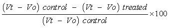
- Preparation of Plant Extract: The leaf-stem of leptadeniahastata was first washed with large amount of water and then dried in a ventilated room, away from dust and direct sunlight. 450grams of the dried leaf-stem was being smashed in 2liters of water. The container was covered with non absorbance cotton wool and placed on a bench in the room to be extracted at room temperature for 72 hours and the contents were properly mixed and filtered, ran through rotary evaporator and kept in a drier until ready for use. Determination of body weight The body weight of each rat was taken before and after the experimental period using a weighing balance. The average weight of each group were taken and recorded.Phytochemical Screening The phytochemical screening was carried out on the leaf-stem extract of leptadeniahastata as per the standard methods to check for the presence or absence of the following chemicals: tannins, anthraquinones, flavonoids, steroids, resins, glycosides, saponins and terpeenoids were qualitatively detected. Mayer’s Test for alkaloids Extract was dissolved in dilute Hydrochloric acid and filtered. The filtrate was treated with Mayer’s reagent (Potassium Mercuric Iodide). Formation of a yellow coloured precipitate indicated the presence of alkaloids (Audu et al, 2007).Modified Borntrager’s Test For cardiac glycosides Extract was hydrolysed with dilute HCl, and then subjected to test for glycosides. The extract was treated with Ferric Chloride solution and immersed in boiling water for about 5 minutes. The mixture was allowed to cool and extracted with equal volume of benzene. The benzene layer was separated and treated with ammonia solution. Formation of rose-pink colour in the ammonical layer indicated the presence of cardiac glycosides (Obasi, et al, 2010). Froth Test for saponinsExtract was diluted with distilled water to 20ml and shaken in a graduated cylinder for 15 minutes. Formation of 1 cm layer of foam indicated the presence of saponins (Harborne, 1998). Test for tannins To the extract, 1% gelatin solution containing sodium chloride was added. Formation of white precipitate indicated the presence of tannins (Harborne, 1998). Test for flavonoidsExtract was treated with few drops of lead acetate solution. Formation of yellow colour precipitate indicated the presence of flavonoids (Audu et al, .2007). Test for terpenoidsExtract was dissolved in water and treated with 3-4 drops of copper acetate solution. Formation of emerald green colour indicated the presence of terpenoids (Obasi, et al, .2010).Test for AnthraquinonesOne gram of the extract was treated with chloroform, then with 5ml sodium hydroxide and 5ml of ammonia. A red or violet or constant yellow colour determined the presence of anthraquinones (Trease and Evans., 1989). Test for Steroidal Rings Two ml of acetic anhydride was added to 0.5ml of the aqueous extract of the sample with 2ml sulphuric acid. Colour changed from violet to blue or green indicated the presence of steroidal rings.Test for Resins An equal volume of copper acetate solution was mixed with the extract solution and shaken vigorously then allowed to separate. A green colour indicated the presence of resins (Sofowora and Adewumi, 1980). All observations were recorded and the number of positive signs indicated the intensity of the reactions that represented the quantity present. Experimental Design Assessment of anti-inflammatory and analgesic activityThis experiment was performed on albino rats (60-70g), obtained National Veterinary Research Institute (NVRI), Vom, plateau state. The animals were acclimatized for 7days before the experiment was performed on them. There were seven groups made up of four albino rats each. Inflammation was induced in all the groups of albino rats with 5% formaldehyde except one group which served as the normal control group. The seven groups were labeled as follows: § Normal Control group: which were fed with normal diet and served as normal control.§ Inflamed control group: Which were induced with formalin and were fed with normal diet.§ Standard control group: Were administered 2mg/kg body weight paraceutamol orally for 7 days in addition to the normal diet and served as the treated control or reference. § Inflamed and treated with 200mg/kg: The animals in this group were administered with 200mg/kg aqueous extract of the leaf-stem of leptadeniahastata for 7 days in addition to normal diet. § Inflamed and treated with 400mg/kg: were administered 400mg/kg aqueous extract of the leaf-stem of leptadeniahastata for 7 days in addition to normal diet. § Inflamed and treated with 600mg/kg: were administered 600mg/kg aqueous extract of the leaf-stem of leptadeniahastata for 7 days in addition to normal diet. § Inflamed and treated with 800mg/kg: were administered 800mg/kg aqueous extract of the leaf-stem of leptadeniahastata for 7 days in addition to normal diet. Determination of Anti-inflammatory Activity of the Leaf-Stem Extract of Leptadenia Hastata.Measurement of paw volume was done by means of volume displacement technique one hour after formalin injection and after the treatment with the leaf-stem extract of leptadeniahastata by the displacement of water. (De Miranda et al., 2000). Briefly, an empty 250ml beaker was first dipped into a 1000ml beaker which contained 500ml of water and the volume of water that was displaced by the empty 250ml beaker was recorded. An albino rat was then placed in the empty 250ml beaker and dipped in the bigger beaker. The volume that was displaced by the empty beaker and the albino rat was also recorded. The volume of the empty beaker was then subtracted from the volume of the albino rat in the beaker. This procedure was repeated for all of the albino rats in the various cages.Reduction in the paw volume compared to the treated control albino rat was considered as the anti-inflammatory response. The percentage inhibition was obtained using the following ratio:
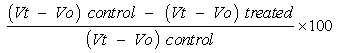 Where Vo represented the average paw volume of the albino rats for each group after inducing with formalin and Vt represented the average paw volume for each group after treatment with the leaf-stem extract (De Miranda et al., 2000).Determination of Analgesic ActivityAn analgesic effect of the plant extract was evaluated by Veerappan et al (2005) method with little modification. It was done concurrently with the determination of anti-inflammatory effect of this plant. After inducing the albino rats with formalin, they were carefully observed for rapid paw licking and paw jumping. Then the leaf-stem extract of leptadeniahastata was administered after one hour and were carefully observed for significant reduction in paw licking and paw jumping after every 30minutes.Estimation of Some Liver enzymes Enzymes used in the assessment of hepatic function include Aspartate transaminase and Alanine transaminase (AST & ALT respectively).Alanine Transaminase: Procedure: Into carefully labeled test tubes, appropriate volumes of the required solution were dispensed as shown in the table below;
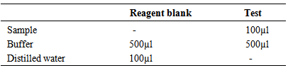 It was mixed and incubated for exactly 30 minutes at 37oC. 500µl of dye reagent was added, mixed and allowed to stand for 20minutes at room temperature. 5.0ml Sodium hydroxide was then added to the reagent blank and the test. It was mixed and the absorbance was read at 546nm of sample against reagent blank after 5 minutes. The ALT activity in serum was obtained from the standard absorbance value table.Aspartate TransaminaseProcedure: Into carefully labelled test tubes, appropriate volumes of the required solutions were dispensed as shown in the table below;
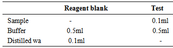 It was mixed and incubated for exactly 30 minutes at 37C. 0.5ml of dye reagent was added to the reagent blank and the test mixed and allowed to stand for 20minutes at room temperature. 5.0ml of Sodium hydroxide was also added to the reagent blank and the test, the absorbance was read at 546nm of sample against reagent blank after 5 minutes. The AST activity in serum was obtained from the standard absorbance value table.Statistical AnalysisThe data were analysed using the SPSS version 15.0. The mean standard errors of means (SEM) of the analysis of samples were calculated. Analysis of variance (ANOVA) was performed to determine significant difference between the means and the P ≤ 0.05s versus the respective control was applied to establish the significant difference.
3. Result
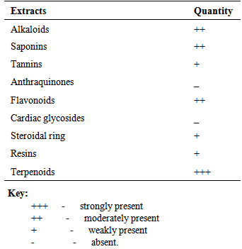
- The leptadeniahastata plant material after extraction with water solvent gave yield of 27.6%. The phytochemical constituents indicated the presence of Alkaloids, Saponins, Tannins, Flavonoids, Steroidal ring, Resins and Terpenoids. It also indicated the absence of Anthraquinones and Cardiac glycosides as shown in table 1.
|
|
|
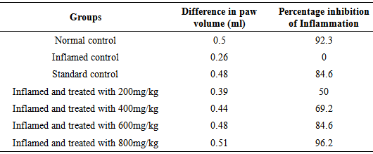
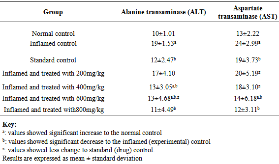
 Where Vo represents the average paw volume of the albino rats for each group after inducing with formalin and Vt represents the average paw volume for each group after treatment with the leaf-stem extract.Analgesic effectAnimal produced paw licking and paw jumping in all groups after inducing with formalin due to the pain. The paraceutamol and the leaf-stem extract of leptadeniahastata at all doses used in the study significantly inhibited the jumping and licking response in mice. The 800mg/kg dose significantly reduced the paw licking and paw jumping response when compared to the reference and the normal control groups.Enzyme activity The results of serum enzyme activity indicated that there was no pronounced elevation of ALT when compared with the normal control group but there was a significant elevation in the inflamed control group. There was a pronounced elevation of AST when compared with the normal control and the reference group. On the hand, the enzyme activity in all the test groups were increased when compared with the normal control experimental groups. This is presented in table 4. However, all the enzymes activities (ALT and AST) were within the acceptable clinical ranges, suggesting that the plant extract is relatively safe for oral medication.
|
4. Discussion
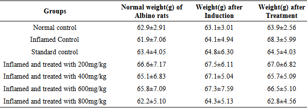
- Results of the phytochemical constituents of leptadeniahastata were presented in table 1. Results obtained showed that terpenoids have the highest phytochemical constituent, followed by saponin, flavonoids, and alkaloids. Steroidal rings, tannins and resins produced the lowest phytochemical constituents. However, the extract was devoid of cardiac glycosides and anthraquinones. The presence of terpenoids, saponin, flavonoids, alkaloids, steroidal ring, tannins and resins indicated that the leave-stem extract of leptadeniahastata possess some medicinal qualities such as anti-inflammatory, analgesic, anti-diabetic and anti-cholesterolemia effect (Aquino et al., 1996). The mean body weight of formalin-induced inflammation in experimental albino rats showed a significant increase when compared with control group as shown in Table 2. This was however reversed after the extract administration. The result for pre-treated groups indicated an initial rapid increase in mean body weight that gradually stabilized with time. Inflammation is characterized by weight loss and body weight loss is associated with increased production of proinflammatory cytokines, such as tumour necrosis growth factor-α (TNF-α) and interleukin-1 (IL-1) (Roubenoff et al., 1997). These cytokines have profound effects on the hormones that govern metabolism and also act directly on the metabolic target organs, such as muscle, liver, gut, and brain (Pomposelli et al., 1988). The result is an increase in resting energy expenditure, a net export of amino acids from muscle to liver, an increase in gluconeogenesis and a marked shift in liver protein synthesis away from albumin and toward production of acute phase proteins, such as fibrinogen and C-reactive protein (Kushner, 1993). Thus the formalin-induced reduction in weight was prevented and it may be due to inhibition of TNF-α and IL-1. The leaf-stem extract of leptadeniahastata are rich in flavonoids. The flavonoids have demonstrated antiproliferative activity, which is found to cause a decrease in the weight and volume of contents of granuloma in inflammation (Koganov et al., 1999).The result of this work showed that the leaf-stem extract of leptadeniahastata significantly inhibited (p ≤ 0.05) the formalin induced inflammation in Albino rats at 200, 400, 600 and 800mg/kg doses. The induced and treated with 800mg/kg test group showed maximum inhibition of inflammation with 96.2% as compared with the reference and normal control group. This could be due to the presence of terpenoids and flavonoids constituents found in this plant. In related development, Nikiema et al (1997) examined triterpenes isolated from Leptadeniahastata latex for their anti-inflammatory activity.Flavonoids as anti-oxidants also have anti-inflammatory properties due to their inhibitory effects on enzymes involved in the production of the chemical mediator of inflammation (Bani et al., 2006). It was discovered that flavonoids have different biological roles. The anti-inflammatory action and analgesic role of flavonoids in vitro or in cellular models involve the inhibition of the synthesis and activities of different pro-inflammatory action mediators such as eicosanoids, cytokines and adhesion molecules and C-reactive proteinGutierrez-Lugo et al., 2004).The presence of prostaglandin in the inflammatory exudates from the injected albino rat has been well demonstrated previously by other workers (Vinegar et al., 1969). The formalin induced inflammation model in albino rats is known to be sensitive to cyclooxygenase inhibitors and has been used to evaluate the effect of non-steroidal anti-inflammatory agents which primarily inhibit cyclooxygenase involved in prostaglandin synthesis (Phadke, 1988). Based on these reports, it is inferred that the inhibitory effect of leptadeniahastata on formalin-induced inflammation in rats in the present day study may be due to inhibition of prostaglandin synthesis. Formalin-induced pain or writhing response in albino rats is a simple and reliable model to evaluate peripheral type of analgesic action of herbal and other drugs rapidly. It was found that leptadeniahastata leaf-stem extract significantly inhibited the formalin induced writhing response at the induced and treated with 800mg/kg test group as compared with the standard control group. It has been reported that the abdominal constriction is related to the sensitization of nociceptive receptors by prostaglandins (Shinde et al., 1999). Therefore, in the present study it is possible that leptadeniahastata leaf-stem extract exerts the analgesic effect probably by inhibiting synthesis or action of prostaglandins.The results of serum liver enzyme activity indicated that there was no pronounced elevation of ALT when compared with the normal control group but there was a significant elevation in the inflamed control group.There was a pronounced elevation of AST when compared with the normal control and the reference group. On the hand, the enzyme activity in all the test groups were increased when compared with the normal control experimental groups. These results clearly indicated that leptadeniahastata leaf-stem extract have a little effect on the liver enzymes of these experimental albino rats.
5. Conclusions
- The phytochemical screening carried out suggested that leptadeniahastata possessed some anti-inflammatory and analgesic effects. The findings of the present study have demonstrated that leptadeniahastata has justified its use in the traditional medicine to treat inflammatory and painful conditions. Based on this, a more detailed study should be carried out for the benefits of the consumers of this local medicinal plant. The plant is relatively safe for medication against inflammations and pains reliever.
6. Recommendations
- 1. There should be increased awareness on the beneficial effects of Leptadeniahastata. 2. Pharmaceutical industry should harness this useful plant to prepare relevant compounds that may be useful in the treatment of a wide variety of other ailments aside inflammation and pain. 3. Clinical and basic researchers are required to investigate more on the pharmacological effects and therapeutic efficacy of Leptadeniahastata.