-
Paper Information
- Paper Submission
-
Journal Information
- About This Journal
- Editorial Board
- Current Issue
- Archive
- Author Guidelines
- Contact Us
Advances in Computing
p-ISSN: 2163-2944 e-ISSN: 2163-2979
2016; 6(1): 6-27
doi:10.5923/j.ac.20160601.02

A New Abnormality Detection Approach for T1-Weighted Magnetic Resonance Imaging Brain Slices Using Three Planes
 Abstract
 Reference
 Full-Text PDF
 Full-text HTML
Mohammed Sabbih Hamoud Al-Tamimi1, 2, Ammar Sabeeh Hmoud Al-Tamimi3, Ghazali Sulong2
1Department of Computer Science, College of Science, University of Baghdad, Baghdad, Iraq
2UTM-IRDA Digital Media Centre (MaGIC-X), Faculty of Computing, Universiti Teknologi Malaysia, Skudai, Johor Bahru, Malaysia
3Department of Computer Engineering, College of Engineering, University of Baghdad, Baghdad, Iraq
Correspondence to: Mohammed Sabbih Hamoud Al-Tamimi, Department of Computer Science, College of Science, University of Baghdad, Baghdad, Iraq.
| Email: |  |
Copyright © 2016 Scientific & Academic Publishing. All Rights Reserved.
This work is licensed under the Creative Commons Attribution International License (CC BY).
http://creativecommons.org/licenses/by/4.0/

Generally, radiologists analyse the Magnetic Resonance Imaging (MRI) by visual inspection to detect and identify the presence of tumour or abnormal tissue in brain MR images. The huge number of such MR images makes this visual interpretation process, not only laborious and expensive but often erroneous. Furthermore, the human eye and brain sensitivity to elucidate such images gets reduced with the increase of number of cases, especially when only some slices contain information of the affected area. Therefore, an automated system for the analysis and classification of MR images is mandatory. In this paper, we propose a new method for abnormality detection from T1-Weighted MRI of human head scans using three planes, including axial plane, coronal plane, and sagittal plane. Three different thresholds, which are based on texture features: mean, energy and entropy, are obtained automatically. This allowed to accurately separating the MRI slice into normal and abnormal one. However, the abnormality detection contained some normal blocks assigned wrongly as abnormal and vice versa. This problem is surmounted by applying the fine-tuning mechanism. Finally, the MRI slice abnormality detection is achieved by selecting the abnormal slices along its tumour region (Region of Interest - ROI).
Keywords: Magnetic Resonance Imaging, Abnormality, Texture features, Threshold, Blocks, Fine-tuning mechanism
Cite this paper: Mohammed Sabbih Hamoud Al-Tamimi, Ammar Sabeeh Hmoud Al-Tamimi, Ghazali Sulong, A New Abnormality Detection Approach for T1-Weighted Magnetic Resonance Imaging Brain Slices Using Three Planes, Advances in Computing, Vol. 6 No. 1, 2016, pp. 6-27. doi: 10.5923/j.ac.20160601.02.
Article Outline
1. Introduction
- Categorically, cancer is the most frightening affliction and brain cancer is the most intricate type to treat. Consequently, good diagnosis and selection of the most appropriate treatment is of great importance. Effective treatments of brain cancers and tumours require the high quality MR images to diagnose the severity of the disease. Lately, MRI has emerged as a useful diagnostic tool for brain and other medical images and it's the most common test for diagnosing and confirming the presence of brain tumour [1-3]. It identifies the tumour location for recommended specialist treatment options [4]. The brain being the most important part of the Central Nervous System (CNS) its structure and function must be thoroughly studied non-invasively by doctors and researchers using MRI imaging techniques [5]. Practically, MR images include both normal and abnormal (defective) slices. Firstly, it must detect the defective or abnormal slices and separate them from the normal slices. To get the true location of the tumour, it is necessary to achieve a precise method for abnormal slices identification by excluding the normal slices. It is customary to discuss the methods for cerebral tissues extraction associated with abnormal slices classification.The experiments were repeated on a set of digitized medical MR images collected from three different standard/challenge dataset is used to fulfill the proposed method, the experiments were repeated with three types of dataset. These three types of dataset comprehensively evaluate the performance of the proposed methods via qualitative and quantitative measures. The first two datasets are obtained via the Internet Brain Segmentation Repository (IBSR) created by the Center for Morphometric Analysis, Massachusetts General Hospital (USA), named IBSR (10Normals_T1) devoid of brain tumour, and IBSR (536_T1) contains brain tumour. These are widely used for brain tumour detection [6-22]. The third dataset named challenge MICCAI (BRATS2012-BRATS-1) is a multimodal Brain Tumour Segmentation (BRATS) challenge that is held in conjunction with the 1st international conference on Medical Image Computing and Computer Assisted Intervention (MICCAI 2012) on October 15th, 2012 in Nice, France. This dataset provides a large number of brain tumour MRI scans in which the tumour regions are manually delineated [23-41]. Moreover, all datasets are free of noise, slice images of each type are 8 bits/pixel greyscale in Digital Imaging and Communications in Medicine (DICOM) file format. They consist of T1-weight sequence in all the three planes (i.e. Axial, sagittal and coronal plane) and contain 4567 MRI slices from 35 patients. For the IBSR (536_T1) and challenge MICCAI (BRATS2012-BRATS-1) datasets the detection of abnormal MRI slices portion ground truth by human experts is available.
2. Related Work
- Radiologists analyse the MR images by visual inspection to detect and identify the presence of tumour or abnormal tissue. The huge number of such images makes this visual interpretation process labour intensive, expensive, and often erroneous. Furthermore, the sensitivity of the human eye and brain to elucidate such images reduces with the increase of number of cases, especially when only a small number of slices contain information of the affected area. Therefore, an automated system for the analysis and classification MR images is essential.Practically, MR images include both normal and defective slices. The likelihood of detecting a premature dementia without using rigid registration of MRI is established [42]. Based on the dissimilarity matrix, a k-Nearest Neighbors (k-NN) classifier is developed. The efficiency and performance of the classifier is tested in a leave-one-out experiment on 58 images. This method achieves an efficiency of 81%. Hybrid techniques consisting of three steps including feature extraction via Discrete Wavelet Transform (DWT), reduce the dimensions size by Principal Component Analysis (PCA) and classification of the outputs using two classifiers are proposed [43]. The Artificial Neural Network (ANN) and k-NN based classifiers are used on dataset comprised of T2-weighted having axial dimension of (256×256) pixels and image size of 70 (with10 normal and 60 abnormal). Remarkably, the number of extracted features is reduced from 1024 to seven using PCA. Accuracy as much as 97% and 98% are achieved from DWT + PCA + ANN and DWT + PCA + k-NN, respectively.In the past, MR brain images are classified using ANN and Support Vector Machines (SVM) method [44]. The pre-processing phase involving DWT is used as input for Neural Network (NN) and SVM. The dataset consisting of T2-weighted, axial, (256×256) pixels MRI, images size 52 with 46 for abnormal (marked by Alzheimer’s disease) and 6 for normal are applied, where 4761 features are extracted. The achieved accuracy of the classifier DWT + Self-Organizing Map (SOM) is 94%, DWT + SVM with linear kernel is 96.15%, DWT + SVM with polynomial kernel is 98.00% and DWT + SVM with radial basis function based kernel is 98.00%. An automatic classification of MR images for normal or abnormal tissues is proposed [45]. This classifier follows two steps such as feature extraction by PCA and classification by the neuro fuzzy. Using an input dataset of size 35 (with 20 as training set and 15 as testing set) the accuracy of 93.33% is achieved. Yet, an accurate differentiation between the abnormal and normal MRI slices requires dedicated research efforts. Thus, it becomes the first objective of the present study.Despite some researcher, achieved high accuracy rate (overall 98%) but the experiments are performed with limited data sets. This paper used standard and challenge dataset to establish a strong basis and constitute a high level of reliability of the proposed net methods.
3. Methodology
- This section demonstrates the new approach to separating the normal slices from the abnormal one in the brain MR images. The proposed method is comprised of four following steps: (1) Division of these tissues into non-overlapping blocks of 8×8 pixels to get a set of features for each block, (2) Determination of the optimal threshold value for splitting the blocks into normal and abnormal types, (3) Detection of abnormal block using the thresholds obtained from the step 2, and (4) Output - detected abnormal slices with its tumour region (ROI).The differentiation of the abnormal MRI slices from the normal one requires the threshold value. The optimal threshold value is obtained from a statistical calculation involving three texture feature parameters such as the mean, energy, and entropy. The abnormal MRI slices can only be obtained once abnormal blocks within the slice are successfully identified. Thus, it is prerequisite to identify the abnormal blocks and refine them in order to obtain an accurate abnormal slice. Figure 1 illustrates the flowchart of the proposed method for the detection of abnormal MRI slices.
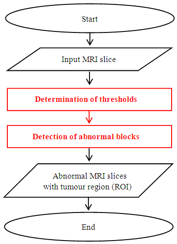 | Figure 1. Flowchart of the proposed method for the detection of abnormal MRI slices |
3.1. Determination of Thresholds
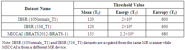
- The optimal threshold values are determined using statistical calculations based on the three parameters: mean, energy and entropy before the abnormality detection began. To get the optimal threshold values, finding the relationship between these features and their distribution in the MRI slice is essential. It is worth noting that all MRI slices have a similar technical specification because all of them for each patient case are obtained from the same MRI device. Furthermore, the optimal threshold value that separates healthy region from the cancerous region is calculated and determined. As mentioned earlier, each MR image consisted of a set of slices. Thus, it is necessary to choose only those slices that contain more information about cerebral tissues (Gray Matter (GM), White Matter (WM), Cerebrospinal fluid (CSF) and tumour (if any)) in the same MR image. To meet such challenges the probability calculation is carried out to select the informative slice in each MRI image, which contained all information about the cerebral tissues. Normally, the GM, WM and CSF exist in all brain slices, but the presence of the tumour is not necessary in all the slices. However, some of them reveal the presence of the tumour in the MR image, where the selection the slice having a high probability of tumour occurrence is prerequisite. The probability distribution (frequency of occurrence) of the tumour in the slice “i” is obtained via the “P” function given by [46], [47],
 | (1) |
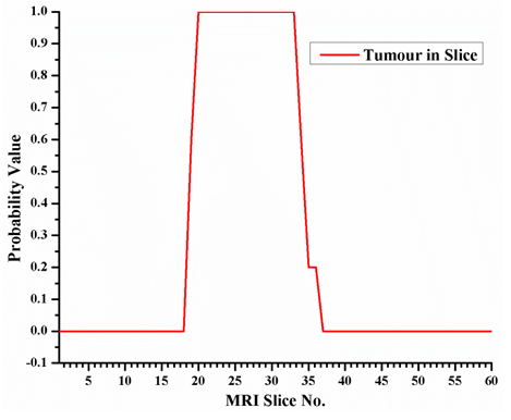 | Figure 2. The probability of the tumour occurrence in MR image slices of IBSR (536_T1) datase |
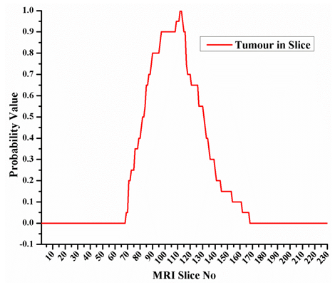 | Figure 3. The probability of the tumour occurrence in MR image slices of challenge MICCAI (BRATS2012-BRATS-1) dataset |
3.1.1. Features Extraction
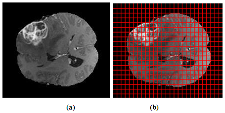
- Firstly, each of the chosen slices is partitioned into non-overlapping blocks of (8×8) pixels. This size of block is considered optimal since is empirically chosen by a set of experiments using various block sizes ranging from (4×4) to (16×16) pixels. Then, for each block, three statistical features mean, energy and entropy, are extracted to determine three threshold values, namely Mean-threshold (T1), Energy-threshold (T2), and Entropy-threshold (T3). Figures 4 and 5 depict two different examples of non-overlapping block division for MRI brain slice using (8×8) block.
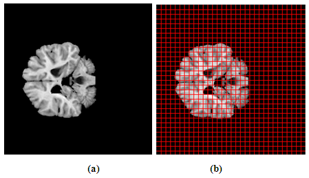 | Figure 4. Non-overlapping block partition of patient Normal_19 of IBSR (10Normals_T1) dataset, (a) Original slice 23, and (b) Non-overlapping block division using (8×8) block size |
 | Figure 5. Non-overlapping block partition of patient BRATS_HG0009 of challenge MICCAI (BRATS2012-BRATS-1) dataset, (a) Original slice 103, and (b) Non-overlapping block division using (8×8) block size |
 | (2) |
 | (3) |
 | (4) |
3.1.2. Mean, Energy and Entropy Distribution
- As mentioned earlier, three features such as mean, energy, and entropy are extracted from each block. A correlation between these features and their distribution in the MRI slice is determined to obtain the optimal threshold values. As previously explained (Section 3.1), the experiment is executed by taking the middle slice from each MR image, where 35 MRI slices from three data sets are selected and manually divided into non-overlapping (8×8) sub-image blocks. Each block was labelled manually either as Normal (N) or Abnormal (A), as illustrated in Figure 6. The next step is to calculate the mean, energy, and entropy for each block using the Equations (1), (2), and (3) respectively.
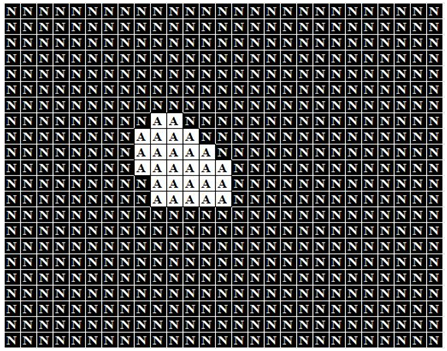 | Figure 6. Manual identification of slice 88 for patient BRATS_HG0015 of challenge MICCAI (BRATS2012-BRATS-1) dataset into Normal (N) and Abnormal (A) regions |
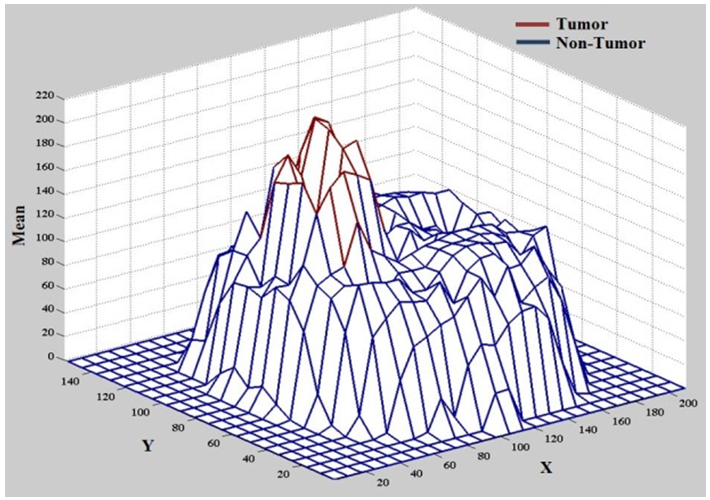 | Figure 7. Distribution of mean of slice 88 for patient BRATS_HG0015 of challenge MICCAI (BRTAS2012-BRATS-1) dataset |
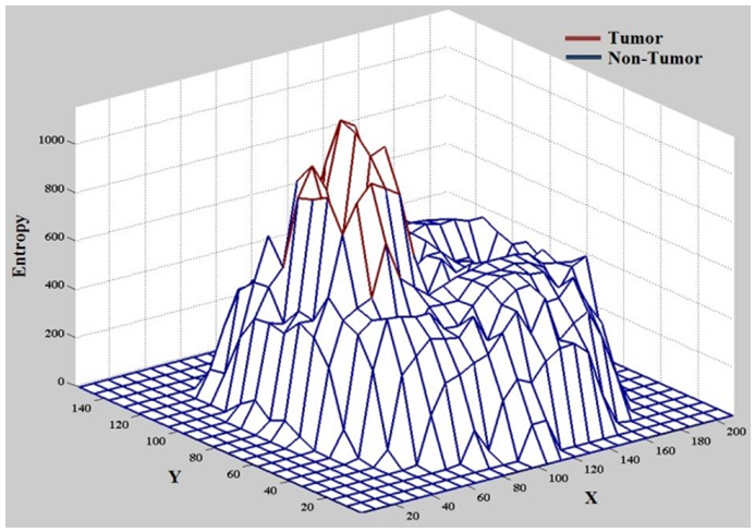 | Figure 8. Distribution of entropy of slice 88 for patient BRATS_HG0015 of challenge MICCAI (BRTAS2012-BRATS-1) dataset |
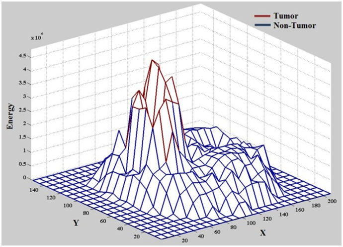 | Figure 9. Distribution of energy of slice 88 for patient BRATS_HG0015 of challenge MICCAI (BRTAS2012-BRATS-1) dataset |
3.1.3. Mean, Energy and Entropy Correlation
- Figure 10 shows the first combination (mean, energy and entropy). The brown stars signify the tumour region and blue one symbolizes the non-tumour zone. The figure clearly shows that when the entropy increases the tumour parts are clearly visible and well separated from non-tumour zones. This indicates that this combination of the three features performs superbly in separating the tumour from non-tumour region – an excellent feature combination for finding the tumour.
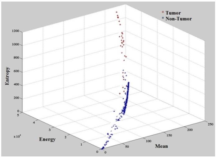 | Figure 10. Relation among mean, energy, and entropy of slice 88 for patient BRATS_HG0015 of challenge MICCAI (BRTAS2012-BRATS-1) dataset |
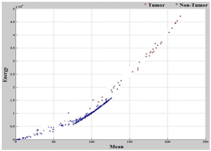 | Figure 11. The relationship between mean and energy of slice 88 for patient BRATS_HG0015 in challenge MICCAI (BRTAS2012-BRATS-1) dataset |
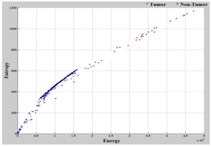 | Figure 12. The relationship between entropy and energy of slice 88 for patient BRATS_HG0015 in challenge MICCAI (BRTAS2012-BRATS-1) dataset |
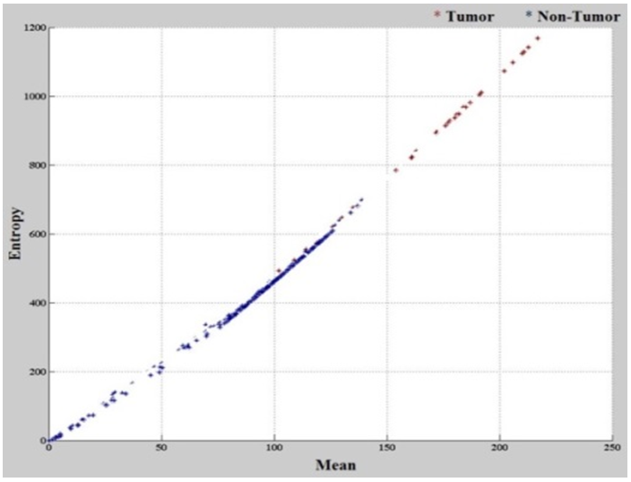 | Figure 13. The relationship between entropy and mean of slice 88 for patient BRATS_HG0015 in challenge MICCAI (BRTAS2012-BRATS-1) dataset |
|
3.2. Detection of Abnormal Blocks
- Once the threshold values have been identified, the following process is to determine abnormal blocks within each MRI slice. It is obvious that if abnormal blocks are found in a particular slice, then the slice is considered as an abnormal slice. Thus, this section utilises the above thresholds, T1, T2 and T3, to detect the abnormal blocks. A block is labelled as abnormal if and only if all the rules are satisfied. The rule is given in Figure 14.
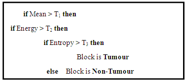 | Figure 14. Abnormal block detection rules |
3.2.1. Fine-tuning Mechanism of Abnormal Blocks
- Undoubtedly, the above abnormality rule is simple yet efficient enough to deliver initial results. However, we cannot confirm the achieved results are 100% correct - some blocks of tumour slice images may be captured as a part of the non-tumour region, and vice versa. It is evident that the inaccuracy in the tumour block affects the results of abnormality detection of the slice. Thus, this study introduces a new technique called fine-tuning mechanism to check, validate and improve the initial abnormal block. The fine-tuning mechanism is based on three types of (3×3) block masks, namely Preceding, Current, and Succeeding Block Mask as displayed in Figure 15.
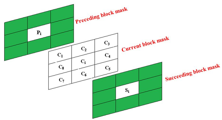 | Figure 15. Three types of block masks with size (3×3) called preceding mask centred at Pi, current mask centred at Ci, and succeeding mask centred at Si. Green colour indicates ignored block |
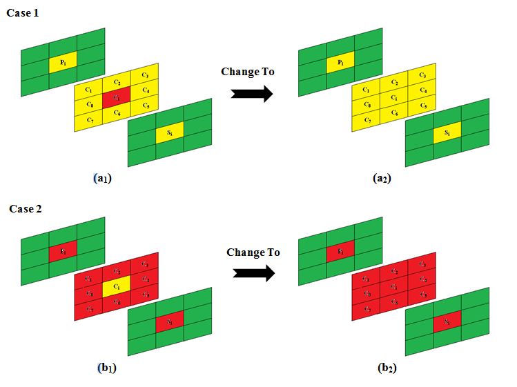 | Figure 16. Fine-tuning of Case 1: (a1-a2), and Case 2: (b1-b2), where the mechanism changes the block Ci based on its neighbours |
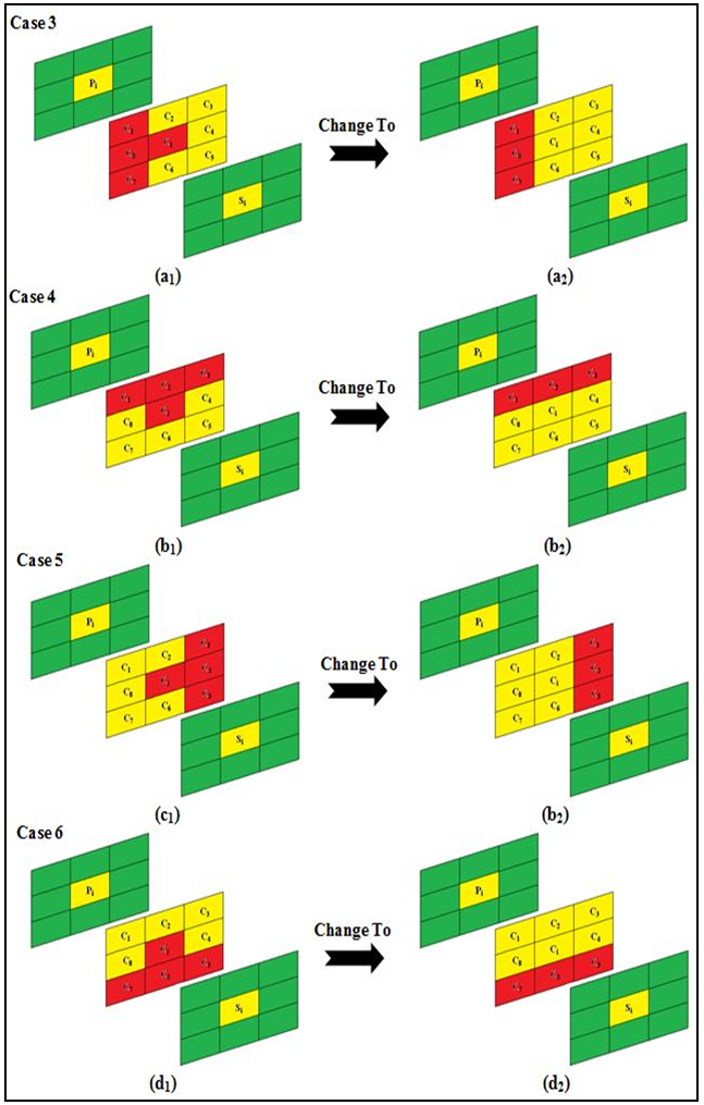 | Figure 17. Fine-tuning of (Case 3 - Case 6), where Ci is changed from tumour to a non-tumour block using the Rule 1 |
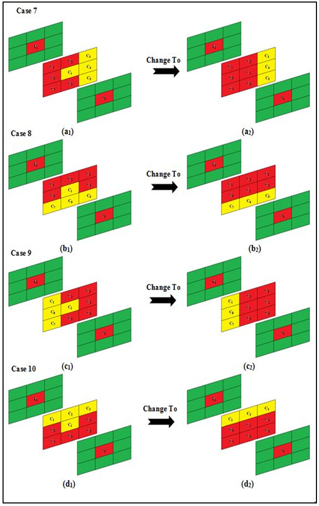 | Figure 18. Fine-tuning of (Case 7 - Case 10), where Ci is changed from non-tumour to a tumour block using the Rule 2 |
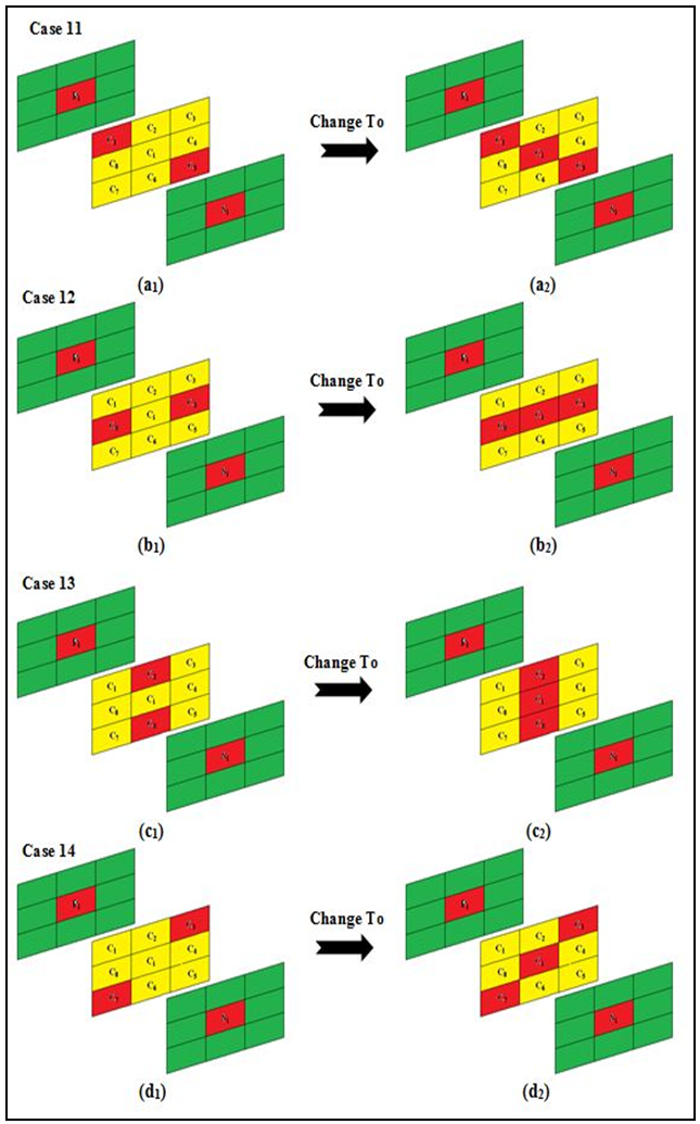 | Figure 19. Fine-tuning of (Case 11 - Case 14), where Ci became a non-tumour block using the Rule 3 |
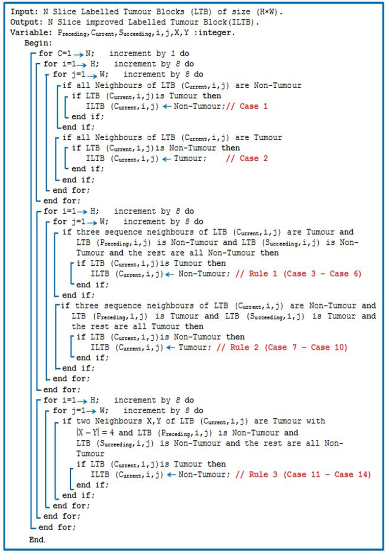 | Algorithm 1. The fine-tuning mechanism |
4. Result and Discussion
- Abnormality detection of MRI slices is performed by conducting a sequence of experiments to evaluate the performance of the proposed abnormal MRI slice detection. The assessment includes: (1) Abnormal block detection (before and after the fine-tuning mechanism), and (2) The abnormal slice detection. The three above-mentioned standard datasets together with the ground truth are used for the evaluation. The performance of the proposed method is measured in both qualitative and quantitatively, and the experimental results are presented hereunder.
4.1. Results of the Abnormal Block Detection before the Fine-tuning
- Three features such as mean, energy and entropy are extracted from each block. The relationship between these features and their distribution in the MRI slice are analysed using Three-Dimensional (3D) graphs. Three different thresholds are established to represent these extracted features. The rules established for the extraction of the ROI tumour region from MRI slice are based on these three thresholds (Section 3.1).The performance of the current method is evaluated with the same visual inspection procedure where MRI slices with different contrast are examined. Figures 20, 21 and 22 shows the experimental results of the block classification using the three data sets, where the left column represents the original MRI slices image, the centre is the (8×8) block division using the proposed method and the right column is block detection result where the blue colour signifies the normal region (non-tumour area) and the other blocks implies the abnormal area (tumour area).
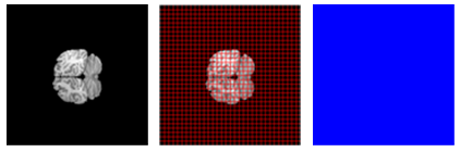 | Figure 20. Abnormal block detection results of IBSR (10Normals_T1) dataset |
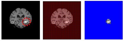 | Figure 21. Abnormal block detection results of IBSR (536_T1) dataset with tumour marked by a red circle |
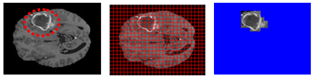 | Figure 22. Abnormal block detection results challenge MICCAI (BRATs2012-BRATS-1) dataset with tumour marked by a red circle |
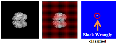 | Figure 23. Misclassified blocks of IBSR (10Normals_T1) dataset without any tumour |
 | Figure 24. Misclassified blocks of IBSR (536_T1) dataset with tumour marked via green circle |
 | Figure 25. Misclassified blocks of challenge MICCAI (BRATS2012-BRATS-1) dataset with tumour marked via green circle |
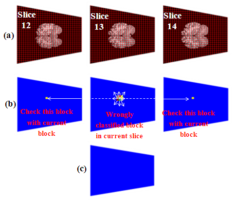 | Figure 26. A fine-tuning A fine-tuning result of tumour block slice 13 of patient Normal_7 of IBSR (10Normal_T1) dataset: (a) The original non-tumour slice 13 (in grid), (b) Fine-tuning of misclassified block of the current slice, and (d) Final result of the questioned block |
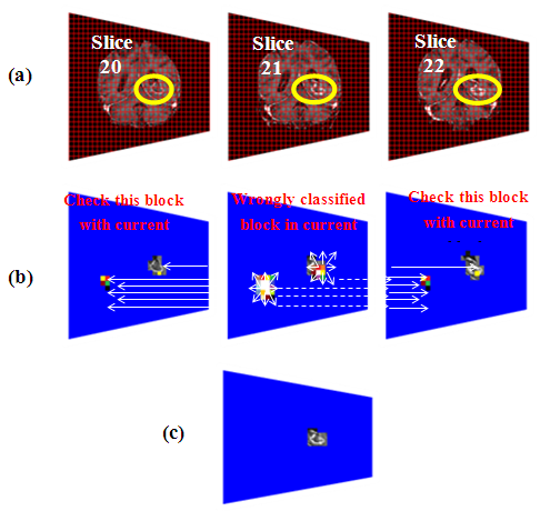 | Figure 27. A fine-tuning result of six tumour blocks (two separate locations) of slice 21 of MRI scan 536_47 from IBSR (536_T1) dataset: (a) The original tumour slice 21 (in grid), (b) Fine-tuning of misclassified blocks of the current slice, and (d) Final result of the questioned blocks |
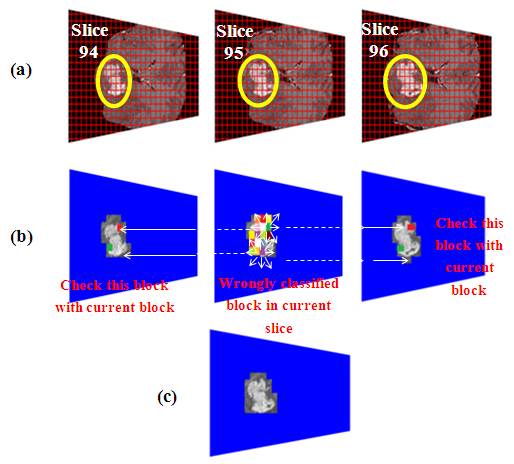 | Figure 28. A fine fine-tuning result of two tumour block of slice 95 of patient BRATS_HG0015 from challenge MICCAI (BRATS2012-BRATS-1) dataset: (a) The original tumour slice 95 (in grid), (b) Fine-tuning of misclassified blocks of the current slice, and (d) Final result of the questioned block |
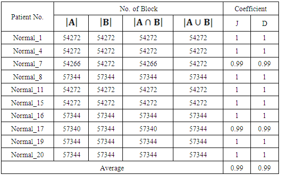
4.2. The Quantitative Assessment of the Tumour Block Detection
- Continuing the above section, discussions are now shifted to quantitative assessment to reaffirm the above findings. For that reason, the same datasets are utilised. Two types of measures, the Jaccard (J) coefficient [50], and the Dice (D) coefficient [51] are used to validate the proposed method. The coefficient of "J" and "D" is used to evaluate the performance of the proposed abnormal MRI slice detection method. The expression for "J" and "D" are given by:
 | (6) |
 | (7) |
|
 | Figure 29. Block abnormality detection results of the IBSR (536_T1) dataset |
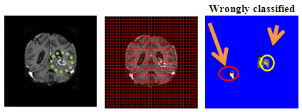
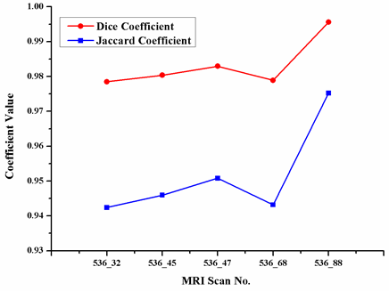
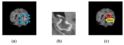 | Figure 30. The misclassified MRI scan 536_45 of IBSR (536_T1) dataset, (a) Original MRI slice 22 with tumour inside the marked blue Square, (b) Zoomed in marked area, and (c) Misclassified blocks, where the red circle indicates wrongly classified blocks and yellow circle represents the tumour area |
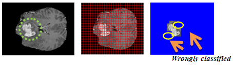
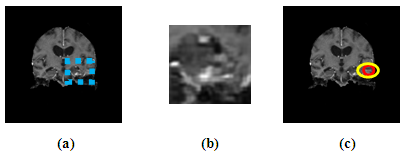 | Figure 31. The misclassified MRI scan 536_47 of IBSR (536_T1) dataset, (a) Original MRI slice 31 with tumour inside marked blue Square, (b) Zoomed in marked area, and (c) Misclassified blocks, where the red circle indicates wrongly classified blocks and yellow circle represents the tumour area |
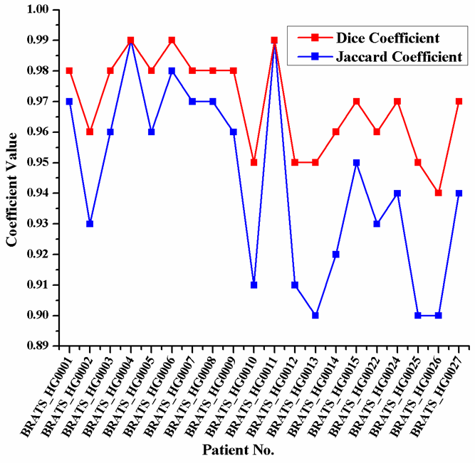 | Figure 32. The Jaccard and Dice coefficients similarity indices for each MRI patient in challenge MICCAI (BRATS2012-BRATS-1) dataset |
4.3. Quantitative Evaluations of Slice Abnormality Detection
- Upon completion of the assessment of the abnormal block detection, further evaluation is required to assess the performance of the proposed abnormal slice detection method. Therefore, a different set of measurements is used, namely sensitivity, specificity and accuracy to evaluate the performance of brain tumour abnormal MRI slice detection method. The formulae are as follows [52], [53]
 | (8) |
 | (9) |
 | (10) |
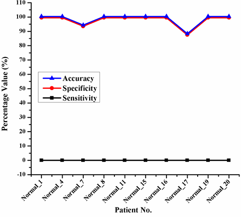 | Figure 33. Experimental results of slice abnormality detection of IBSR (10Normals_T1) dataset |
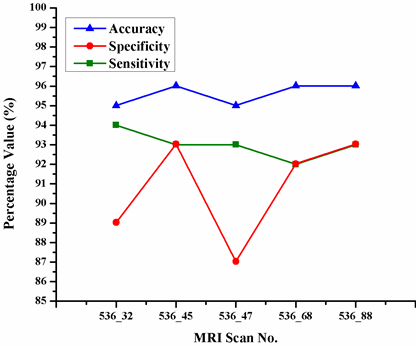 | Figure 34. Experimental results of abnormality detection by slice obtained from the IBSR (536_T1) dataset |
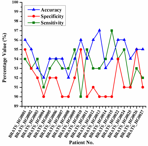 | Figure 35. The sensitivity, specificity and accuracy of each MRI patient obtained from challenge MICCAI (BRATS2012-BRATS-1) dataset |
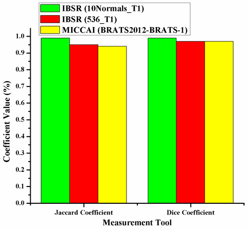 | Figure 36. The measured Jaccard coefficient and Dice coefficient for the three distinct datasets: IBSR (10Normals_T1), IBSR (536_T1), and challenge MICCAI (BRATS2012-BRATS-1) |
5. Conclusions
- This study summarizes the above findings of the abnormal block detection, as displayed in Figure 36, to evaluate the overall performances of the proposed method. The figure reveals that the proposed method performed extremely well in all circumstances regardless of datasets.Based on the above findings, it can be summarized that the proposed abnormal MRI slice detection method, which include abnormal block detection, has performed superbly regardless of datasets (see Figure 37).
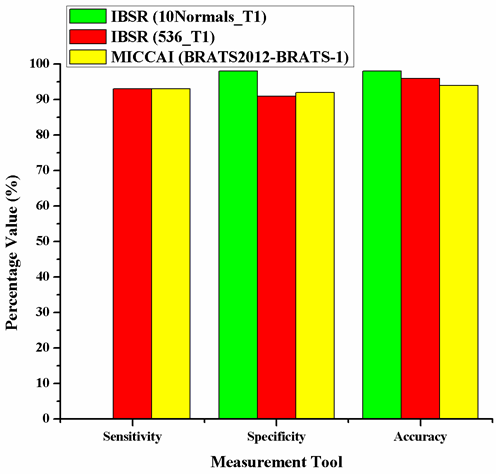 | Figure 37. The Results for the sensitivity, specificity and accuracy among the three used datasets |
ACKNOWLEDGEMENTS
- The author(s) would like to thank the Ministry of Higher Education and Scientific Research-Iraq and University of Baghdad-Iraq for providing the financial support and facilities for this research.