Advances in Analytical Chemistry

p-ISSN: 2163-2839    e-ISSN: 2163-2847

2018;  8(2): 15-21

doi:10.5923/j.aac.20180802.01


Synthesis, Characterization and Antimicrobial Activity of V(IV), Ag(I) and Cd(II) Complexes with Mixed Ligands Derived from Sulfamethoxazole and Trimethoprim

Maysoon M. Abdul Hassan1, Adnan H. Abbas1, Eman H. Abed1, Entisar E. Abodi2

1Ministry of Science and Technology, Environment and Water Directorate, Baghdad, Iraq

2Department of Chemistry, College of Education for Pure Science (Ibn El-Haithm), University of Baghdad, Baghdad, Iraq

Correspondence to: Maysoon M. Abdul Hassan, Ministry of Science and Technology, Environment and Water Directorate, Baghdad, Iraq.

Email:

Copyright © 2018 The Author(s). Published by Scientific & Academic Publishing.

This work is licensed under the Creative Commons Attribution International License (CC BY).
http://creativecommons.org/licenses/by/4.0/

Abstract

A series of new metal complexes were prepared by mixing 4-amino-N-(5-methylisoxazole-3-yl)-benzene -sulfonamide (L1) as a chelating ligand in the presence of the co-ligand trimethoprim (L2), with Vanadium (V), Cadmium (Cd) and Silver (Ag) ions in alcoholic medium. The complexes were characterized in solid state by using flame atomic absorption, elemental analysis C.H.N.S, FT-IR, UV-Vis Spectroscopy, conductivity and magnetic susceptibility measurements. Tetrahedral geometry was suggested for CdL1L2, AgL1L2 complexes, while VL1L2 complex has a square pyramidal. The ligand L1, with the metal ions, was clearly behaving as a bidentate through O and N atoms of sulfonylamid, while L2 behaves as a bidentate ligand through N and N atoms for all the complexes. Conductivity has shown that all the preparations complexes are ionized, and the nature of bonding between the metal ion and the donor atoms of the ligands was demonstrated by calculating the ligand field parameters by using suitable Tanaba-Sugano diagrams. Biological activity of V(IV), Cd(II), Ag(I), TMP, and SMX, with different concentrations (50, 100, 250, 500, and 1000)ppm, were studied respectively, complexes with chelating ligand and co-ligand were evaluated against Staphylococcus aureus, Pseudomonas aeruginosa and Escherichia coli, after incubating for 24hr at 37ºC. Results showed that L1 and L2 enhance effect of (V) against growth of (Staph. aureus and E. coli) with concentration (50, 100, 250, 500, and 1000)ppm, while Ps. aeruginosa was sensitized to (Cd) and (Ag) with concentration more than (250)ppm, after incubated for 24hr at 37ºC. Whereas, the SMX is exposed to a failure in its efficiency against Staph. aureus, Ps. aeruginosa, and E. coli.

Keywords: Sulfamethoxazole, Trimethoprim, Chelating agent, Antibacterial activity

Cite this paper: Maysoon M. Abdul Hassan, Adnan H. Abbas, Eman H. Abed, Entisar E. Abodi, Synthesis, Characterization and Antimicrobial Activity of V(IV), Ag(I) and Cd(II) Complexes with Mixed Ligands Derived from Sulfamethoxazole and Trimethoprim, Advances in Analytical Chemistry, Vol. 8 No. 2, 2018, pp. 15-21. doi: 10.5923/j.aac.20180802.01.

1. Introduction

Sulfamethoxazole (SMX) (4-amino-N-(5-methylisoxazol -3-yl)-benzenesulfonamide) belongs to the sulfonamides group of chemotherapeutics, with formula C10H11N3O3S and molecular weight 235.3gm/mol (Fig. 1) (Barragry, 1994), while Trimethoprim (TMP) 5-(3, 4, 5-trimethoxybenzyl) pyrimidin-2,4-diyldiamine, with formula C14H18N4O3 (TMP) and molecular weight 290.3gm/mol (Fig. 2) (Goodman and Gilman, 1980). The medicinal use of SMX is for treatment of tuberculosis, malaria and urinary tract infections (Katzung, 1998), while TMP is a synthetic broad spectrum antibiotic used in treating urinary tract infections, respiratory infections and middle ear infections (Brogden et al., 1982; Bax et al., 2000; Margassery and Bastani, 2002). SMX was inhibited dihydrofolic acid bacterial synthesis by competing with paraaminobenzoic acid, whereas TMP blocks the production of tetrahydrofolic acid from dihydrofolic acid by binding to and reversibly inhibiting dihydrofolate reductase. The synergistic activity of a combination of sulfamethoxazole and trimethoprim against a wide variety of gram-positive and gram-negative bacteria has been well established (Soheilian et al., 2005). The presence of donor atoms (N,S,O) at various positions in sulfamethoxazole and trimethoprim molecules enable them to behave as multidentate ligands and thus form chelates of diverse structural types with a wide range of metal ions (Rothova et al., 1993). The chemistry of metal complexes with heterocyclic compounds containing nitrogen, sulfur, and /or oxygen as ligand atoms has attracted increasing attention which exhibit enhanced bactericidal, fungicidal, herbicidal, and insecticidal activities in addition to their application as potential drugs (Srivastava, 1989).
The research goal to study antibacterial activity of Vanadium, Cadmium and Silver complexes with ligands derived from Sulfamethoxazole and Trimethoprim.
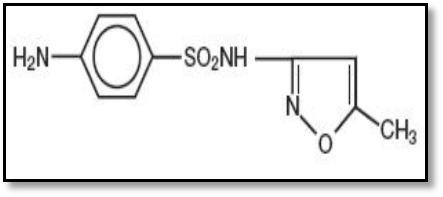
Figure (1). Structural formula of (SMX)
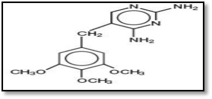
Figure (2). Structural formula of (TMP)

2. Experimental

Physical measurement
Elemental C.H.N.S analysis were carried out on a EM-017mth instrument, the FT-IR spectra in range (4000-400 cm-1) and (400-200 cm-1) were recorded by using KBr and CsI respectively, discon IR-Prestige-21, Single beam path Laser, Shimadzu Fourier Transform infrared Spectrophotometer, UV-Visible spectra were measured using UV-1650PC Shimadzu, range (200-1100)nm. The magnatic susceptibility values of the complexes were obtained at room temperature using Magnatic Suscceptibility Balance of Johanson mattey catalytic system division, while atomic absorption measurements were obtained by using Shimadzu Atomic Absorption at (680) Flame Spectrophotometer. The conductivity values of the complexes were measured by using 0.001M DMF as a solvent, (WTW) Conductometer. Melting point apparatus of Barnstead Electrothermal BI was used to measure melting points for all the compounds.
Materials and methods synthesis of metal complexes
An ethanolic solution of the sulphamethoxazole as a primary (L1)1mmole (0.2533)gm and 1mmole (0.29032)gm of trimethoprim (L2) as a co-ligand, were added slowly, into warm ethanolic solution of metal salts 1mmole [VOSO4.H2O (0.1809gm), AgNO3 (0.16987gm) and Cd(NO3)2.4H2O (0.3084gm)]. The mixture solution was heated and refluxed with stirring for about (2.5-3)hr. The colored precipitates were filtered, washed several times with ethanol and finally ether, and dried using desiccators. The ligands and their metal complexes were characterized by physical and analytical methods such as flame atomic absorption, elemental analysis C.H.N.S, FT-IR, UV-Vis Spectroscopy, conductivity and magnetic susceptibility measurements.
Antimicrobial assay
The antimicrobial activity of the ligands and synthesized complexes were studied using the agar diffusion technique. The surface of Moller-Hinton agar in a petri dish (90mm diameter) was uniformly inoculated via spreading 100μl of bacterial suspension (Staphylococcus aureus, Pseudomonas aeruginosa and Escherichia coli) (24hr old), with concentration (1×10⁹) cells/ml, approximately (Macfarland solution). Then 0.1 ml of each concentration (50, 100, 250, 500 and 1000)ppm of (L1), (L2), and complexes with metals (V), (Cd), and (Ag) preparing by DMSO were submerged into the wells (5mm diameter), the plates were leaved for 30min before incubation at 37°C for 24hr, then inhibitory zone (mm) were measured indicating to antibacterial activity.

3. Results and Discussion

Chemistry
Stable complexes were isolated in all cases based on the metal analysis, spectroscopic data, molar conductance and magnetic susceptibility studies. The general formula of the complexes can be depicted as:
[VOL1L2]SO4. H2O,[AgL1L2](NO3). H2O and [CdL1L2] (NO3)2. H2O.
The analytical data together with some physical properties of the complexes are summarized in (Table 1).
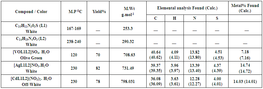
Table (1). Some analytical and physical data of primary ligand (L1) with co-ligand (L2) and their metal complexes
FT-IR spectra of ligands (L1), (L2), and its metal complexes
The relevant infrared for the primary ligand (SMX) and the secondary ligand (TMP) together with metals as a metal complexes (Table 2). (SMX) is a potentional ligand which may act as a bidentate or tridentate as illustred by its structure so it expected that IR measurements are highly indicating with respect to the complexetion behivour with various metal ions. Infrared spectrum of the free ligand of (L1) and (L2) shows two strong bands at (3466 and 3377)cm–1 (3468 and 3317)cm–1 corresponding to the asymmetric and symmetric stretching vibrations, respectively of the aromatic amino group [11]. The medium and strong band which appeared for (L1) and (L2) at (3298 and 3118)cm–1, respectively are due to the presence of asymmetric sulfonamide (-NH) and a weak band at (3143)cm–1 as symmetric frequency. Another band observed at (1622)cm–1 is related to methoxazole ring stretching vibration for (L1). Others two bands appeared at (1382 and 1157)cm–1 are due to asymmetric and symmetric stretching frequencies of sulfonyl group (Huheey et al., 1993). The stretching frequencies of the (C-N) band of sulfonamide are exhibited in the (1311)cm–1 region. The bands related to aromatic amino group undergoes as positive shift to higher or lower frequencies at (3469, 3450 and 3435)cm–1 in positions of V (IV), Cd (II), and Ag (I) complexes because of resonance contribution and also H-bonding (Stuart, 2004) indicating bonding of this ion to aniline N-atom (Nakomato, 2009). The multiband and shifting of sulfonamide (-NH) in the spectra of the prepared complexes, indicating the involvement of this group in chelation with central metal ion by nitrogen of this group according to the data reported in literature (Melina et al., 2013). The band related to methoxazole ring stretching vibrations suffered a very slight shift in range of (±5)cm-1 in the spectra of the Cd (II), and Ag (I) complexes indicating that the methoxazole moiety is not participation in coordination with metal ions (Robert et al., 2015). The bands corresponding to asymmetric of sulfonyl group undergoes a shift toward higher frequencies which observed at (1384-1386)cm–1 in Ag (I), Cd (II) complexes, but V (IV) a shift toward lower frequencies at (1365)cm–1 while a small bands of the symmetric stretching suffered of a shift toward higher frequencies about (3-18)cm–1 in all complexes except V (IV) complex undergo lower frequency at (2)cm-1. According to these results, the coordination mode of this ligand with metal ions is clearly predicted as a bidentate through the (O,N) atoms of sulfonylamid group for all complexes more evidence new bands which appeared in the range (414-443)cm-1 and (482-551)cm-1 due to the stretching frequency of (M-O) and (M-N) bonds, respectively. The coordination of the secondary ligand is indicated by the positive shift of (C=C), (C=N) ring stretching frequencies (Casanova et al., 1993; Barragry, 1994). The position of this band has been completely changed in the spectra of all the complexes and confirming the coordination nature of co-ligand, the band at (424)cm-1 observed of TMP (C-C) out of plane bending shifts to higher frequency and splits into two components in the complexes which again confirms the coordination of secondary ligand through two nitrogen (Shakir and Azim, 2005), the new bands were recorded at (260-265)cm–1 in the spectra of all complexes are attributed to (M-NTMP) bond indicating participation two nitrogen coordination with metal ion (Majeed et al., 2010).
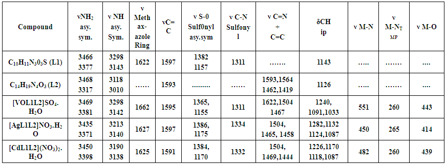
Table (2). The most diagnostic FTIR bands of the mixed ligand SMX as primary ligand (L1) and TMP as co-ligand (L2) and their metal complexes in (cm-1)
Electronic spectral and magnatic moment studies
The electronic spectrum of the primary ligand (L1) exhibited two absorption bands in the ultraviolet region, the band at (212)nm assigned to the (π→ π*) transition for the intera ligand aromatic system (C=C) and a strong absorption band at (270)nm which refer to (n→π*) transition for oxygen atom of (S=O) group or nitrogen atom of amine moiety and imine (N=C-) group, respectively (Gulcan et al., 2012). The Electronic spectrum of (L2) shows two main bands the first appeared at (249.45)nm due to inter a ligand (π-π*) transition located on (C=C) group. The second absorption appeared at (290.5)nm arises from (n-π*) transition may be located on nitrogen atom of imine (-N=C-) group (Table 3) (Figgis and Hitchman, 2000).
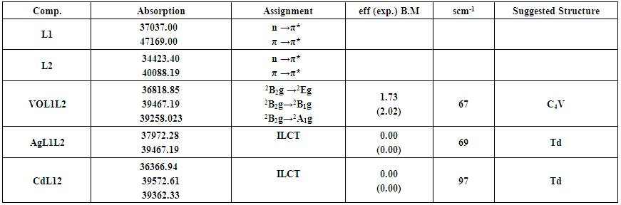
Table (3). Electronic spectra, conductance in DMF solvent and magnetic moment (B.M) for the prepared ligand L1, co-ligand L2 and their metal complexes
Electronic spectrum of V(IV) complex
The spectrum of V (IV) complex shows three bands, at (36818.85, 39467.19 and 39258.02)cm-1 which corresponds to (2B2g→2Eg, 2B2g→2B1g and 2B2g→2A1g) transitions, respectively. These are in accordance with the proposed square pyramidal geometry of V (IV) (Gulcan et al., 2012). The magnetic moment of this complex is (2.02) B.M. This is in correlation with the spin only magnetic moment obtained for this complex with a single unpaired electron (Figgis and Hitchman, 2000). The magnetic moment value is higher than the spin value of vanadium ion only. This result indicates a higher orbital contribution which is in accordance with the published data for square pyramid vanadium complexes (Hashim and Alais, 2012). Conductivity measurements showed the ionic behavior of this complex.
Electronic spectrum of Ag (I) complex
In this work, diamagnetic of the silver complex according to the electronic spectrum of Ag (I) complex, no (d-d) transition is located as it belongs to (d10). The exhibited bands at (37972.28 and 39467.19)cm-1 may be due to charge transfer from the donor atoms of ligand to the silver ion (L→AgCT), tetrahedral geometry can be suggested. The conductivity measurement for this complex showed to be ionic.
Electronic spectrum of Cd (II) complex
According to the electronic spectrum of Cd (II) complex, no (d-d) transition is located as it belongs to (d10) (Julia et al., 2015). The prepared complex is off white in color with the diamagnetic being expected. The ultraviolet-visible spectrum of this complex shows a relative change in the bands position compared to that of free ligands due to charge transfer between Cd (II) ion and ligands. The conductivity measurements for the prepared complex showed to be ionic. From spectroscopy and this information (Td) geometry can be suggested (Fig. 3).
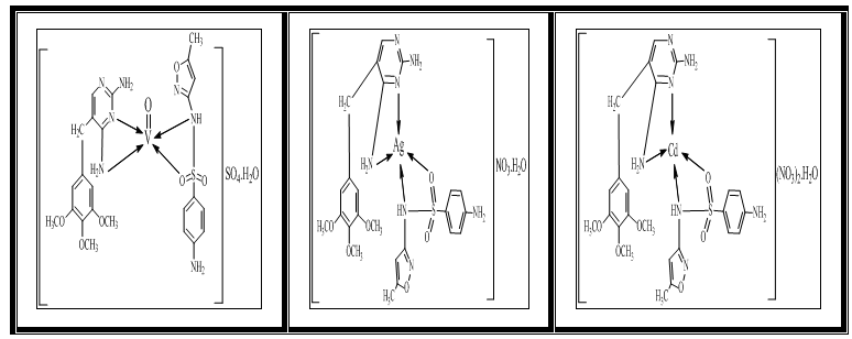
Figure (3). Suggested structure of the prepared complexes

4. Antimicrobial Activity

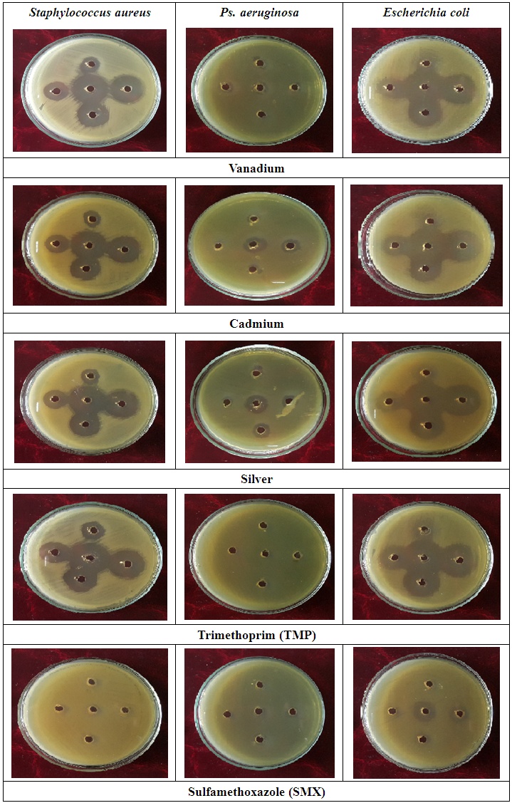
Antibacterial activity of ligands and there metal complexes vanadium, cadmium, and silver, complexes have been tested by disc diffusion technique. The various gram positive bacteria (Staph. aureus) and gram negative bacteria (Escherichia coli and Ps. aeruginosa), were used for antimicrobial activity. Inhibition zone (mm) were measured after incubating for 24hr at 37°C. Results showed that (SMX) is exposed to a failure in its efficiency against Staph. aureus, Ps. aeruginosa, and E. coli (Fig. 4).
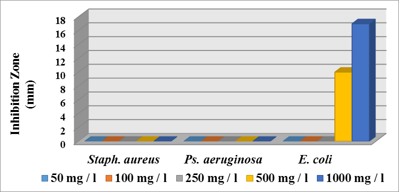
Figure (4). Effect different concentration of SMX on the various genus of bacteria
Whereas, (L1) and (L2) enhance effect of (V), (Cd), and (Ag) against growth of (Staph. aureus and E. coli) with concentration (50, 100, 250, 500, and 1000)ppm, while Ps. aeruginosa was sensitized to (Cd) and (Ag) with concentration more than (250)ppm, after incubated for 24hr at 37ºC (Fig. 5, 6, 7, and 8).
Figure (5). Effect different concentration of materials on the growth of Staph. aureus
As in the case of many other elements, small amounts of vanadium are beneficial to the growth and development of animals and, according to the suggestion of Nilsen and Uthus (1990), also human body, although there is no strong evidence that vanadium is an essential trace element for human. Its deficiency in mammals inhibits growth, impairs the generative functions, thyroid metabolism and bone mineralization, and disturbs the lipid and car- bohydrate balance. Hence, vanadium is a necessary ingredient of the daily diet (Moskalyk and Alfantazi, 2003). On the other hand, larger doses taken as a variety of compounds are toxic and noxious, causing irritation of the eyes and mucous membranes of the upper respiratory duct, coughing, fatigue and depression.
Figure (6). Effect different concentration of materials on the growth of E. coli
Many antibacterial agents inhibit steps that are important for the formation of peptidoglycan, the essential component of the bacterial cell wall. The cadmium salts themselves are poisonous, but the stability of the complexes prevents the liberation of (Cd) ions in the test media. Additionally, cadmium salts are also poisonous for human being, whereas ligands and complexes are not (Foni et al., 2014).
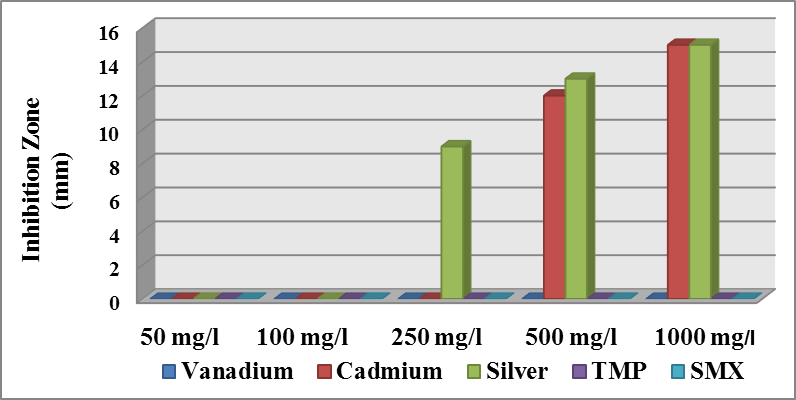
Figure (7). Effect different concentration of materials on the growth of Ps. aeruginosa
Silver was also proposed to act by binding to key functional groups of enzymes. Silver ions cause the release of K+ ions from bacteria; thus, the bacterial plasma or cytoplasmic membrane, which is associated with many important enzymes, is an important target site for silver ions (Schreurs and Rosenberg, 1982). In addition, they inhibited cell division and damaged the cell envelope and contents of bacteria (Richard et al., 1984). Bacterial cells increased in size, and the cytoplasmic membrane, cytoplasmic contents, and outer cell layers all exhibited structural abnormalities. Finally, silver ions interact with nucleic acids, they interact preferentially with the bases in DNA rather than with the phosphate groups, although the significance of this in terms of their lethal action is unclear (Thurman and Gerba, 1989).
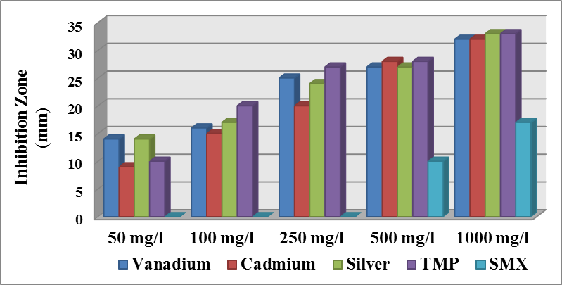
Figure (8). Inhibition zone (mm) of metal ions complexes with (L1) and (L2) against bacteria

References

[1]  Barragry, T.B. (1994). Sulfonamides. Veterinary Drug Therapy. Lea & Febiger, Philadelphia, Baltimore, Hong Kong, London, Munich, Sydney, Tokyosayfa Chapter 12: 297.
[2]  Bax, R.N., Mullan, and Verhoef, J. (2000). Int. J. Antimic. Agents 16; 51-59.
[3]  Brogden, R.N., Carmine, A.A., Heel, R.C., Speightand, T.M. and Avery, G.S. (1982). Trimethoprim: a review of its antibacterial activity, pharmacokinetics and therapeutic use in urinary tract infections. Drugs, 23: 405-430.
[4]  Casanova, J., Alzuet, G., Ferrer S., Borrás, J., García- Granda, S,. and Perez-Carreño, E. (1993). Metal Complexes of Sulfanilamide Derivatives. Crystal Structure of [Zn(Sulfathiazole)2]·H2O, J. Inorg. Biochem., 51, 689.
[5]  Figgis, B.N., and Hitchman, M.A. (2000). Ligand field theory and its Application. Wiley-VCH, New York, Singapore, Toronto.
[6]  Foni, B.B., Tapashi, G.R., Atiar, R., and Talha, B.E. (2014). An in vitro antibacterial and antifungal effects of cadmium(II) complexes of hexamethyltetraazacyclotetradecadiene and isomers of its saturated analogue. Asian Pac. J. Trop. Med. 7(Suppl 1): S534-S539.
[7]  Goodman and Gilman, (1980). The Pharmaceutical Basis of Therapeutics. New York, 1116-1119.
[8]  Gulcan, M., Sonmez, M., and Berber, I. (2012). Synthesis, characterization and antimicrobial activity of a new pyrimidine Schiff base and its Cu(II), Ni(II), Co(II), Pt(II) and Pd(II) complexes. Turkey Journal of Chemistry, 36: 189-200.
[9]  Hashim, C.S., and Alais, M.F. (2012). Synthesis, spectroscopicstudy of Pt (IV), Au (III), Rh (III), Co (II) and V (IV) complexes with sodium[5-(p-nitro phenyl)-/4-phenyl-1,2,4-triazole-3-dithiocarbamatohydrazide] and cytotoxicity assay on rhabdomyosarcoma cellline of heavy metals. J. Baghdad Sci., 9(4).
[10]  Huheey, J.E., Keiter, E.A., and Keiter, R.L. (1993). Inorganic Chemistry, Principles of Structure and Reactivity, 4th ed. New York: Harper Collins College Publishers.
[11]  Julia, H.B.N., Raphael, E.F., Alexander, C., Wilton, R.L., and Pedro, P.C. (2015). Silver complexes with sulfathiazole and sulfamethoxazole: Synthesis, spectroscopic, crystal structure and antibacterial assays. Journal Polyhedron 85: 437-444.
[12]  Katzung, B.g. (1998). Basic and Clinical Pharmacology. Appleton and Lange, 7th ed. Lange Medical Books"; Mc, Graw-Hill New York: 761-762.
[13]  Majeed, A., Yousif, E., and Farina, Y. (2010). Synthesis and Characterization of Transsition MetalComplexes of 2-Thioacetic Acid Benzothiazolle Ligand. J. Al-Nahrain University Science, 13(1): 36-42.
[14]  Margassery, S. and Bastani, B. (2002). Trimethoprim/ Sulfamethoxazole in the Treatment of Bacteriuria in Women. J. Nephrol, 14: 410-414.
[15]  Melina, M., Fernando, P., Paula, C. de Souza, Clarice, Q., Leite, J.E., Otaciro, R., Nascimento, G.F., María, H., and Torre. (2013). Study of A Series of Cobalt(II) Sulfonamide Complexes: Synthesis, Spectroscopic Characterization, and Microbiological Evaluation Against M. Tuberculosis. Crystal Structure of [Co(Sulfamethoxazole)2(H2O)2]·H2O, J. Mol. Struct., 1036: 180-187.
[16]  Moskalyk, R.R., and Alfantazi, A.M. (2003). Processing of vanadium: a review”, Miner. Engineer., Vol. 16: 793-805.
[17]  Nakomato, N. (2009). Infrared and Raman Spectra of Inorganic and Coordination Compounds, 6th ed., John Wiley and Sons, Inc., New Jersey.
[18]  Nielsen, F.H., and Uthus, E.O. (1990). The essentiality and metabolism of vanadium”, In: N.D. Chasteen (Ed.): Vanadium in Biological Systems, Kluwer Academic Publishers. Dordrecht, The Netherlands, pp: 51-62.
[19]  Richards, R.M.E., Odelola, H.A., and Anderson, B. (1984). Effect of silver on whole cells and spheroplasts of a silver resistant Pseudomonas aeruginosa. Microbios 39: 151-158.
[20]  Robert, M., Silverstein, F.X. Webster, and David, J.K. (2015). Spectrometric Identification of Organic Compound, 7th ed. Jon-wiley. New York.
[21]  Rothova, A., Meenken, C., Buitenhuis, H.J., Brinkman, C.J., Baarsma, G.S., Boen-Tan, P.T., Klaassen-Broekema, N., Schweitzer, C.M., and Timmerman, Z. (1993). Am.T.N. J. Ophthalmol. 115(4):517-23.
[22]  Schreurs, W.J., and Rosenberg, H. (1982). Effect of silver ions on transport and retention of phosphate by Escherichia coli. J. Bacteriol., 152: 7-13.
[23]  Shakir, M., and Azim, Y. (2005). Metal Directed Synthesis of Hexaazamacrocyclic Complexes of Co (II), Ni (II), Cu (II) and Zn (II) and Their Physico-Chemical Studies, Polish J. Chem., 79: 1115-1121.
[24]  Soheilian, M., Sadoughi, M.M., Ghajarnia, M., Dehghan, M.H., Yazdani, S., Behboudi, H., Anisian, A., and Peyman, G.A. (2005). Ophthalmology. 112(11): 1876-82. Epub. Sep 19.
[25]  Srivastava, R.S. (1989). Inorganic Chemistry, Acta, 56(1): 65.
[26]  Stuart, B. (2004). Infrared Spectroscopy, John Wiley and sons Ltd, England.
[27]  Thurman, R.B., and Gerba, C.P. (1989). The molecular mechanisms of copper and silver ion disinfection of bacteria and viruses. CRC Crit. Rev. Environ. Control, 18: 295-315.