Advances in Analytical Chemistry

p-ISSN: 2163-2839    e-ISSN: 2163-2847

2015;  5(1): 8-16

doi:10.5923/j.aac.20150501.02

A Validated Stability Indicating RP-HPLC Method for Simvastatin, Identification and Characterization of Forced Degradation Products of Simvastatin Using LC-ESI-MS/MS

Sanjay Sawant, Minal Ghante

Sinhgad Technical Education Society’s Smt. Kashibai Navale College of Pharmacy, Maharashtra, India

Correspondence to: Sanjay Sawant, Sinhgad Technical Education Society’s Smt. Kashibai Navale College of Pharmacy, Maharashtra, India.

Email:

Copyright © 2015 Scientific & Academic Publishing. All Rights Reserved.

Abstract

The objective of the present study was to report the stability of Simvastatin (SMV), a HMG-CoA reductase inhibitor, based on forced degradation studies. SMV was subjected to forced hydrolytic (acidic, alkaline and neutral), oxidative, photolytic and thermal stress in accordance with the ICH guideline Q1A (R2). SMV was found to degrade significantly in all stress conditions except photo degradation. Resolution of the drug and degradation products was achieved on a Hi-Q Sil C-18 column (4.6 × 250 mm, 5 μm) utilizing acetonitrile, methanol and phosphate buffer (65:25:10% v/v/v) of pH 4 at a flow rate of 1.2 ml/min and at the detection wavelength 237 nm. The major acidic stress degradation product was characterized by LC-ESI-MS/MS and its fragmentation pathway was proposed. Validation of the liquid chromatographic (LC) method was carried out in accordance with ICH guidelines. The method met all required criteria and was applied for analysis of commercially available tablets viz. Simvas 40.

Keywords: Simvastatin, Stress degradation, Stability indicating assay method, HPLC, LC-MS

Cite this paper: Sanjay Sawant, Minal Ghante, A Validated Stability Indicating RP-HPLC Method for Simvastatin, Identification and Characterization of Forced Degradation Products of Simvastatin Using LC-ESI-MS/MS, Advances in Analytical Chemistry, Vol. 5 No. 1, 2015, pp. 8-16. doi: 10.5923/j.aac.20150501.02.

1. Introduction

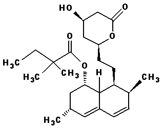
Stability testing is now-a-days the key procedural component in the pharmaceutical development program for a new drug as well as new formulation. Drugs undergo physicochemical degradation upon storage. Pharmaceutical companies perform forced-degradation studies (stress testing) during preformulation to help in selection of compounds and excipients for further development, to facilitate in salt selection or formulation optimization, and to produce samples for developing stability-indicating analytical methods. Thus, stability testing of a drug under various temperature and humidity conditions is indispensable during the drug development process. A stability-indicating method is “a validated quantitative analytical procedure that can detect the changes with time in the pertinent properties of the drug substance and drug product”. A stability-indicating method accurately measures the active ingredients, without interference from degradation products, process impurities, excipients, or other potential impurities. Stability testing guidelines issued by International Conference on Harmonization (ICH) and other international agencies [1–5] require the reporting, identification and characterization of degradation products (DPs). Tandem mass spectrometry (MSn) and LC coupled with mass spectrometry (LC-MS, LC-MS/MS) are becoming the most versatile techniques for characterization of pharmaceutical DPs and impurity profiling [6]. Simvastatin (SMV) is HMG-CoA reductase inhibitor used for the treatment of dyslipidemia and the prevention of cardiovascular diseases [7]. It is chemically known as (1, 3, 7, 8, 8a) -8-{2-[(2R, 4R)-4-hydroxy-6-oxotetrahydro2H- pyran-2yl] ethyl}-3, 7-dimethyl-1, 2, 3, 7, 8.8a-hexahydronaphthalen-1-yl-2, 2-dimethyl butanoate as shown in Figure 1.
Figure 1. Chemical structure of Simvastatin
SMV is a prodrug which is converted into its β- hydroxy acid form which inhibits HMG CoA reductase enzyme (3-hydroxy-3-methylglutarylcoenzyme), a rate limiting enzyme in the synthesis of cholesterol in liver [8]. The drug is officially listed in US Pharmacopeia, British Pharmacopeia and European Pharmacopeia. SMV can be estimated by UV spectrophotometry [9-25], RP-HPLC [24-40] HPTLC [41-45] and LC-MS/MS [46-53] alone or in combination with other drugs. Till-date, two official methods utilizing HPLC Gradient methodology are reported in European Pharmacopoeia (EP) [54] and United State Pharmacopoeia (USP) [55]. Literature survey reveals that, till date, there are many stability indicating RP-HPLC [24-41] methods and determination of SMV in biological samples by LC-MS [46-53] however the identification of the degradation compounds of SMV found during forced degradation studies needs to be carried out. The present manuscript describes the (i) degradation behavior of SMV under hydrolysis (acid, base and neutral), oxidation, photolysis and thermal stress conditions, (ii) optimization of LC conditions to separate the drug and its DPs on a reversed-phase C18 column, (iii) method validation, (iv) characterization of DPs and (v) proposed fragmentation pathway of DPs using LC-ESI-MS/MS.

2. Experimental

2.1. Chemicals and Reagents

Simvastatin was supplied by Gen Pharma Ltd; Pune, India. Acetonitrile (HPLC grade) was procured from Thomas Baker Ltd; India and used without purification. Analytical reagent grade (AR) hydrochloric acid, sodium hydroxide pellets, hydrogen peroxide solution (30%) were purchased from Omkar Traders (Mumbai, India). Ultrapure water was obtained from water purification unit Elga option Q/OQ007XXm1 (Elga Ltd., Bucks, England). Buffer materials and all other chemicals were of AR grade. The marketed pharmaceutical tablet dosage form of SMV i.e. SIMVAS 40 by Micro Labs, India was purchased from local market.

2.2. Instrumentation

An HPLC system used for analysis of stressed samples consisted of quaternary pump (PU-2089), solvent mixing module (MX-2080-31), multi-wavelength PDA detector (MD-2018), interface box (LC-NET ΙΙ/ADC), rheodyne manual injector (7725i, USA) with 20 μl capacity, chromNAV data system software 1.8.1.6 version (all from Jasco, Tokyo, Japan) were used.
In all studies, separations were achieved on Hi-Qsil C-18 (4.6 × 250 mm, 5 μm) column (Thermo Scientific, Japan). Carousel six stage reaction station (Radleys Tech, UK) was used for generating hydrolytic DPs. The thermal degradation study was performed using a high precision hot air oven (Pathak Electrical Works, Mumbai, India) capable of controlling temperature within ± 2℃. Photo degradation study was carried out in a photostability chamber (Labin, Mumbai, India).
A pH meter (Equip-tronics, Mumbai, India) was used to check and adjust the pH of buffer solution. Also sonicator (UCB40, Spectra-lab, Mumbai, India) and precision analytical balance (ME 204, Mettler Toledo Group, India) were used in the present studies.
The HPLC-MS analyses were carried out on an Agilent 1200SL Series liquid chromatographic system interfaced to an Agilent 6410B Triple Quad LC-ESI-MS/MS system (Mass HunterData Acquisition, Qualitation and Quantitation soft-ware, USA) equipped with an Agilent XDB-C column (1.8µm, 4.6×50mm) at a column temperature of 40°C was used for the confirmation of atomic mass number of unknown compound/s formed during forced degradation studies in acidic and oxidative stress conditions in mobile phase containing a mixture of acetonitrile, methanol and phosphate buffer (65:25:10% v/v/v) of pH 4 adjusted with ortho-phosphoric acid, pumped at flow rate of 1.2 ml/min.
MS (500-MS IT) system consisted of direct infusion mass with positive as well as negative APCI ionization (+APCI and -APCI) modes, mass ranging from 50-2000 m/z. The system was controlled by 500-MS Workstation software. In LC-MS studies the separation was carried out on Hypersil Gold C-18 (4.6 x 250 mm, 5 µm).

2.3. Stress Decomposition Studies

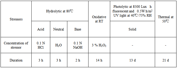
Forced degradation studies of bulk drug and drug formulation included appropriate solid state and solution state stress conditions in accordance with regulatory guidelines. The stressors, choice of their concentration and preparation of samples were based on published guidelines (56). As the drug was insoluble in water, it was dissolved in mobile phase; acetonitrile, methanol and phosphate buffer (65:25:10% v/v/v) of pH 4 to a final concentration of 2 mg/ml. The stock was diluted 50:50 (v/v) with the stressor (e. g. HCl, NaOH, H2O2 and water). All hydrolytic studies were conducted at 60℃. The oxidative study was carried out in 3% (v/v) H2O2 at 60℃. For thermal stress testing, the drug was sealed in glass vial and placed in a thermostatic block at 50℃ for 21 days. Photolytic studies on the drug in the solid and solution state were carried out by exposure to a UV lamp in a chamber set at accelerated conditions of temperature and humidity (40℃/75% RH). Parallel blank set was kept in dark for comparison. After subjecting to stress, samples were withdrawn at appropriate time interval. The optimized stressed conditions are enlisted in Table 1.
Table 1. Optimized stress conditions for the drug
     

2.4. Sample Preparation for HPLC and LC-MS Analysis

The stressed samples of acid and base hydrolysis were neutralized with NaOH and HCl, respectively to obtain 1000 μg/ml solutions. Neutral hydrolysis, thermal and photolytic samples were diluted with mobile phase to obtain concentration at 1000 μg/ml solutions. The oxidative stress sample was diluted with mobile phase to obtain 100 μg/ml solution. All the prepared samples were passed through 0.45 μm membrane filter before HPLC and LC-MS analyses.
Characterization of Degradation Products
The stressed solutions, in which sufficient amounts of products formed, were subjected initially to LC-PDA and further to LC-MS analyses for characterization of DPs.

3. Results and Discussion

3.1. LC-MS Conditions

The main aim of present research work was to separate SMV and its DPs. An Hi-Q-sil C-18 column (4.6 × 250 mm, 5 μm) was found to be suitable for this analysis after having tried with different columns. During the optimization process on above-mentioned column, several conditions with various mobile phases like methanol/water and acetonitrile/water in different proportionalities were tried in an isocratic mode. The peaks corresponding to DPs did not resolve completely and tailing was observed. To get acceptable separation between the drug and its DPs, potassium dihydrogen ortho-phosphate buffer was used. Further, studies were carried out using varied proportions of acetonitrile (A), methanol (B) and potassium dihydrogen ortho-phosphate buffer (C). The pH of the buffer, flow rate and composition of the mobile phase were systematically varied to optimize the method. To detect drug and DPs with sufficient peak intensity, the wavelength at 237 nm was chosen. Finally, a mobile phase consisting of A, B and C (pH 4.0; 0.01M) (65:25:10% v/v/v) at a flow rate of 1.2 ml/min and PDA detection at 237 nm, in an isocratic mode gave good separation of drug and its DPs. The advantage of the method was simple and rapid. Validation of the optimized LC method was done with respect to various parameters outlined in ICH guideline Q1A (R2) [56] and was extended to LC–MS studies.

3.2. LC-MS Studies

LC-MS/MS studies were carried out in +APCI ionization mode in the mass range of 50-2000 amu. High purity helium was used as carrier gas and nitrogen was used as nebulizer. Mass parameters were optimized to the following values: Rf loading: 80%; capillary voltage: 80 volts; syringe volume: 250 μL; spray chamber temperature: 50℃; nebulizer pressure: 35psi; drying gas temperature: 300℃; drying gas pressure: 10 psi; vaporizer gas temperature: 350℃; vaporizer gas pressure: 20 psi; spray shield voltage: ± 600.0 volts.

3.3. Evaluation of Method Validation Parameters

3.3.1. Specificity
Specificity is the ability of the analytical method to measure the analyte concentration accurately in presence of all potential DP. The specificity was determined by subjecting SMV to stress degradation under various conditions. The DP was well separated, peak purity assessment was carried out on the stressed samples of SMV by using diode-array-detector and the specificity was also established by subjecting the degradation sample to LC–MS analysis using same method. The mass detector also showed an excellent mass purity for SMV and its DP which unambiguously proves the specificity of the method.
3.3.2. Linearity
Linearity test solutions were prepared from stock solution at six concentration levels of analyte (20, 40, 60, 80, 100 and 120 μg/ml). The peak area versus concentration data was performed by least squares linear regression analysis. The calibration curve was drawn by plotting SMV average area for triplicate injections and the concentration expressed as a percentage. Linearity was checked over the same concentration range for three consecutive days. Good linearity was observed in the concentration range from 20 to 120 μg/ml of SMV. The data was subjected to statistical analysis using a linear regression model; the linear regression equation and correlation coefficient (r2) were y = 28528x + 99441 and 0.9999 respectively. These results indicate estimable linearity. The LOD and LOQ for SMV were estimated at a signal-to-noise ratio of 3:1 and10:1, respectively. The LOD and LOQ were 6.80 μg/ml and 20.61 μg/ml respectively.
3.3.3. Precision
Precision of the method was verified by repeatability and intermediate precision studies. Repeatability studies were carried out by analyses of three different concentrations of the drug in hexaplicate on the same day. Intermediate precision of the method was checked by repeating the studies on three different days. Additionally, the developed HPLC method was checked through separation studies on the mixture of reaction solutions on a different chromatographic system on a different day. The results of repeatability and intermediate precision experiments are shown in Table 2. The developed method was found to be precise as the RSD values for repeatability and intermediate precision studies were < 1.5. Separation of the drug and its DP in a mixture of stressed samples was found to be similar when analyses were performed on a different chromatographic system on different days.
Table 2. Precision studies
     
3.3.4. Accuracy
Accuracy of the method was assessed employing the standard addition method at three different levels (80%, 100%, 120%). The mixtures were analysed in triplicate and the percentage of added drug obtained from difference between peak areas of unfortified and fortified samples of SMV. The HPLC area responses for accuracy determination are depicted in Table 3. Good recoveries (99.46± 0.23) of the spiked drugs were obtained at each added concentration, indicating that the method was accurate.
Table 3. Recovery studies
     
3.3.5. Robustness
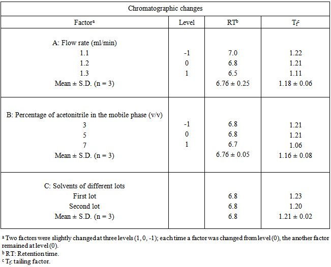
To determine the robustness of the method, experimental conditions were purposely altered. Three parameters selected were flow rate, composition of mobile phase and solvent from different lots. The mobile phase flow rate was 1.2 ml/min which was changed to 1.1 and 1.3 ml/min and the effect was studied. The effect of mobile phase composition was analyzed by use of acetonitrile, methanol and potassium dihydrogen ortho-phosphate buffer (pH 5.0; 0.01M) and in ratio of 64:24:12 (v/v/v) and 66:26:8 (v/v/v). Also acetonitrile and methanol of different lots from same manufacturer was used. For all changes in conditions, the sample was analyzed in triplicate. When the effect of altering one set of conditions was tested, the other conditions were held constant at the optimum values. In all the calculated varied chromatographic conditions, no significant change in retention time and tailing factor of SMV was observed. The summary of results is shown in Table 4.
Table 4. Robustness studies
     
3.3.6. System Suitability Test
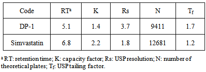
The system suitability parameters with respect to theoretical plates, capacity factor, resolution factor, tailing factor were calculated and are given in Table 5. It could be seen from table that all the peaks were well resolved.
Table 5. HPLC system suitability parameters
     
3.3.7. Degradation behaviour
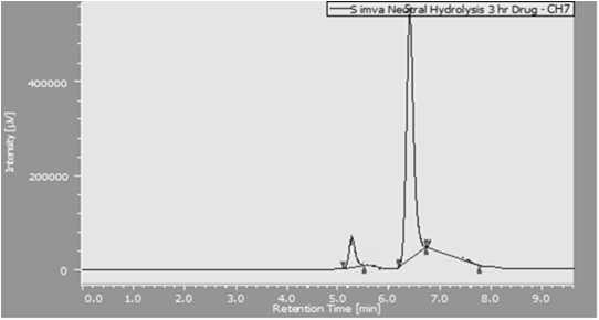
The drug degraded into DP under all stress conditions except photo degradation. There was insignificant photo degradation (< 0.45%) with no degradation product. The respective extent of degradation for acid, base, oxidative, neutral and thermal was 53.16, 44.65, 44.25, 53.13 and 38.93% respectively under these conditions. The chromatograms of the degraded samples in stressed is shown in Figure 2.
Figure 2. Peak of marketed formulation
3.3.8. Study of the Stability of Commercial Tablets
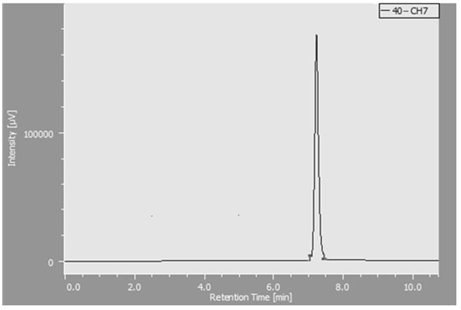
The assay content of SMV, commercially available marketed formulation was analyzed by the proposed method after exposure to accelerated storage condition (i.e. 40℃/ 75% RH). The peak at retention time 6.8 min for the drug was observed in the chromatogram of the drug samples extracted from tablets and no additional peak was found Figure 3. Experimental results of the amount of SMV in tablets, expressed as percentage of label claim were in good agreement with the label claims as reported in Table 6 thereby suggesting that there is no interference from any excipients, which are normally present in tablets and packaging material is of good quality which is reported in.
Figure 3. Peak of marketed formulation
Table 6. Study of stability of commercial tablets (n = 3)
     
3.3.9. Mass Fragmentation Pathway of the Drug
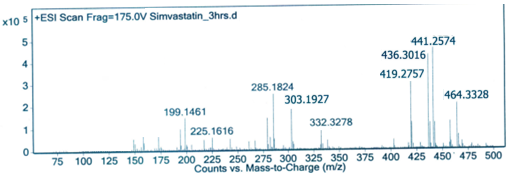
SMV is a lactone based compound and hence the mass fragmentation pathway of the drug was established from results of LC/APCI/MS in positive ion mode and MS/MS analysis using optimized mass parameters. In all stress conditions except photo degradation, SMV showed common degradation peak around the retention time 5 min, only drug solution under acidic conditions was considered for identification of that common degradation compound with the help of ESI-MS/MS. Peak at m/z ratio of 419.2754 amu (M+1)+ while the most intense peak at m/z ratio of 441.2573 (M+Na+) and at m/z ratio of 436.30 (M+NH4)+ as parent ion or molecular peak ions may be due to adduct ionization for SMV same as observed in other reported methods of LC-MS and LC-MS/MS [40,43,45 & 47]. The major daughter ions or fragmentation ion peaks at m/z ratio of 303.1927, 285.1823 & 199.1462amu were obtained corresponds to loss of ester side chain from SMV (m/z 303 amu) and then subsequently loss of water molecule (m/z 285 & 199amu) as shown in fragmentation pattern, Figure 4 [43, 47].
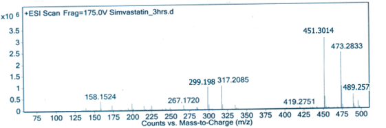
Figure 4. Mass spectrum of degradation compound in acidic stress conditions
3.3.10. Postulated Structure of the Degradation Products
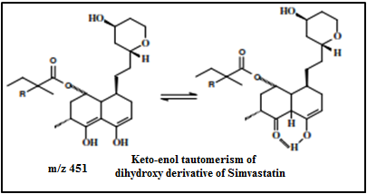
Fig 5 shows typical fragmentation pattern for common degradation compound found in acidic stress conditions for SMV. It was observed that promising ion peak at m/z ratio 451amu in acidic condition showed an increase in 32amu than that of SMV (m/z 419.27). This data indicated an insertion of two oxygen atoms suggesting that there might be keto-enol tautomerism, which is in agreement with m/z 451, attributed to dihydroxylated SMV derivative, Figure 5. Proposed compounds refer to keto-enol tautomerism - a chemical equilibrium between a keto form (a ketone or an aldehyde) and an enol. The interconversion of the two forms involves the movement of a proton and the shifting of bonding electrons; hence, the isomerism qualifies as tautomerism.
Figure 5. Mass spectrum of degradation compound in acidic stress conditions
Figure 6. Keto enol tautomerism

4. Conclusions

It was possible in this study to develop a stability-indicating RP-HPLC assay method for SMV by subjecting the drug to ICH recommended stress conditions. DP was formed in all conditions except photo degradation and was separated in a single run, by an isocratic LC method. The method proved to be simple, accurate, precise, specific and robust. It was successfully employed for the analysis of marketed formulation stored for three months under accelerated conditions of temperature and humidity. The developed method was extended to LC-MS/MS for characterization of DPs. The degradation product was identified as 2 dihydroxy derivative of SMV. The complete degradation pathway of the drug and mechanism of the formation of DP-1 is proposed. The proposed method can thus be used for routine analysis, quality control and for studies of pharmaceutical tablets containing this drug.

ACKNOWLEDGEMENTS

Authors are thankful to Prof. M. N. Navale, Founder President, Sinhgad Technical Education Society, for constant encouragement and support.

References

[1]  International Conference on Harmonization (ICH), Q1A (R2): Stability testing of new drug substances and products, IFPMA, Geneva, 2003.
[2]  World Health Organization (WHO): Draft stability testing of active pharmaceutical ingredients and pharmaceutical products, Geneva, 2007.
[3]  Committee for Proprietary Medicinal Products (CPMP): Note for guidance on stability testing: stability testing of existing active substances and related finished products, EMEA, London, 2002.
[4]  Therapeutic Products Directorate (TPD): Guidance for industry stability testing of existing drug substances and products, Health Canada, Ottawa, ON, 2003.
[5]  M. Bakshi and S. Singh, 2002, J. Pharm. Biomed. Anal., 28, 1011.
[6]  Y. Wu, Biomed. 2000, Chromatogr., 14, 384.
[7]  K. D. Tripathi, Essentials of Medical Pharmacology (6 th Ed.), Jaypee Brothers Medical Publications, New Delhi (India), 2008, pp 615.
[8]  John H. Block and John M. Beale, Wilson and Gisvold’s textbook of Medicinal and Pharmaceutical Chemistry (11 th Edn.) Lippincott Williams and Wilkins, Philadelphia (USA), 2004, pp 662-63.
[9]  Chavhan V; Reddy K; Ahhirao K., 2014, Development of UV Spectrophotometric Methods and Validation for Estimation of SMV in bulk and Tablet Dosage Form by Absorbance Maxima and Area under the Curve method. J App. Pharm. 6(1), 55-64.
[10]  Chavhan V; Naghbhidkar N; Shukla M; Singh V. 2014UV Spectrophotometric Method Development and Validation for estimation of SMV in bulk and tablet dosage form using Mixed Hydrotropy solubilisation technique. Inter. J. Adv. Pharma. Sci. 5(1), 1740-1750.
[11]  Chavhan V; Lokhande S; Warimani SC; Bapat G; Sayed Mustafa., 2014, UV Spectrophotometric method for simultaneous estimation of Sitagliptin Phosphate and SMV in bulk and dosage form by Dual wavelength method. Inter. J. Pharma. Integrated Life Sci. 2(2), 1-18.
[12]  Arayne M. S., Sultana N., Hussain F. & Ali S. A., 2007, Validated spectrophotometric method for quantitative determination of SMV in pharmaceutical formulations and human serum. J. Anal. Chem. 62(6), 536-541.
[13]  Jain N; Jain R; Swami H; Pandey S; Jain DK., 2009, Spectrophotometric method for simultaneous estimation of SMV and Ezetimibe in bulk drug and its combined dosage form. Inter. J. Pharma. Pharma. Sci. 1(1), 170-175.
[14]  Rajput SJ; Raj HA., 2007, Simultaneous spectrophotometric estimation of Ezetimibe and SMV in tablet dosage forms. Indian J. Pharma Sci. 69(6), 759.
[15]  Balaji S; Sunitha A., 2010, Development and validation of spectrophotometric method for simultaneous determination of SMV and Ezetimibe in tablet formulations. Pakistan J. Pharm. Sci. 23(4), 375-378.
[16]  Mane VB; Babar S; Kulkarni N., 2011, Development of UV Spectrophotometric method for the simultaneous estimation of SMV and Ezetimibe in tablet dosage form by simultaneous Equation and Absorbance ratio method. Development, 3(3), 1459-1466.
[17]  Bhatia NM; Deshmukh DD; Kokil SU; Bhatia MS., 2009, Simultaneous spectrophotometric estimation of SMV and Ezetimibe in tablet formulation. J. Chem, 6(2), 541-544.
[18]  Singla V; Bhaskar R; Bhaskar R., 2003, Simultaneous Estimation of SMV and Metformin Hydrochloride in Bulk and Solid Dosage Forms. Rasayan Journal, 3, 507-513.
[19]  Palabiyik IM; Onur F; Yardimci C; Özaltin N., 2008, Simultaneous spectrophotometric determination of Ezetimibe and SMV in pharmaceutical preparations using chemometric techniques. Química Nova, 31(5), 1121-1124.
[20]  Thamake SL, Jadhav SD, & Pishawikar SA., 2009, Development and validation of method for simultaneous estimation of Atorvastatin Calcium and Ramipril from capsule dosage form by first order derivative spectroscopy. Asian J. Res. Chem. 2(1), 52-53.
[21]  Moussa BA; Mohamed MF; Youssef NF., 2010, Derivative spectrophotometric method for simultaneous determination of Ezetimibe and SMV in combined tablets. European J Chem. 1(4), 348-351.
[22]  Joshi HV; Patel JK; Kothapalli L., 2010, Simultaneous derivative and multicomponent spectrophotometric determination of SMV and Ezetimibe in tablets. Der Pharma Chemica. 2(2), 152-156.
[23]  Phaneemdra D; Venkatesh V; Ramarao N. 2012, Simultaneous estimation of SMV and sitagliptin by using different analytical methods. Inter. J Adv. Pharma. Anal, 2(1), 19-23.
[24]  Bonde P; Sharma S; Kourav N; Attar AM., 2010, Development and validated UV spectrophotometric and RP-HPLC methods for the estimation of SMV and Ezetimibe in combined pharmaceutical dosage form. Inter. J Curr. Trends Sci Tech, 1(3), 135.
[25]  Sharma S., 2012, Development of UV Spectrophotometry and RP HPLC Method and Its Validation for Simultaneous Estimation of Sitagliptin Phosphate and SMV in Marketed Formulation. Inter J Pharma. Biol. Arch. 3(3), 673-678.
[26]  Gupta A; Devu S Srinivasan KS; Gupta RS; Semwal VP., 2012, Development and Validation of Stability Indicating RP-UPLC Method for Simultaneous Determination in Fixed Dose Combination of Ezetimibe and SMV. J. Chroma. Sep. Tech. 3, 131-133.
[27]  Lakka NS; Goswami N; Balakrishna, P; Sailaja, V., 2011 Development and validation of a stability-indicating RP-HPLC for the simultaneous determination of Atorvastatin Calcium and SMV in pharmaceutical solid dosage forms. Int. J Res Pharm Sci, 2, 608-615.
[28]  Kavitha KY, Geetha G, Hariprasad R, Venkatnarayana R, Subramanian G., 2012, Development and validation of stbility indicating RP- HPLC method for the simultaneous estimation of Sitagliptin phosphate and SMV. Inter. Res J. Pharma. 3(12), 123-127.
[29]  Chaudhari BG; Patel NM; Shah PB., 2007, Stability-indicating reversed-phase liquid chromatographic method for simultaneous determination of SMV and Ezetimibe from their combination drug products. J. AOAC Inter. 90(5), 1242-1249
[30]  L. Guzik, W. Mrozik and W. Kamysz., 2004, Determination of SMV in Pharmaceutical Dosage Forms by optimized and validated method using HPLC/UV. Chrot. Chem. Acta. 83(4), 371-377.
[31]  Jain N, Jain R, Swami H, Jain DK., 2008, RP-HPLC Method for Simultaneous Estimation of SMV and Ezetimibe in Bulk Drug and its Combined Dosage Form. Asian J. Res. Chem. 1(1), 29-31.
[32]  Shivshanker K, Sreekanth N, Harikrishnan N, Roosewelt C, Rao GS, Gunasekaran V., 2007, Validated simultaneous estimation of SMV and Ezetimibe by RP-HPLC in pure and pharmaceutical dosage form. Asian J. Chem. 19(6), 4303-4308.
[33]  Samaa JR., Kalakuntlab RR, Rao VS, Reddannaa P., 2010, Simultaneous estimation of SMV and Ezetimibe in pharmaceutical formulations by RP-HPLC method. J. Pharm. Sci. Res. 2(2), 82-89.
[34]  Hefnawy M, Al-Omar M, Julkhuf S., 2009, Rapid and sensitive simultaneous determination of Ezetimibe and SMV from their combination drug products by monolithic silica high-performance liquid chromatographic column. J. Pharm. Biomed Anal. 50(3), 527-534.
[35]  Madan J, Thakkar V, Dwivedi AK, Singh S., 2007, Ion-pairing RP-HPLC analytical methods for simultaneous estimation of SMV and its hydroxyl acid. J. Sci. Indust. Res. 66, 371-376.
[36]  Nagaraju P., 2010, A Validated Reverse Phase HPLC Method for the Simultaneous estimation of SMV and Ezetimibe in Pharmaceutical dosage forms. J Global Pharm. Tech. 2(4).
[37]  Kumar DA, Sujan DP, Vijayasree V, Rao JV., 2008, Simultaneous determination of SMV and Ezetimibe in tablets by HPLC. J. Chem. 6(2), 541-544
[38]  Sultana N, Saeed Arayne M, Naz Shah S, Shafi N, Naveed S., 2010, Simultaneous determination of prazosin, atorvastatin, Ezetimibe and SMV in API, dosage formulations and human serum by RP-HPLC. J. Chinese Chem. Soc. 57(6), 1286.
[39]  Rahman MU, Parveen G, Nyola NK, Khan S, Talegaonkar S, Yar MS, Khar RK.,2010, Simultaneous estimation of SMV and Ezetimibe in pharmaceutical tablet dosage forms by RP-HPLC: A review. Int. J. Pharm. Res. Dev. 2(9), 8.
[40]  Neelima B, Kumar PR, Krishna MM, Bindu VH, Prasad YR., 2008Simultaneous estimation of SMV and Ezetimibe by RP-HPLC in pure and pharmaceutical dosage form. Orient. J Chem. 24(1), 195-200.
[41]  Dixit RP. Barhate, C. R., & Nagarsenker MS., 2008, Stability-indicating HPTLC method for simultaneous determination of Ezetimibe and SMV. Chromatographia 67(1-2), 101-107.
[42]  Chaudhari BG., Patel NM. & Shah PB., 2007, Determination of SMV, pravastatin sodium and Ezetimibe calcium in tablet dosage forms by HPTLC. Indian J Pharm Sci. 69(1), 130.
[43]  Dhaneshwar SS; Deshpande P; Patil M; Vadnerkar G; Dhaneshwar SR., 2010 Development and validation of a method for simultaneous Densitometric analysis of SMV and Ezetimibe as the bulk drugs and in the tablet dosage form. Acta Chromatogr. 20(1), 71-79.
[44]  Rathinaraj S., 2010, Development and Validation of A HPTLC Method for the Estimation of SMV and Ezetimibe. Inter. J. Pharma. Biol. Arch. 1(4).
[45]  Rathod Sonali; Patil Pallavi; Chopade Vittal. , 2012, Development and Validation of HPTLC method for the estimation of Sitagliptin Phosphate and SMV in bulk and Marketed Formulation, Int. J. Drug Dev. Res. 4(3), 292-297.
[46]  Barrett B; Huclova J; Bořek-Dohalský V; Němec B. & Jelinek I., 2006, Validated HPLC–MS/MS method for simultaneous determination of SMV and SMV hydroxy acid in human plasma. J. Pharma. Biomed. Anal. 41, 517-526.
[47]  Khaled M. Alakhali. , 2013, Method Validation for Analysis of SMV in Human Plasma Using Liquid Chromatography Tandem Mass Spectrometry (LC-MS-MS), J. Clin. Dign. Res, 7(12), 2739-43.
[48]  Alejandro Álvarez-Lueje, Christian Valenzuela, Juan Arturo Squella, Andluis Joaqu N N Ez-Vergara. Stability Study of SMV under Hydrolytic Conditions Assessed by Liquid Chromatography. 1631-36.
[49]  Wang, Jing, Shijia Liu, and Wenzheng Ju., 2007, Determination of SMV in human plasma by LC/MS/MS. China Pharma. 5, 013.
[50]  Tamer A. Ahmed, Jamie Horn, John Haysli, Markos Leggas, 2012, Validated LC–MS/MS method for simultaneous determination of SIM and its acid form in human plasma and cell lysate: Pharmacokinetic application. J. Pharma. Anal. 2(6), 403-11.
[51]  Amy Y. Yang, Li Sun, Donald G. Musson, Jamie J. Zhao., 2005Application of a novel ultralow Elution volume 96-well solid-phase extraction method to the LC/MS/MS Determination of SMV and SMV acid in human plasma, J. Pharma. Biomed. Anal. 38(3), 521.
[52]  Haitao Yang, Yan Feng, Yiwen Luan., 2003, Determination of SMV in human plasma by liquid chromatography–mass spectrometry, Journal of Chromatography B,785(2), 369-375.
[53]  Mohammed Jemal, Zheng Ouyang, Mark L. Powell, 2000, Direct-injection LC–MS–MS method for high-throughput simultaneous quantitation of SMV and SMV acid in human plasma, J. Pharma. Biomed. Anal. 23(2-3) 323-340.
[54]  United States Pharmacopoeia, USP30-NF25, Pharmacopeial Forum. 2002, 32(1), 3179.
[55]  European Pharmacopoeia. 4th Edition, Council of Europe, Strasburg Cedex. 2002.
[56]  S. Singh and M. Bakshi, Pharm. Tech. on-line, 1, 2000.
[57]  International conference on Harmonization (ICH), Q2 (R1): Validation of Analytical Procedures- Text and Methodology, Geneva, Switzerland, 2005.