-
Paper Information
- Next Paper
- Previous Paper
- Paper Submission
-
Journal Information
- About This Journal
- Editorial Board
- Current Issue
- Archive
- Author Guidelines
- Contact Us
Nanoscience and Nanotechnology
p-ISSN: 2163-257X e-ISSN: 2163-2588
2016; 6(1A): 55-61
doi:10.5923/c.nn.201601.10

FEM Investigation of Coated Magnetic Nanoparticles for Hyperthermia
 Abstract
 Reference
 Full-Text PDF
 Full-text HTML
Samir Taloub 1, Farida Hobar 1, Iordana Astefanoaei 2, Ioan Dumitru 2, Ovidiu Florin Caltun 2
1Laboratory of Microsystems and Instrumentations (LMI), Electronic Department, Faculty of Science Technology, Constantine 1 University, Constantine, Algeria
2Laboratory of Magnetic Materials for Technological Applications (LMAT), Faculty of Physics, AlexandruIoanCuza University, Iasi, Romania
Correspondence to: Samir Taloub , Laboratory of Microsystems and Instrumentations (LMI), Electronic Department, Faculty of Science Technology, Constantine 1 University, Constantine, Algeria.
| Email: |  |
Copyright © 2016 Scientific & Academic Publishing. All Rights Reserved.
This work is licensed under the Creative Commons Attribution International License (CC BY).
http://creativecommons.org/licenses/by/4.0/

Magnetic nanoparticles (MNPs) are of particular interest for biomedical application such as single molecule detection, drug release or magnetic hyperthermia treatment. The concept of hyperthermia is to use MNPs to heat a region of the body affected by cancer to temperatures between 42°C to 48°C. At these temperatures, the cancerous cells can be destroyed. In this paper, it was modeled the heating process of a single MNP inserted in a biological tissue under an external appliedmagnetic field. Using the finite element analysis in COMSOL Multiphysics software, it was analyzed the thermal response of MNPs with different shapes: sphere, cube, rod and core-shell structure materials and/or thickness. The results demonstrate the impact of nanoparticle shape and surface coating in temperature dissipation in and around the nanoparticle.
Keywords: Magnetic nanoparticles, Surface coating, Hyperthermia, Core-shell structure, COMSOL Multiphysics
Cite this paper: Samir Taloub , Farida Hobar , Iordana Astefanoaei , Ioan Dumitru , Ovidiu Florin Caltun , FEM Investigation of Coated Magnetic Nanoparticles for Hyperthermia, Nanoscience and Nanotechnology, Vol. 6 No. 1A, 2016, pp. 55-61. doi: 10.5923/c.nn.201601.10.
1. Introduction
- Magnetic hyperthermia is a novel method of cancer treatment. In the domain of oncology therapeutics, hyperthermia is a general term used for describing the increasing of the temperature of tissue above the normal physiologic level within targeted cancerous cells without damaging the surrounding healthy tissue [1-4]. Magnetic nanoparticles have been recognized for potential use in hyperthermia, and the treatment consists in the introduction of ferromagnetic or super-paramagnetic particles into the tumor tissue [5]. Depending on the size of MNP these can be found in the following magnetic states [6]: superparamagnetic, single-domain or multi-domains ferro or ferromagnetic. The alternating magnetic field produces the heating of MNP by three major mechanisms [6]: hysteresis loss, Neel and Brownian relaxation. The heating of multi-domain MNP in AC magnetic field occurs mainly due the hysteresis loss (magnetization lags in time behind the applied magnetic field). Superparamagnetic MNP with small size (less than 20 nm for Fe3O4) are the single-domain MNP that have not hysteretic behavior, so the power generates by relaxation process. Eddy current heating is assumed negligible due to the small size of the particles [7]. The heatingprocess depends on particle size, shape and nature, but also onthermal characteristic of tissue as well on magnitude and frequency of the applied magnetic field.In the design of MNPs, with the selection of a suitable magnetic core, fine tuning of surface coating materials for functionalization and biocompatibilization represents a major challenge for the practical use of MNPs in clinical applications. The coating can consist of long-chain organic ligands or inorganic/organic polymers, noble metals (gold, silver), etc. This surface coating is important for i) prohibiting agglomeration (clustering) of MNPs due to the interparticle interactions and eventually providing the colloidal stability of water/organic solvent based suspensions / solutions (ferrofluids) prepared with MNPs ii) providing biocompatibility of MNPs by preventing any toxic ion leakage from magnetic core into the biological environment iii) serving as a base for further anchoring of functional groups such as biomarkers, antibodies, peptides, etc. [8, 9].The researchers just recently are aware of the distinct properties of amorphous and /or crystalline core-shell structure and the related potential medical applications. The synthesis of gold nanoshells usually involves gradual deposition of small gold colloids onto the surfaces of cores grown by the Stöber method [10]. The gold particles then grow and coalesce, from isolated islands to incomplete irregular coating, finally form a continuous complete shell covering the core. In this paper, we report a comparative study of the heat generation of a single nanoparticle (MnFe2O4) with various shapes (sphere, cube and rod) and the same volume (Vsphere=Vcube=Vrod) encased in a spherical cell or tissue region. The proposed model describes the spatial-temporal temperature distribution and the thermal effects due to heat propagation in the tumor cell during the treatment. After that, we study effect of different coating materials; as polymer and gold, the shell thickness of the chosen materials play an essential role on the temperature response of the system (augmentation/diminution) through the influence of thethermal characteristics of the used materials. It was simulated the incomplete coating surface with different amount of the nanoparticles attached to the surface of the magnetic core with related hyperthermia behavior. Finite element simulations of the heating process of nanoparticles were carried out using COMSOL Multiphysics (heat transfer module). The simulations analyses the temperature profile (heat dissipation) in and around the nanoparticle.
2. Methods
- It was considered an individual magnetic particle inside a spherical domain of tissue with the radius of 0.5 µm (Fig.1.). The spatio-temporal temperature distribution given by the MNP was analyzed in COMSOL Multiphysics [11].
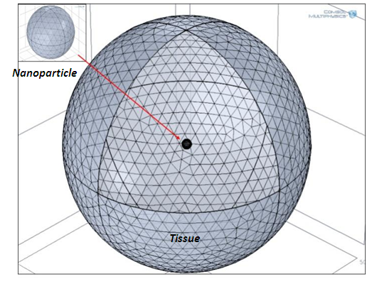 | Figure 1. The geometry discretization |
 | (1) |
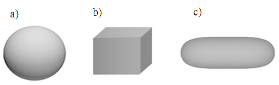 | Figure 2. The different shapes used in simulation: a) Sphere, b) Cube and c) Rod |
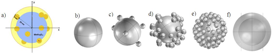 | Figure 3. Incomplete shell structure simulated in COMSOL: (a) the radius of naked magnetic core was chosen to be 20 nm, gold nanoparticles with 4 nm in radius was attached to the surface of the magnetic core (complete shell thickness 8 nm). (b) Magnetic core, (c) (d) (e) 10, 40, 80 gold nanoparticles attached on the surface respectively, f) complete shell |
|
3. Results and Discussion
- 3.1. The time evolution of temperature and thermal equilibrium of the tumoral cell determined by the heating of the MNP with different shapes is presented in the Figure 4.
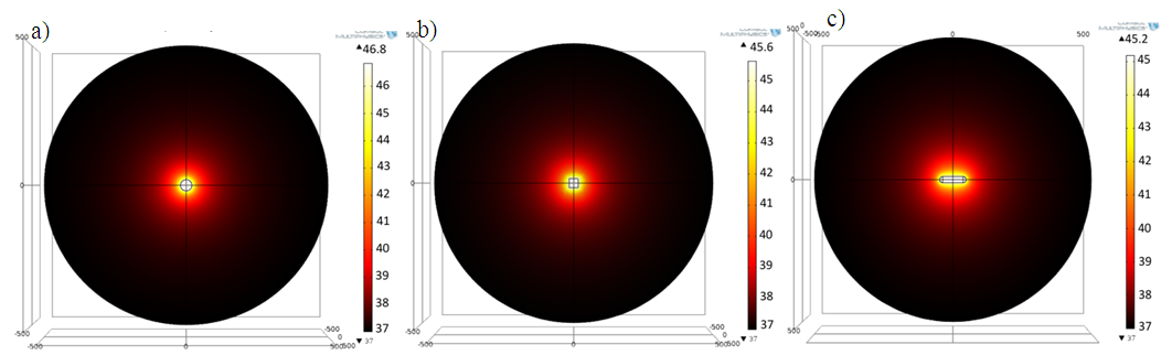 | Figure 4. The 2D spatial temperature distribution on z-y direction for 1μm tumoral cell of a) sphere, b) cube and c) rod, after 3μs from the beginning of heating process |
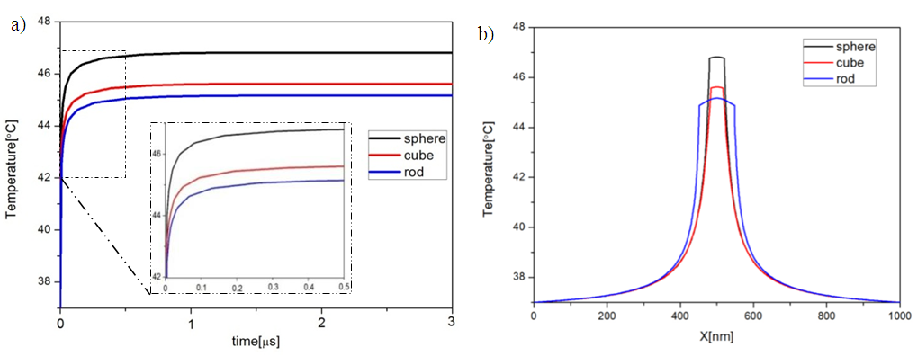 | Figure 5. The spatial and temporal evolution of the temperature for the three different NPs shapes (Sphere, Cube and Rod). (a) Temporal evolution in the point x=0 (center of the heat source); (b) the radial temperature distribution after 3μs from the beginning of heating process |
 | Figure 6. a) The maximum temperature achieved by following surface coating thickness (5, 10, 20, 30 and 40 nm): gold (black line) and polymer (red line). b) simulated core-shell structure |
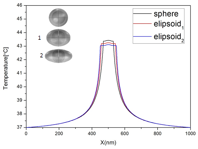 | Figure 7. a) The radial temperature distribution for different form of surface coating, sphere 20 nm in radius, elipsoid1: 25-25-43.2 nm, elipsoid2: 22-25-49 nm. Vsphere = Velipsoid1 = Velipsoid2 |
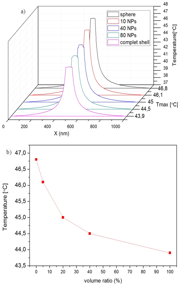 | Figure 8. a) The radial temperature distribution for different amount of surface coating after 3μs from the beginning of heating process. b) The temperature evolution relative with the volume ratio (%) |
4. Conclusions
- In our simulations was used a magnetic nanoparticle consisting of only one magnetic domain of MnFe2O4 ferrite since it offer high magnetization values that are important for hyperthermia applications. The study can be extended to different kind of magnetic materials and type of shells. In a simple way we have demonstrated that the temperature achieved by same volume of magnetic materials is maximum for a spherical particle comparing with cubic or rod shaped nanoparticles. Because was used the same volume and specific absorption rate particle the thermal field covers more central volume in the case of rod shape particles while, the thermal equilibrium is reached at the same time of 1µs from the beginning of the heating process for all the three simulated shapes.Thickness and shape uniformity was studied and we note that the temperature profile related on the rapport material/thickness of surface coating, the thermal properties of shell defined the aspect of temperature evolution (increase/decrease). Shell thickness uniformity enables some change in maximum temperature achieved by the magnetic core but still insignificant (less than 0.5°C).The uncompleted surface coating effect was studied, it was simulated the thermal response of different covering volume ratio of gold shell, and was shown that the open surface left through the shell growth process influence significantly on the temperature profile evolution.The use of nanoparticles which are composed of a magnetic core surrounded by a functionalized biocompatible surface shell, depend on the selection of the suitable materials (core-shell), surface coating thickness and surface uniformity.
ACKNOWLEDGMENTS
- Samir Taloub acknowledge the financial support of Constantine 1 University for his research stage at AlexandruIoanCuza University of Iasi.Ioan Dumitru acknowledge the support given by Romanian CNCS-UEFISCDI under project PN-II-RU-TE-2012-3-0449.